The Structure of the Bill and Hairs of Ornitho - rhynchus paradoxus; with a Discussion of the Homologies and Origin of Mammalian Hair.
|
|
|
- Mervyn Mosley
- 5 years ago
- Views:
Transcription
1 BILL AND HAIRS OF OTtNITHORHYNOHUS PARADOXUS. 143 The Structure of the Bill and Hairs of Ornitho - rhynchus paradoxus; with a Discussion of the Homologies and Origin of Mammalian Hair. By Edward B. Poulton, M.A., F.R.S., Ac, Hope Professor of Zoology in the University of Oxford. (With Plates 14, 15, and 15A.) CONTENTS. PAGE I. INTRODUCTION 144 II. THE PUSH-RODS 147 III. THE GLAND-DUCTS OP THE BILL AND THE STRUCTURES ASSOCIATED WITH THEM 152 IV. THE HAIES OP OLD AND YOUNG ORNITHORHYNCHUS 1. Historical, by W. Blaxland Benham, D.Sc. (Lond.), Hon. M.A. (Oxon.), Aldrichian Demonstrator in Comparative Anatomy in the University of Oxford General Structure and Arrangement of Hair.. 15S 3. Comparison between the Hairs of Old and Young Animal Minute Structure and Formation of Hair and its Sheaths 1G4 5. Mode of Succession of the Hairs Recapitulation of Essential Peculiarities of Hair of Ornithorhynchus as compared with that of Higher Mammalia 169 V. THE HOMOLOGIES AND ORIGIN OP MAMMALIAN HAIR 1. Historical, by W. Blaxland Benham Conclusions derived from the Study of Ornithorhynchus 180 YOL. 36, PART 2. NEW SEE. L

2 144 EDWARD B. POULTON. I. INTRODUCTION. IN the summer of 1884 I made a communication "On the Tactile Terminal Organs and other Structures in the Bill of Ornithorhynchus " to the meeting of the Physiological Society at Oxford. A short account, containing however the chief results, was published in the Proceedings of the Society ('The Journal of Physiology/ 1884, pp. 15, 16). In searching for the terminations of the abundant medullated nerves I found ' two remarkable structures in the epidermis of the bill: (1) Epithelial rods which appear to convey the effect of surface pressure to a group of nerve end organs resembling Pacinian bodies. These rods, from their obvious analogy to the push of an electric or, better still, pneumatic bell, may be conveniently called " push-rods." (2) Other epithelial rods along the axis of which passes the duct of a gland. These rods resemble in certain respects a shortened and truncated hair. The interpretation of these latter rods as modified hairs was disputed at the meeting by Dr. Klein, but supported by Professor Schafer. This controversy and the obvious interest of the inquiry led me to undertake further work, which included an investigation of the hairs covering the body of this animal. From time to time this work has been continued from 1884 up to the present date (December, 1893), and many drawings have been made and discussed with friends, although until now I have published nothing more upon the subject. During the summer of last year (1892) I worked for some weeks in the laboratory of my friend Professor Lankester, at which time the drawings were arranged and the description of PI. 14 written. Within the last few days the recent description of the pushrods by Professor J. T. Wilson and Mr. C. J. Martin (' Macleay Memorial Volume,' pp ) has reached me. I was prepared to find that their investigation would have rendered any further publication on this point unnecessary, inasmuch as fresh material had been available, the want of which had confronted me throughout, and especially in the attempt to
3 BILL AND HAIRS OF ORNITHORHYNCHUS PAEADOXUS. 145 trace the endings of the nerves. The authors have, however, relied almost entirely upon photomicrography for their illustrations, with the result that the figures upon two (xxiv and xxv) out of the three plates are utterly worthless, and convey a most inadequate conception of the appearances presented by the sections. It is evident from the descriptions that the sections relied upon were adequate, and perhaps the negatives and the first impressions were also satisfactory: but in the form in which the paper reaches the public there is something inconsistent in the confident references made in the text to the representation of detail in a figure which turns out on inspection to be the merest smudge (see especially fig. 5, pi. xxiv, which, among all figures ever published in scientific papers, must surely take the palm for obscurity). My sympathies are entirely with those who have been misled into trusting a process which for this purpose appears to be entirely useless. The authors have, however, made some drawings, the photographic representations of which are infinitely better than those of the sections. Fortunately, too, these figures, which are all to be found on the last plate (xxvi), deal with points of structure which needed the fresh tissues for their adequate treatment, and are therefore unrepresented among my illustrations. I therefore propose to publish my figures together with their explanation (in the "Description of Plates") exactly as I wrote them in 1892, except for mere verbal corrections, &c. Thus my description of the push-rods is entirely independent of that recently published in Australia, although it will be found that the two accounts are in substantial agreement. The gland-ducts and their associated hair-like and nervous structures are not alluded to by the above-mentioned authors. This is to be regretted, inasmuch as many of the most important points can only be decided by the use of the fresh tissues. A detailed description of these, so far as it is possible with the material at my disposal, is also given below, and the same is true of the hairs covering the body of the young and mature animal. The material upon which all the work described below has
4 146 EDWARD B. POULTON. been done consisted of (1) parts of the bill hardened in chromic acid by the late Professor Moseley in 1874, at the same time that he prepared the tongue which I have described in this journal (see * Quart. Journ. Micr. Sci.,' July, 1883, p. 453, for further details concerning this material), and the horny teeth (see ' Quart. Journ. Micr. Sci./ July, 1888, pp. 31, 32); (2) a specimen kept many years in spirit in the Oxford University Museum, the ovary of which was described in this journal (January, 1884, p. 118) ; other similarly preserved specimens in the Museum were also employed; (3) the young Ornithorhynchus of which I was enabled to describe the true teeth through the kindness of the late Professor W. K. Parker ('Quart. Journ. Micr. Sci./ July, 1888, pp. 10, 11). This latter specimen was 83 cm. long in the curled-up attitude in which I received it, and the larger hairs had alone appeared above the skin. Since the above was written I am enabled, through the kindness of Professor W. N. Parker, to supply figures of the natural size of the young specimen of Ornithorhynchus referred to above. The appearance from the left side is seen in Plate 15A, fig. 1, from the ventral aspect in fig. 2. The latter has been already published, reduced one half, by the late Professor W. K. Parker. The drawings from which PI. 15A was prepared were made by Mr. M. P. Parker. The considerable amount of work which has been already published upon material from the single specimen here figured is indicated in the following list of memoirs : G. B. HOWES. ' Journ. Anat. and Physiol./ 1886, xxi, p Describes and figures the shoulder girdle. G. B. HOWES. ' Journ. Anat. and Physiol.,' 1892, xxvii, p Describes the pelvis, and figures a section of it. The late W. K. PARKER. "Mammalian Descent," London, Describes some of the external characters (p. 48, et seq.), and figures the ventral view of the specimen one half the natural size (p. 25). This same view is represented the fnll size in PI, 15A, fig. 2, accompanying this
5 BILL AND HAIRS OF ORNITHORHYNOHUS PARADOXUS. 147 W. N. PAEKEK. 'Brit. Association Reports,' 1891, p Compares the snout with that of the young Echidna. W. N. PAKKEH. "On some Points in the Structure of the Young of Echidna aculeata," ' Proc. Zool. Soc. Loud./ read January 16th, 1894, and to be published shortly. Compares with young Echidna, especially as regards " Jacobson's organ," a section of which is figured. E. B. POULTON. ' Proc. Roy. Soc,,' 1888, xliii, p Describes the true teeth, and figures a section of one. E. B. POULTON. Quart. Journ. Micr. Sci,,' 1888, vol. xxix, p. 9. Describes the true teeth, and figures their appearance, in sections, and in minute structure. R. WIEDBBSHEIM. ' Zeit. f. wiss. Zool., 1 liii, 1892, suppl., p. 43. Describes and figures the pelvis. This paper having been prepared for the press at a time of great pressure, my friend Dr. Benham has very kindly relieved me of the labour of writing the historical part of the two latter sections. I also wish to thank my friend Professor Howes for kindly suggesting certain lines of recent research which bore upon the subject. So far as the earlier sections are concerned, with the exception of Wilson's and Martin's recent paper there is no other record worthy of mention. II. THE PUSH-RODS. It will not be necessary to describe the histological details of these structures at any great length. Reference to PI. 14 and to the full descriptions will be sufficient. I shall therefore chiefly dwell upon the points which are not touched by Wilson and Martin, or in which my interpretation differs from theirs. The proportion of these structures to the gland-ducts varies in different parts. Thus on the presumably highly sensitive ridges within the lower jaw the former are the more numerous, as may be seen by glancing at fig. 5, where the pushrods are seen in section as circles. On the external surface of the bill, however, these proportions are usually reversed. In relation to the sensitive condition of the bill we learn with great interest that it is, in the living state, covered with "smooth, soft, and humid skin" ("Anatomy of the
6 148 EDWAED B. POULTON. Muzzle of the Ornithorhynchus," by Professors J. T. Wilson and C. J. Martin; 'Macleay Memorial Volume,' p. 180). In this country, where we see only dry or spirit-preserved specimens, it has generally been looked upon as a tough, horny structure. The glands to be described below are doubless responsible fox the humidity, although they may also, as I have suggested ( f The Journal of Physiology/ 1884, p. 16), secrete a substance which protects the skin from the prolonged action of water. Wilson and Martin describe structures in the horny teeth which they believe to represent push-rods which have undergone cornification (1. c, p. 192). In offering this interpretation I believe that they have mistaken for push-rods the columns of cells which extend from the summits of the numerous papillae towards, and often as far as, the surface. These cells are much less cornified than those around, and stain readily; furthermore it may be said that the columns " show a series of imbricated superposed cells," although the arrangement differs in various details from that of the push-rods. The fact that a column invariably extends from the summit of a papilla, while a push-rod is invariably surrounded by two or more papillae, is sufficient proof that the two structures are not homologous. The structure of the cell-columns and their relation to the papillae are clearly shown in my paper already quoted (' Quart. Journ. Mic. Sci.,' July, 1888, PI. IV, figs. 4, 5, 8, 10, 11). Speaking of the dermal sheath which surrounds the lower part of each push-rod, the above-quoted authors regard the rod as an epithelial downgrowth which causes " a depression in the summit of a dermal papilla" (1. c, p. 193). Inasmuch as two or more distinct papillae arise from the upper edge of the dermal sheath (to be seen in section in PI. 14, fig. 3, accompanying this paper; and, by the eye of faith, in pi. xxiv, fig. 6, of the ' Macleay Memorial Volume '), I should prefer to regard the rod as a modified interpapillary process, with the surrounding papillae united into a continuous sheath below, while they remain free above.
7 BILL AND HAIKS OF OENITHORHYNOHUS PAEADOXUS. 149 The shaft of a push-rod consists of four concentric layers of cells, which are rendered quite distinct by their unequal staining (figs. 3 and 4). Wilson and Martin regard the outermost of these layers as belonging to the general epidermis, and forming " a kind of follicle" round the rod proper. I took the same view in 1884, speaking of the third layer as " followed by concentrically arranged fusiform cells belonging to the general epidermis" (1. c, p. 16). The innermost imbricated cells, constituting the first layer, are looked upon by Wilson and Martin as hollow truncated cones made up of three or more cells. This interpretation is certainly supported by the teased preparation represented in their pi. xxvi, fig. 17. I could never make out any cell outlines in this layer when studied in transverse sections of the rods, although three or more nuclei were often seen (fig. 3). Longitudinal sections, on the other hand, show the outlines of successive cells very distinctly (fig. 4). My observations to this extent support Wilson's and Martin's conclusions as to the nature of the innermost cell-structures. As to the second layer of imbricated cells, and the larger far less sloping cells constituting the'third layer, we are in entire agreement, and my fig. 4 would, in this respect, serve for the illustration of Wilson's and Martin's description. It should be noted, however, that the nuclei of the cells of all the layers are surrounded by pigment masses and granules. The central group of fine filaments occupying the axis of the rod, together with the ringof similar structures placed between the first and second layers of imbricated cells, are very conspicuous, and early attracted my attention (1884,1. c, p. 16). They are clearly seen in figs. 3 and 4. Longitudinal sections show that they pursue a parallel course along the rod except at the convex lower extremity, where I could just make out, in the most favorable preparations, that the filaments of the central group diverge in a brush-like manner (fig. 1). The most careful examination, under very high powers, of the best material I could command, displayed a structure which is represented in fig. 4. Each filament appeared to be
8 150 EDWARD B. POULTON. made up of short sections corresponding in length to the thickness of the adjacent jmbricated cells. From this structure I formed the conclusion that the filaments are probably built up of units contributed by the cells which lie outside them. Wilson and Martin describe them as made up of bead-like varicosities, each of which is placed between a pair of cells (simple or compound) in vertical succession (1. c, pi. xxvi, fig. 15, for a diagrammatic representation). At the meeting of the Physiological Society (1884) I suggested that the filaments might be nerve terminal organs, and that the imbricated arrangement of cells around them might result in pressure whenever the free surface of the rod was in contact with external objects, thus effecting the stimulation of the nerve. Professor Schafer pointed out, in the discussion, that the highly refringent character of the filaments is opposed to the view that they are terminal jierve-fibrils. I have often tried to find a connection with the abundant nerves below the base of the push-rod, but such an investigation needs the fresh tissues. Wilson and Martin now describe the filaments as naked axis-cylinders prolonged from the nerve-fibres below the rod, which suddenly lose their medullary sheaths soon after entering the epithelium. Although their drawings from gold-stained preparations (pi. xxvi, fig. 21) appear to leave no doubt that the filaments are connected with nerves and form some kind of terminal organ, it is obviously erroneous to speak of them as " naked axis-cylinders" (p. 196) or "nerve-fibrils" (p. 197). They are highly refringent and have none of the characters of these nerve structures. It is noteworthy, too, that although the filaments are represented in pi. xxvi, fig. 22, as black varicose threads, the authors accurately state in the description (p. 200) that the fibrils " are not black, but only highly refracting.'' Furthermore, if the photomicrographs establish nothing else, they certainly prove that this conclusion as to the nature of the filaments is erroneous; neither naked axis-cylinders nor nerve-fibrils could have caused the appearances seen in transverse section in pi. xxv, fig. 13, and in longitudinal section in fig. 8. These authors state (p. 196) that I failed to recognise the
9 BILL AND HAIfiS OF ORNITHORHYNOHUS PABADOXUS. 151 nervous character and connections of the filaments. I am in entire agreement with this statement: " the nervous character" does not exist, and the nervous connections could not by any possibility have been seen in the tissues with which I was supplied. Although I did not recognise connections which could not have been seen, I certainly inferred them, as the title of my communication indicates (" On the Tactile Terminal Organs," &c, ' Journ. of Physiology,' 1884). The filaments must be regarded as remarkable terminal organs, entirely distinct in histological nature from the axiscylinders which terminate in them ; and Wilson and Martin's fig. 25 seems to suggest that the change in nature takes place at or close to the point at which the medullary sheath disappears. The appearance presented by the filaments is very clearly shown in figs. 3 and 4 accompanying this paper. They must be looked upon as a new and interesting form of nerve terminal organ, probably epithelial in origin. As regards the Pacinian-like bodies, Wilson and Martin support my previous account, but they also describe certain larger forms of these structures, rather more deeply placed than those immediately below the push-rods. Bodies similar to those beneath the rods occur in the mouth, and were described and.figured in some detail in my paper on " The Tongue of Ornithorhynchus" in this Journal ('Quart. Journ. Micr. Sci./ July, 1883, PI. XXXII, fig. 5). The constant occurrence of a group of Pacinian-like bodies at the base of each push-rod (figs. 5 and 6) is of great physiological interest, as it strongly supports the view widely held but as yet unproved that the function of this form of nerve end-organ is to aid the nervous system in the appreciation of pressure. The obvious use of the push-rods is, as I stated in 1884, " to supply specially moveable areas yielding to surface pressure, which is thus communicated to the terminal organs below." Another interesting end-organ described by these authors is placed among the epidermic cells of the base of the rods. In these "lenticular bodies" the axis-cylinder is described as
10 152 EDWARD B. POULTON. ending in a disc which separates " two clear vesicular cell-like structures." This form of ending evidently needs the fresh tissues for its demonstration. At first sight the push-rods appear to be modified hairs, but the examination of their minute histological details does not support this comparison. It is possible, however, that they may be found to have some bearing upon the recent suggestion that the Mammalian hair corresponds to an epidermic tactile organ of the lower Vertebrata. However this may be, the resemblance to the Mammalian hair as it now is, in my opinion, is far less close than that of the epidermic structures associated with the ducts of glands which open on the surface of the bill. Souza Fontes, in 1879 {' Beit. z. Anat. Kenntniss der Hautdecke des Ornithorhynchus/ Inaug. Dissert., Bonn), mentioned and figured these structures and the gland-ducts described below, but the paper is quite unworthy of mention. Indeed, the principal feeling evoked by a glance at the Plate is one of surprise at the system which can confer a University Degree for such a production. III. THE GLAND-DUCTS OF THE BILL AND STRUCTURES ASSOCIATED WITH THEM. The gland-tubes of the bill, and, indeed, of the general body surface, closely resemble the Mammalian sweat-glands, the secretory part of the tubule being wider than the duct, and lined with short columnar cells surrounded by a longitudinal layer of smooth muscle-cells (fig. 8,gld.). The wall of the duct is composed of nucleated, probably polyhedral, cells, indistinctly marked off from one another in my sections. These cells are separated from the lumen by a cuticle, represented as a row of thin, deeply staining, plate-like structures resembling nuclei (fig. 8, d', transverse section; below d longitudinal section). Externally the tubes are surrounded by a membrana propria, in which nuclei are especially distinct in the transverse section of the duct (d'). The existence of such typical structures in the most primitive mammal indicates that sweat-glands are among the most
11 BILL AND HAIRS OP OJ&NITHORHYNOHUS PARADOXUS. 153 ancestral features of the Class. Although the hairs of Ornithorhynchus will be shown below to present many peculiarities which are, as I believe, ancestral, the sweat-glands are essentially similar to those of Mammalia generally. In the bill, the deeply placed coiled gland-tube is succeeded by a coiled duct which, as in many other mammals, enters the base of an epidermic downgrowth the interpapillary process (fig. 8). The epidermic process itself, however, is by no means typical, but presents many special peculiarities, some of which support the conclusion that it is a modified hair, sharply cut off above at the level of the uppermost epidermic layer, shortened below by retraction of the hair-bulb, so that the latter descends but a short distance beneath the lowest layer of the epidermis (figs. 8, 13 16). Nevertheless, in the young Ornithorhynchus the bulb-like part of the structure extends to a somewhat deeper level (compare fig. 16). These hair-like structures were briefly described in 1884 (1. c, p. 16, where, however, in line 11 from bottom, the words "hair papilla" are obviously intended for "hair-bulb"). The epidermic processes are, like the push-rods, surrounded below by a continuous dermal sheath, the upper edge of which gives rise to several papillary upgrowths (figs. 8, for longitudinal, figs for transverse, section). The process is continued upwards through the stratum corneum as a cylinder either straight or with s-like curves, which remains perfectly distinct from the epidermis around, being separated by a downgrowth of cells so marked that their direction becomes vertical. In this respect the structures in question resemble the push-rods (compare fig. 1 with fig. 8). At its upper free end this cylinder is sharply truncated so as to be flush with the surface of the bill. But in favorable examples it is surrounded by a distinct circular depression, and it may even project a little above the general surface (figs. 8, 13, and 15). At the posterior part of the upper bill the upper ends of the cylinders are remarkably expanded, so that their outline becomes funnel-shaped (fig. 14). The S-like curves into which many of the cylinders are thrown
12 154 BDWABD B. POULTON. in their, passage to the surface are exceedingly characteristic in appearance (figs. 13 and 15). The cylinder itself is hair-like in structure, being composed of elongated fusiform cornified cells in which traces of a nucleus, surrounded by pigment, can be detected (fig. 8). The lumen of the duct traverses the axis of the corneous cylinder, and is star-like in transverse section (figs. 9 and 10). The cells near the cylinder are disposed in concentric circles round it (fig. 9). Tracing the cylinder downwards into the interpapillary process, we find many points of resemblance to a hair. The general epidermis is continued over it as a sheath, which strongly suggests the outer root-sheath of a hair, and between it and the cylinder itself a line of separation tends to appear (fig. 8, o.r.s., and also at level c; see also fig. 10). Below, this sheath forms the outer part of the bulb, and is separated from the inner part by a space containing small branched cells, the nature of which could not be determined in my material (fig. 8, sp., and fig. 13). The cells of this outer sheath are richly pigmented, like, or even more than, those of the lower layer of the general epidermis with which they are continuous (figs. 8 and 10). Within this sheath the cylinder is surrounded by a distinct layer composed of flattened cells, shown by transverse sections to be two or more deep, but varying in thickness in different parts (fig. 10, fig. 11, c. j compare also fig. 8, c). This layer may represent the inner root-sheath, or the cuticle of the hair, or both of these together. It is continued over the inner part of the bulb, and separates the latter from the space described above (fig. 8, sp.). Within this layer the cylinder consists, as described above, of fusiform corneous cells arranged longitudinally; below, these elements pass into polyhedral cells, staining in carmine, &c. The inner part of the bulb is made up of these latter, and presents the strongest possible resemblance to the bulb of a hair (figs. 8 and 11). In certain slender cylinders which occur intermixed with the others, this part of the bulb appears to be wanting (fig. 15). In the upper part of the bulb, and for a variable distance above it, the duct is lined by a single layer of distinct cubical
13 BILL AND HAIRS OF ORNITHORHYNCHTJS PABADOXUS. 155 cells (fig. 8), and it coils in a very characteristic manner in the neck and upper part of the bulb (fig. 13; in fig. 8 the coiling is only slightly marked; figs. 14 and 15 represent peculiar forms in which this character is not present). The coiled duct passes downwards into a straight section resembling the gland duct in the dermis with which it is continuous below the bulb (fig. 8). It was, however, impossible to establish the existence of a cuticle to the duct in the bulb, although the inner part of the cells immediately round the lumen stained far more deeply than the outer part (fig. 11, d'.). Immediately below the whole bulb the duct is invariably surrounded by a large ganglion containing abundant medullated nerve-fibres showing Ranvier's nodes, and large ganglioncells (figs. 8 and 12). This ganglion is surrounded by a fibrous sheath which appears to be prolonged from the dermal sheath (corresponding to the hair-sac) of the hair-like cylinder. Large nerves are seen entering the ganglion, the sheath of which is continuous with their epineurium (fig. 8). The duct which pierces the ganglion is separated from it by a distinct fibrous sheath, clearly shown in transverse section (fig. 12) but very thin in the longitudinal (fig. 8). This sheath appears also to be derived from the layer corresponding to the hair-sac, and to be continuous with its inner part, while the ganglionic sheath is continued from its outer (fig. 8). This, at least, appears to be the probable interpretation of the appearances represented in fig. 8; but the whole structure of these hair-like epidermic cylinders, and the nervous tissues evidently associated with them, is so remarkable and complex that the fresh tissues are required for their satisfactory elucidation and for the discovery of the nerve terminations which we must believe to exist in connection with the apparatus. I believe that this account represents all that can be ascertained by the careful examination of the available material, and that it supports the conclusion I suggested in 1884 ^that the gland ducts of the bill reach the surface by entering the bulbs and by advancing along the medulla of shortened and degenerate hairs. Such an opinion is further confirmed by the
14 156 EWARD B. POULTON. fact which will be established below that the corresponding glands of the general body surface of this animal bear a constant relationship to the larger hairs. IV. THE HAIRS OF OLD AND YOUNG ORNITHORHYNCHUS. 1. Historical, by W. Blaxland Benham, D.Sc. (Lond.), Hon. M.A.. (Oxon.), Aldrichian Demonstrator in Comparative Anatomy in the University of Oxford. The fact that Ornithorhynchus possesses two kinds of hair, larger and smaller, was known to Blumenbach (1) and to Home (2). The latter also recognised the peculiar character of the larger hairs. He writes (p. 69), " The hair is made up of two kinds : a very fine thick fur half of an inch long, and a very uncommon kind of hair three-quarters of an inch long; the portion next to the root has the common appearance, but for a quarter of an inch towards the point it becomes flat, giving it some faint resemblance to very fine feathers." Later, Glockner (3), in a very brief note describes the larger hair in somewhat similar terms. In 1823 van de Hoeven (4) reproduced Peron's (5) figures of these hairs; the figures are very small, but are the first published, and show the characteristic flattening, the narrow stalk, and pointed free end. In his monograph, Meckel (6) makes no addition to our knowledge of these structures. In 1859 Leydig (7), in his classic paper on the Mammalian coat, is the first to record the fact that the smaller hairs are in bundles several in each follicular neck and points out that each hair has nevertheless its own special follicle opening into the bottom of the common pit. He also states that the large spiny hair is surrounded by the bundles of small ones, and he gives a figure (pi. xx, fig. 7) of the arrangement, and shows the sebaceous glands to each hair and the sweat gland accompanying the large one. These facts are confirmed by Welcker (8), a few years later, who gives measurements of the hairs, the smaller being -007 mm. in diameter, the larger ones *045 mm..across. He too
15 BILL AND HAIBS OF ORNITHORHYNCHUS PARADOXTJS. 157 gives a figure (pi. 1, fig. 5) showing the grouping of the hairs, and points out the fact that the roots of the small hairs diverge, that the hairs then converge in the common follicular neck, whence they issue in a bundle (p. 69). Whilst Leydig describes only four or five hairs in a bundle, Welcker found fifteen to thirty of them. No author gives any histological details of the structure of the hair till Waldeyer (9) in 1884, who only describes and illustrates by photographs the difference in the character of the medulla and cortex in the various parts of the larger hair (pi. vii, figs ). According to him (p. 100), the "bristle hairs " commence basally in a rounded shaft of moderate diameter with a thick medulla; then follows a more flattened but very slender "isthmus," which further on widens out considerably to form the main part of the hair; this narrows again at the tip to a point. The isthmus is noumedullated, and the medulla of the broad part differs from that in the basal shaft. The paper dealing with the skin of Ornithorhynchus, by Souza Fontes (10), consists of little but quotations from Leydig. It contains nothing worth recording. These authors give no information as to the bulb, papilla, and root-sheaths, nor any other histological details of the hair itself. As to the arrangement of the hair, Meijere (11), who has recently examined the arrangement of the hair throughout the Mammalia, gives a figure on p. 49 showing the hairs on the back of Ornithorhynchus in more or less irregular rows the large hairs, represented as round in section, being separated from each other by three or four bundles of small hairs: on the ventral surface, however, the number of the latter is greater. His measurements closely agree with those given by Welcker, being -048 mm. diameter for the large ones, 008 mm. for the small hairs. 1. BLVMENBAOH. "Anatomical Observations on the Structure of Ornithorliynchus paradoxus," 'Philosoph. Mag.,' xi, 1802, p HOME. "A Description of the Anatomy of Ornithorhynchus paradoxus," 'Phil. Trans.,' 1802, p GIOCKNEE. " Ueber die Haare des Ornithorhynchus," 'Isis,'!819, p. 651.
16 158 EDWARD B. POULTON. 4. VAN DE HCEVEN. "Memoire sur le genre Ornithorhynque," 'Nova Aota'(Bonn), PERON and LESUEUR. ' Voyage de decouvertes aux Terres australes pendant les annexes ' 6. MECKEL. ' Ornithorhynchi paradoxi descriptio anatomica,' LEYDIG. " Ueber die ausseren Bedeckungen der Saugethiere," ' Arch. f. Mikr. Anat.,' WELCK.EE. " Ueber die Entwickelung und der Bau der Haut und der Haare bei Bradypus," ' Abliand. Naturf. Gesell. zu Halle,' ix, 18G6. 9. WALDEYEE. ' Atlas den Menschlichen und Tierischen Eaare,' SOTJZA FONTES. 'Beit. z. Anat. Kenntniss der Hautdecke des Oniithorhynchus,' Dissert. Inaug., Bonn, MEIJEEE. 'Over de Haren der Zoogdiere,'Inaug. Dissert., Amsterdam, General Structure and Arrangement of the Hair. The hair-like appearance of the epidermic cylinders in the bill led me to investigate the hairs of the body generally, and although the results seem to have no special bearing on the significance of the former structures they are of much value and interest on their own account. This research was undertaken in the winter of , when all the figures (except fig. 24) represented on PI. 15 were drawn. Since then it has been renewed from time to time, special attention having been devoted to the subject in Professor Lankester's laboratory dnring the summer of Over the general surface of the body and head of Ornithorhynchus we meet with large hairs, each of which is attended by bundles of small hairs. Furthermore, on the dorsal area at least, each large hair is attended by a constant number of four bundles of small ones. The latter commonly vary in number in the dorsal region from seven to twelve in a bundle, and all emerge in a sheaf from a common follicular mouth. The four bundles are arranged on each side of and behind the larger hair, and, as all slope obliquely backwards, the shafts of the small hairs lie beneath that of the large one belonging to the same group. The protective large hairs are evidently subject to much wear and tear, and succeed each other very rapidly; the new
17 ftfll AND HAtBS OJ? OBNITHOEHYNOHUS PAEADOXUS. 159 successional hair, which is to be met with iu nearly every section, emerging from the same follicular mouth in front of, and therefore overlapping, the base of the old one. The succession of smaller hairs is less rapid, but one or more younger, growing hairs are to be seen in every bundle. The exact relationship in two instances can be made out by ascertaining the proportion of dark circles (sections of young hairs) to the smaller unpigmented circles (sections of bases of old hairs) in each of the four bundles in figs. 17 and 18 of PI. 15, which contains all the figures illustrating the structure of the hair of Ornithorhynchus. The proportions in the four bundles of three other groups were as follows: Bundle I.» II. III. IV. Bundle I. II. III. IV. FlKST GEOUP young hair to 9 or 10 old. hairs to 8 hair to 7 hairs to 5 SECOND GROUP.. 1 young hair to 8 old.. 2 hairs to 8. 1 hair to 6. '6 hairs to 7 Bundle I. II. III. IV. THIRD GROUP.. 2 young hairs to 7 old.. 1 hair to hairs to 10.. The proportion of young hairs was ascertained in each case by counting the numbers of dark and light circles in a section taken sufficiently near the surface of the skin to bring all the hairs of a bundle into p a common follicular neck, as in fig. 17. These figures, together with the number shown in fig. 17, prove that, at any rate iu the dorsal region, Leydig's estimate of the number of small hairs in a bundle is too small, while that of Welcker is far too large. Each group of hairs is attended by a single gland exactly VOL. 36, PART 2. NEW SKE. M
18 160 EDWARD B. POULTON. resembling those of the bill of the same animal or the sweatglands of mammals generally. On the head and back, the duct invariably opens just in front of the large hair, so that it is the most anterior member of the whole group. Such a group is distinctly represented in transverse section immediately beneath the skin in fig. 17, where the young succession al hairs, both large and small, are at once distinguished by the presence of pigment from the colourless bases of the shafts of older hairs. At this level the gland-duct (d.) is seen in section in front of the large hair. At a lower level, as shown in fig. 18, the duct is replaced by a secretory tube of typical structure (g.), which is often placed, as in this figure, between the large hair and one of the bundles of smaller hairs. The latter, at this level, have become separate, but their outer root-sheaths are continuous peripherally, forming a single epithelial mass. Furthermore, the two masses on each side have approached and tend to coalesce (compare figs. 17 and 18), soon doing so completely. At this level, and just below it, the small unpigmented bases of the older hairs come to an end (one is thus ending in each of the right-hand bundles in fig. 18). The younger growing shafts, with their outer rootsheaths no longer continuous, descend much deeper, gradually converging to form a single bundle, which lies in the middle line under the follicle of the large hair. The growing small hairs then end in bulbs at various levels, but the most deeply placed do not, as a rule, descend so far as the bulb of the large hair. The large hairs terminate superiorly in large, although narrow, flattened leaf-shaped expansions borne by a shaft long enough to carry them just beyond the ends of the small hairs. As the neck of this shaft is comparatively thin, it is probable that in the living state the terminal shields tend to fall over, and lie flat on the finer hair beneath, their tips pointing backwards and overlapping the basal part of the shields behind them. The tip of the shield is beautifully formed and free from pigment. Looked at from the side or in section, the pigment
19 BILL AND HAIRS OP ORNITHORHYNOHUS PABADOXUS. 161 is seen to be more developed on the under side, and a broad unpigmented margin is always found along the upper surface. This is in part due to the cuticle of the upper surface, which is much thicker than that of the under, as can be well seen in sections (fig. 18). The degree to which the pigment extends towards the upper surface differs, however, in individual hairs. Passing along the centre of the shield is a very distinct medulla containing abundant gas-bubbles, entering as a result of the contraction of soft protoplasmic cells, which are present and stain in logwood, &c, in the shields which have not yet appeared above the surface (fig. 18). Traces of this medulla can be followed nearly to the tip of the structure. The pigment is sometimes so far restricted to the under side of the shield as to be entirely beneath the level of the medulla in the middle line, but it extends further at the sides (fig. 18). Below the shield there is a constricted neck in which the medulla is apparently wanting and the pigment scanty, but immediately below, at the beginning of the long shaft, the medulla is strongly developed suddenly and the pigment assumes the characteristic ladder-like appearance, being arranged in bands alternating with spaces containing colourless shrunken cells and gas bubbles. The arrangement is not, however, so regular as that of the corresponding part of the smaller hairs. Below, the shaft passes into the somewhat more slender unpigmented base in which a medulla is wanting. Hence in one of the larger hairs we can distinguish a shield, neck, shaft, and base. The small hairs can be divided into similar regions. The free tip is unpigmented, and is followed by a section corresponding to the shield, with the same longitudinal arrangement of small granules and large fusiform masses of pigment. Traces of a medulla marked by gas bubbles are to be seen in places. Then follows the part which represents the neck, not, indeed, more slender, but with the same diminution in the amount of pigment and cessation of the medulla. To this succeeds the shaft with an extremely uniform alternation of medullary dark and light bands, the latter containing distinct
20 162 EDWARD B. POULTON. gas bubbles. As in the larger hairs, the shaft passes into a pigmentless and, in this case, far more slender base (fig. 18, in which the sections of the colourless bases of small hairs are seen to be much smaller than the dark circles which represent the sections of shafts and upper parts). There is no trace of either pigment or medulla in the bases of both larger and smaller hairs (see figs. 17 and 18 for sections of bases). Such is the appearance and arrangement of the groups of large and small hairs which apparently cover the whole surface of the body and head. On the upper surface of the tail the large hairs become stiff and bristly, but still flattened. They probably correspond to the shields only of the large hairs already described. Passing from the back on to the tail the small hairs become stfort and scanty, and towards the tip disappear altogether. The successional hair emerges as on the body, and the overlap is the same, but the gland duct opens beneath and behind instead of in front of the hair. The under surface of the tail in a male individual not quite fully grown was covered with short flattened large hairs set very obliquely, in fact almost horizontally. They appear to want the medulla and the hair pigment. In several full-grown individuals this part of the tail was more or less bare, but the bases of large hairs could be detected, together with patches of hairs having frayed and worn ends. The manus and pes, except for the bare palmar and plantar surfaces, are covered with hairs very similar to those last described, but even shorter. Everywhere the groups of large and small hairs, or the large hairs alone, appear to be set in irregular rows, transverse to the long axis of the body which they clothe. 3. Comparison between the Hairs of Old and Young Animal. Very interesting results follow from the comparison of the hairs of the mature animal with those of a young one in which
21 BILL AND HAIRS OF ORNITHORHYNCHUS PARADOXUS. 163 only the tips of the larger hairs had appeared above the surface, while the smaller ones were still at some distance below it (see PI. 15A). The parts of the large hairs which had been formed evidently corresponded to the terminal shields of the adult, from which they differed in their greater thickness, although the differentiation of an upper surface with a greatly thickened cuticle from a lower surface in which the pigment was concentrated, was equally marked (figs. 19, 20, and 23). The gland, which was quite short in the young animal, opened in front of the large hair, as in the adult. The small hairs differ from those of the adult and in such a manner as to indicate the probable origin of the four bundles emerging from common mouths. When longitudinal sections or successive transverse sections of the young skin are examined, each large hair is found to be accompanied by four tubes, exactly like the ducts of glands,opening on the surface. These tubes correspond in position to the four bundles of small hairs in the adult. Tracing them down wards, each tube gives rise to a bundle containing a much smaller number of hairs than in the adult.' Each bundle nearly always contains a single hair which is specially prominent, and it is this latter which occupies the lumen of the tube itself, the apex being however, in animals of this stage of growth, a considerable distance below the surface of the skin. In some cases, however, the four bundles of a group appear to contain only three chief hairs between them, in others as many as five or six. Four tubes appear to be always present, evidently representing the four common follicular necks and mouths of the adult. There is great disparity in size between the chief hair in a bundle and the smaller ones which are grouped around it. The hairs of the four sheaves terminate in bulbs, placed, as in the adult, at very different depths below the surface. From this structure it may be inferred that each of the four bundles is, in the course of development, at first represented by a single hair (the chief one), formed in a follicular
22 164 EDWAED B. POULTON. tube which is open to the surface, and that the smaller hairs are developed in follicles which are outgrowths from some part of this open tube, and which do not tend to coalesce as in the mature animal. The distinction between chief and smaller hairs is subsequently lost, probably in the hairs which succeed those described above. In an animal of this age, however, the first-formed hairs have not approached their full size, and no trace of any successional hair either large or small is to be seen. The existence of a distinct lumen opening on the surface at some distance above the apex of the hair is of such great morphological importance and interest in relation to the origin of hairs and their homology with feathers and scales, that the observation was confirmed again and again, by examining both longitudinal and transverse sections, until there could be no doubt about the matter. I was then anxious to ascertain whether the same fact held true with the larger hairs. The general surface of the body was valueless for this inquiry, as the animal was rather too old, and the tips of the large hairs were visible. On the under side of the tail, however, many, at any rate, of the large hairs (here unaccompanied by small ones) had not reached the surface, and there was an open tube above them, as in the case of the small hairs elsewhere. The probable bearing of this and many other peculiarities will be discussed at the end of the paper. The hairs upon the tail bear the same relation to those on the body as that already described in the adult. 4. Minute Structure and Formation of Hair and its Sheaths. The hair is developed from a bulb composed, as in other mammals, of polyhedral cells of the rete mucosum connected with the superficial epidermis by an outer root-sheath. Protrusions of the latter give rise to apparently typical sebaceous glands, situated, in the smaller hairs, at the level at which the bundle unites to enter a common follicular neck. The upper
23 BILL AND HAIRS OF ORNITHORHYNCHUS PARADOXUS. 165 part of the outer root-sheath often contains cystic growths like those described in other mammals. The bulb is entered by a dermal papilla which is, at any rate in the large hairs of the young animal, of enormous size and length, extending beyond the bulb far into the base of the hair proper, where the cells are fusiform, pigment abundant, and the cuticle well denned (see fig. 23, in which, however, the two halves have fallen apart, so that the hair appears to be thicker than the bulb). The bulbs and papillae are especially large in the tail. A very long, well-marked papilla also penetrates the long narrow bulbs of the smaller hairs. The relations of the adult bulb and papilla appeared to be very similar, but were made out with greater difficulty than in the young animal, which was chiefly employed for the histological side of this inquiry. From the tip of the papilla, at any rate in the larger hairs, an axial rod of soft protoplasmic cells, deeply staining in reagents, is continued (fig. 18, mature; figs. 19 and 24, young). This, when dried and shrivelled, admits the air and forms the characteristic medulla. Around the papilla the inner zone of cells of the bulb forms the hair proper with its cuticle, the structure and mode of formation being typical except for the bilateral symmetry of the larger hairs indicated by their shape, thickened upper cuticle, and predominant pigment on the under surface (fig. 18, mature; figs. 19, 20, and 23, young). External to this zone the cells form the inner root-sheath, while the outer root-sheath appears to be continuous with the lower part of the bulb. The latter sheath is probably always typical, but it was extremely hard to make out in certain sections of the young animal (figs. 19, 20, 23), although in others it was perfectly distinct and of normal appearance (figs. 21, 22, 24). This discrepancy is due to the facts that the thickness of the sheath varies greatly at different levels, and that the animal was not prepared for histological investigation. The inner root-sheath is always present in the developing hair, and is a structure of great importance, throwing much
24 166 EDWABJD B. POULTON. light upon the corresponding sheath as it is described in other mammals. As in the latter, the inner root-sheath surrounds that part of the hair which is enclosed in the follicle, but growing less rapidly it does not extend to the neck through which the hair protrudes; hence we do not find it at all in sections of the upper part of the follicle (figs. 17 and 18). It is far thicker and more important in the hairs of the young animal, and especially in those of the tail (figs. 21 and 22, i. r.s.). In structure it consists of a network of corneous fibres enclosing fusiform meshes with a longitudinal direction, through which the outer root-sheath can be seen (fig. 24). Hence in certain cases Henle's original description of his inner root-sheath as a fenestrated membrane is certainly true of Ornithorhynchus. Henle's account is also followed by Mertsching (' Archiv f. Mikr. Anat.,' 1888, p. 32), who shows in pi. v, fig. 8, that this sheath in the human hair possesses a fenestrated structure. In transverse section the corneous fibres of Ornithorhynchus are seen to be polyhedral and irregular in outline (figs. 19, 21, 22, i. r. s.), and if the section be taken at some little distance above the bulb, distinct spaces appear between them (figs. 19 and 21). Round the small hairs, on the other hand, only a uniform layer of the proportions usually found in Mammalia could be detected (fig. 19). In some of the longitudinal sections of larger hairs I could make out a thin internal layer exhibiting a serrated edge, with teeth the reverse of those on the hair cuticle. This evidently represents the so-called cuticle of the inner rootsheath, and it is probably shown in section in fig. 21. Well above the bulb (fig. 21) this thin internal lamina was the only trace of a separation of the sheath into layers, and even this could be detected only occasionally; and I gained the impression that it is not a distinct and definite layer, but merely the condensation, as it were, of the innermost part of the inner root-sheath upon the exterior of the hair and the moulding of its surface by contact with the cuticle of the latter. But at a lower level, just above the bulb, there is seen what I believe to be the homologue of Huxley's and Henle's layers. Thus in fig. 22 the fibres of the inner zone have not become corneous and take the stain readily,
25 BILL AND HAIRS OF OKNITHORHYNCHUS PARADOXUS. 167 while still nearer the bulb in fig. 20 the same inner layer shows distinct traces of cells with their nuclei. In both cases this inner zone probably represents Huxley's layer, but it does not, at least in OrnithorhynchuSj imply any differentiation of the sheath into layers, and when we consider the immense development of the structure in this animal, it seems probable that the distinction can hardly be sustained throughout mammals generally. The appearance which has led to the separation of the sheath into two layers is, I believe, due to the fact that the inner and outer zones do not arise from the cells of the bulb at one and the same horizon, but that the inner zone rises at a higher level than the outer. It therefore follows that, in sections at a certain horizon, the cells of the bulb will have undergone complete transformation into the corneous substance of the sheath in the outer zone, while they will still remain unchanged, and at a rather higher level only partially changed, in the inner zone. At any rate, I am convinced that this is the explanation of the two layers in figs. 20 and 22. In certain sections the inner part of the sheath appears to be made up of fibres and the outer part homogeneous (fig. 18), but in the thickest and best developed sheaths (fig. 21) the fibrous structure is distinct in the outer as well as the inner part (with the exception of the thin "cuticle"). 5. Mode of Succession of the Hairs. The succession of the hairs already alluded to presents some features of great interest. The appearance of two hairs in one follicle is spoken of as an occasional appearance in other mammals. In the large hairs of Ornithorhynchus it is the invariable rule, and the succession follows the bilateral symmetry of the hair itself, the new hair always overlapping the anterior surface of the old. Hence a section of the neck of the follicle commonly shows the terminal shield of the emerging young hair anteriorly, and the circular or oval base of the old hair opposite the middle of its posterior surface (fig. 17). We do not find two shafts in one neck, because the "old hair is doubtless shed by the time th^t the new shield has risen above the surface,
26 168 EDWARD B. POTJLTON. In deeper sections it is found that the base of the old hair, becoming irregular in outline, first enters (fig. 18), and then soon comes to an end, in the posterior wall of the outer root-sheath, terminating as a knob, a rounded end, or in diverging lines of corneous cells, which spread among the cells of the sheath; while the new hair passes far deeper and ends in a bulb. When, similarly, successively deeper sections of a bundle of small hairs are examined, it is found that the pigmentless bases of old hairs soon come to an end, together with their follicles, while the much larger coloured shafts of the still growing hairs pass down far deeper to end in bulbs. These appearances are confirmed by the examination of longitudinal sections, which show that the extreme ends of the bases thin away, become sinuous, and sometimes branch among the cells of the outer root-sheath. The probable interpretation is as follows : All the parts of a hair which possess the typical structure are developed from a bulb, which is nearly exhausted when the hair has almost attained its full length. The hair is, however, further protruded by the development and growth of a solid and pigmentless base possessing none of the typical features of a hair, from the outer root-sheath, which is thus used up, the whole follicle being shortened and retracted towards the surface. When the base of the old hair has reached a certain height the lower blind end of the outer root-sheath begins to descend again, forms a new bulb and a new hair, which therefore ascends and passes the base of the old one. It is perhaps significant that each fresh hair which develops during the life of the individual should originate in an epidermic downgrowth, thus repeating the development of the first-formed embryonic hair. It is, I think, improbable that the outer root-sheath which forms the base of the old hair, and not the typical structure of the shaft, should itself possess the power of originating a new bulb. It is more probable that the new bulb is developed from cells which are the genetic descendants of the old one, and which retain similar potentialities.
27 BILL AND HAIRS OF OB.NITHORHYNCHUS PA1UD0XUS. 169 In the case of the small hairs, however, we do not meet with two in any of the single follicles; and here the old hair is shed before the tip of the new one reaches its base, or, as I think more probable, a new follicle is formed, perhaps from the point which represents that at which the earliest follicles were budded out from the first single tube. 6. Recapitulation of Essential Peculiarities of Hair of Ornithorhynchus as compared with that of Higher Mammalia. In order to draw the conclusions which, as I believe, follow from the investigation of these structures in the lowest mammal, it will be convenient to recapitulate, making a short statement of the points in which the hairs differ from those of other Mammalia. In the GROUPS OF HAIRS we notice a marked bilateral symmetry (fig. 17), and a definite relation to the bilateral symmetry of the body itself. In the LARGE HAIRS there is (1) a most distinct bilateral symmetry in shape, in structure (differentiation of upper and lower surface), and in succession, and a definite relationship to the bilateral symmetry of the body. The terminal shields are scale-like, especially in the tail, where they become sessile. (2) The inner root-sheath is of immense size (especially in the tail), and possesses a definite and peculiar structure, being formed of longitudinal corneous fibres united into a network. (3) The bulb and papilla are extremely large, the latter penetrating the base of the hair. (4) The hair is developed in a tube, which is open to the surface, in a tubular and not a solid downgrowth from the epidermis. The SMALL HAIRS are (1) arranged in bundles, which are bilaterally disposed in relation to the large hair, and have a definite relationship to the bilateral symmetry of the body, although the shape and structure of the individual hairs is not
28 170 EDWABD B. POULTON. bilaterally but radially symmetrical, while the succession is probably asymmetrical. In these characters and in the general appearance the individual smaller hairs are not scale-like, but resemble those of Mammalia generally. (2) The inner root-sheath has about the same relative size and the same appearance as that of Mammalia generally. (3) The bulb and papilla are both very long and narrow, but apparently not so remarkable as those of the large hairs. (4) The hair is developed in a tube open to the surface. (5) The fact that the bundles of small hairs reach the surface by a common follicular mouth is probably shown by the mode of development to be a comparatively recent feature of no ancestral significance. Comparing these features of the large and small hairs, we may omit the last-mentioned characteristic of the latter, which is probably unimportant. There is similarity in the most important point of all development in open tubes; but in every other respect there is divergence, and always of such a kind as to bring the small hairs nearer to those of mammals generally. Furthermore, the characters which separate the large hairs from those of other mammals are, as I hope to show in all cases, characters which suggest homology with one or other of the epidermal structures of the higher Vertebrate Classes. V. THE HOMOLOGIES AND ORIGIN OF MAMMALIAN HAIR. 1. Historical; by W. Blaxland Benham, D.Sc, &c. There are two main suggestions as to the relation of hairs to other Vertebrate epidermal structures : (1) The first is that hairs are homologous with the scales of reptiles and the feathers of birds. (2) The second regards them as not homologous with these structures. In the latter case two other epidermal structures have been suggested as the ancestors of hairs: (a) Maurer would derive hairs from epidermal sense-organs of fishes and Amphibia; (b)
29 BILL AND HAIRS'OF OSNITHORHYNCHUS PARADOXUS. 171 Emery regards hairs as "substitution-derivatives" of certain elements of the placoid scales of fishes; whilst (c), finally, there are authors who have contented themselves with contesting the hair-feather-scale theory, but have not attempted to homologise hairs with anything else, appearing to regard them as structures sui generis. All authorities appear to be in agreement that scales and feathers are homologous structures. The mode of origin is closely similar in the two cases the first forecast being an upgrowth of the corium giving rise to a papilliform projection carrying the epidermis outwards; the cells of the epidermis then proliferate to a slight extent, and later become horny. In the scale the papilla is more or less flattened, with its free edge (apex) directed backwards, and the cornification becomes more marked on the upper (outer) surface than on the lower (inner) surface of the papilla. 1 In the case of the feather, the papilla becomes more or less cylindrical and more upright; further, its base sinks down into the corium at an early age, so that the root of the feather comes to lie in a follicle. The axis of the papilla (corium) gives rise to the pith of the feather axis; the enveloping cornified epidermis to the barbs. But the most superficial coat of the papilla (the original outermost layer of epidermis) forms a sheath which closely surrounds the feather forecast. As the latter grows it breaks through the apex of the sheath, which then dwindles and is ultimately cast off. This feather-sheath consists of two layers of cells a more superficial cornified layer, and a deeper layer of granular cells. The feather, then, may be derived from a scale by supposing that the original papilla, after growing outwards for a time, has sunk downwards into the corium so as to give rise to a follicle and a root-sheath, for the purpose of better support and nutrition. Those authors who take the view that the hair is homologous with the feather, believe that this process of sinking has gone 1 It is noteworthy that this statement is in every detail an exact description of the formation of the large hairs of Ornitborhjnchus, save that they are developed as an upgrowth at the bottom of an open pit. E. B. P.
30 172 fidward B. POULfON. on further, and that it takes place at an earlier stage in the ontogeny of the hair; while the proliferation of the epidermis also commences much earlier than in the feather-forecast, and is more extensive and rapid. On the other hand, it is argued that if the hair is homologous with the feather, there should be a more or less close agreement in (a) the final structure of the two, (b) their mode of development, and (c) their arrangement on the body. It is unnecessary here to compare the structure of hair and feather in detail. In both there is a (1) root (embedded in the skin) embracing a vascular papilla, which is of much greater extent in a feather than in a hair; and (2) a projecting shaft of horny cells (which in the feather is flattened like a scale, but bears secondary processes upon it, while in the hair it is cylindrical or flattened). With regard to the arrangement of these structures, nothing need, I think, be said here. But in dealing with the development we are at once met with the fact that two divergent accounts have been given with regard to the very first forecast of the hair. Some authors, amongst them Gotte (2), Reissner (1), Studer (4), Kerbert (3), describe the first trace of a hair as showing itself in the form of a projection of the corium into the epidermis, just as in the feather and scale. This is the view which is taken by all those who regard feathers and hairs as homologous (Claus, Wiederheim, Hertwig, and others). It may be well to refer to the observations of Davies (5) on this point, who finds the earliest forecast of a hair or rather a spine, for he studied the matter in the hedgehog marked out by a proliferation of " intermediate cells," and not by the deeper columnar cells. He carefully describes the mode of origin of feather and spine, and regards both of them as descendants from a common scale-like structure. He holds that the "scales" covering the feet of birds are not homologous with Reptilian scales; the former frequently carry feathers, just as the "scales" of Dasypus carry hairs, and he argues that if the reptilian scale is equivalent to a feather and to a hair, then two, of these homologues cannot be superposed. These scales
31 BILL AND HAIRS OF ORNtTHORHYttCHUS PARADOXTJS. 173 in birds are merely secondary thickenings round the base of the feather. He compares the hair rather with the definitive feather, which arises from the root of the down papilla, and he emphasises the fact that in this case, as in the hair, the feather forecast has to make its way up through the neck of a solid follicle of degenerating cells (cf. his figs ), as on the appearance of the definitive feathers there has been an active downgrowth of the primary feather follicle. The spine of the hedgehog is, of course, derived from a more simple hair, and is not to be compared with the quill of a feather. On p. 629 he summarises his ideas on the mode of origin of the feather: 1. A simple thickening of the skin. 2. A radially symmetrical knob. 3. A backwardly directed papilla whose horny layers become thickened round the apex. 4. A backwardly directed papilla whose point ended in a short, thick, hair-like process. 5. A longer hair-like structure, which consisted of a firmer cortical layer and a looser axial tissue, and whose base became sunken with the cutis papilla into the skin. 6. By the bursting of the wall of the freely projecting part of this structure the enclosed tissue became free, and, separating into distinct filaments, gave rise to the primitive " down." Bat even admitting the general similarity in the mode of their development, one of the earliest to draw attention to the striking difference between hairs and feathers was Gegenbaur, who contrasted the early outgrowth of the feather forecast, in which the greatest share is taken by the corium papilla, with the early downgrowth of the hair forecast, due to active proliferation of the epidermic constituents. This doubt as to the strict homology of feather and hair is accentuated by the statements made by other observers Kolliker, Romer (12), Maurer (9), &c. that the corium papilla is not the first to become defined, but that the epidermis is for some time the only representative in the hair forecast. This statement, which appears from Maurer's work (1892) to hold true for a variety of forms, may be explained in either of two ways:
32 174 EDWARD B. POTJLTON. the upholders of the hair-feather homology interpret it merely as a precocious development of the epidermic constituent, owing to the greater importance of the root in the hair, while the corium papilla, which only becomes of importance for nutritive purposes at a later stage of development, is correspondingly delayed. Such blearing of the record is not unknown in ontogeny; indeed, the fact that the follicle of the hair commences as a solid structure with no morphological communication with the exterior, and only later shows a differentiation into outer and inner root-sheaths as the hair pushes its way through the superficial layers of epidermis may be, and has been, explained by invoking the same causes precocious and retarded development. But another meaning has recently been given to the early activity of the epidermis in the formation of the hair. This and the various other divergences from the process occurring in feather-formation have been regarded as pointing to an entirely different ancestor for the hair, viz. to the epidermal senseorgans of fishes and Amphibia. Maurer (1892) is the parent of this ingenious theory, and discusses the question in a very complete and thorough manner. In his earliest paper he contrasts the development of hair, on the one side, with that of the feather and scale on the other. He has examined the matter for himself, and finds what he regards as very essential differences between them. He describes the early stages in the development of the hair in a variety of mammals, cat, mouse, mole, hedgehog, Dasyurus, and Perameles; and though the details may differ, he finds that invariably the first trace of a hair is expressed by the elongation of the cells of the deepest layer of the epidermis; so that the forecast is sharply marked off from the surrounding epidermis. These elongated cells may, as in Talpa, reach the surface, which is here slightly pitted (giving an appearance exceedingly similar to that of an early stage of an Ichthyopsid sense-organ). Usually, however, the superficial flattened cells of the epidermis are continued over the tops of the elongated cells. The modification of cells indicating a corium papilla
33 BILL AND HAIRS OF ORNITHOKHYNOHUS PARADOXUS. 175 appears distinctly later in Dasyurus very much later. Papillae of the corium are scattered about, in many of the cases studied, quite independently of hair forecasts; and Maurer believes that these papillae, even if they happen to underlie such forecasts, are not the true root papillae of hairs. In the development of feather (or scale) he finds very marked differences; the papillae projections of corium are distinctly the first to appear, and the epidermis covering a papilla is for a considerable time not different from the surrounding epidermis. There is no elongation of cells of the Malpighian layer; the deepest cells are cubical, as elsewhere. The papilla, it is well known, as it grows outwards becomes oblique, so that upper (outer) and lower (inner) surfaces are distinguishable; the epidermis is scarcely changed, but is thicker on the outer side, and this thickening is due to proliferation of cells in the middle layers not of the deepest layers. With regard to the rootsheath of a hair, suppose, he says (1893, b), that an epidermic scale or feather, with its papilla, were to sink downwards into the corium (as a hair forecast does) then the resulting sheath will be simple. A deep-lying position does not necessitate a complex sheath enclosing the horny structures ; so that an epidermal structure, borne by a deep-lying papilla and possessing a complex sheath (hair), is not directly explained as a papilla which has sunk downwards (feather). Further, although the feather-sheath is fairly complex, it is very early cast off after the feather forecast has ruptured the apex of it; whereas the root-sheath of a hair persists throughout life and grows so long as the hair continues to grow. He gives (1893, c) an explanation of this feather-sheath he compares it with the moulted cuticle of reptiles. In the case of these animals, more or less extensive portions of the stratum corneum are from time to time cast off; but before this occurs the underlying epidermis has formed a new "cuticle " and new stratum corneum, so that in section the old stratum corneum does not rest directly upon the new one, but is separated from it by a layer of granulecontaining cells; below this is the new "cuticle" and then the new stratum corneum. The feather-sheath consists of an VOL. 36, PART 2. NEW SER. N
34 176 EDWAED B. POULTON. outer layer of cornified cells and a deeper layer of granulecontaining cells, immediately surrounding the feather forecast. This sheath, then, that is shed later on, he believes to be the same thing as the old "cuticle;" instead of a new "cuticle" below it, there has been formed the feather. He thus explains this feather-sheath in much the same way as the believers in the feather-hair theory might explain the inner root-sheath. But, he points out, there is no such periodical shedding of the stratum corneum in mammals, preceded by the formation of definite layers below; there is a gradual transition from the deepest layer to the most superficial cells which drop off from time to time. Having in this way contested the supposed homology between hair and feather, he proceeds to elaborate his theory as to the homology between a hair and an epidermal sense-organ of fish and Amphibia. He believes that there is no essential difference between an "Endknospe" and a "Nervenhiigel;" each is essentially a collection of nerve end-cells (which may or may not traverse the whole depth of the epidermis) surrounded by elongate supporting cells, which separate the organ from the general epidermis. He describes (1892, a) these in various fish from different parts of the body. In the Perennibranchiate Amphibia and in several Caducibranchiate (e. g. Triton) they exist throughout life; while in others (Salamandra) and in Anura they are present only in the larva. In the newt, after the metamorphosis, these sense-organs have a definite relationship to the wart-like papillse which have made their appearance. At the top of the wart, in a cuplike depression, lies the sense-organ, which is protected by the overhanging lips of the cup, formed of horny epidermal cells. This removal of the sense-organ from the surface and its loss of function are evidently related to terrestrial life, and differ from the sinking of this same organ in fishes, where it still retains its function. In Menopoma, Menobranchus, and Cryptobranchus, though aquatic, the sense-organ is similarly removed from the surface; this he explains by the suggestion that these
35 BILL AND HAIES OF OENJTHOBHtfNOHUS PARADOXUS. 177 forms were originally terrestrial and have returned to an aquatic life. Now in Cryptobranchus the condition of the sense-organ and its relations are very interesting. The epidermis contains a considerable number of layers, the three or four outer ones being horny. The sense-organ at the apex of the wart-like papillae is oblique to the surface; there is a corium papilla below it, on the apex of which the sense-cells are grouped. The supporting cells are greatly increased in number, and leave only a very narrow channel between them leading to the centrally-placed sense-cells. After describing these organs, he proceeds to compare the various parts with those of a hair. The axially-placed sensecells, having no longer any purpose to serve, have disappeared, and with them the nerve; nevertheless, the cells are represented by the medulla of the hair. The supporting cells have given rise to the cortex; the outermost, or covering cells, as he terms them, become the cuticle, while the protecting cells, overhanging the sense-organ in Triton, &c, are represented by the inner root-sheath in the hair. This gives what in Maurer's opinion is the only intelligible explanation of this inner root-sheath. In reply to this theory as to the origin of the Mammalian hair, Leydig (8) admits that there is no doubt a remarkable similarity in the mode of development of the two structures, but points out other structures which might with greater probability be taken into consideration in dealing with its phylogeny, viz. the " perle-organe " of certain Cyprinoid fishes and the " femoral glands " of Lizards, which he had previously described (7). He rejects a view which he was tempted at one time to hold, that hairs may be derived from horny projecting papillseon the skin of Amphibia which are mere local thickenings of the epidermis. Maurer again (c) takes up the cudgels in favour of his theory, and describes the results of his own examination of these two organs, which he has made with an open mind, putting himself, as it were, in the position of a sceptic of his own theory. But he finds no reason to alter his previous opinion, indeed is strengthened in it: he adduces confirmatory evi-
36 178 EDWARD B. POULTON. dence and launches other arguments against the feather-hair theory. Still more recently, Emery (15) has given in a preliminary note another view as to the origin of hair, promising further details later. Hairs are not homologous with scales and feathers, but all three are descended from the placoid scales of fishes by the substitution of horny material for bony substance. In other words, the horny part of these structures in the higher animals represents the enamel, and the underlying papilla (whether ossified or not) represents the basal (cement) plate of the placoid scale. Starting with this assumption, for which he at present brings forward no justification, he seeks to show that he is supported by the relation of the hairs to the scales in the scale-bearing mammals, for he finds, asdidromer for Dasypus, that hairs arise on the papillae or forecasts of scales, and not between them, as Weber believes. In cases where hairs are, in the adult, situated between scales (as on parts of the body of Dasypus, Chlamydophorus, and other mammals) Emery supposes that the scales which originally bore the hairs have been crowded out of existence by the greater development of neighbouring scales, which in their turn may lose the hairs; these, however, may commence to develop, as Romer has shown in the case of Dasypus. With reference to the "scales" of certain mammals (Manis, Dasypus) it has been stated, though without sufficient foundation, that they are " fusions of hairs." There is, however, not the slightest foundation for this view, as Max Weber (13), Romer (12), and others have pointed out. They are true scales, similar to those of reptiles 1 and having a similar developmental history, the chief difference being the shedding of the scale in reptiles and its permanence in mammals. Both the abovementioned writers agree in regarding the scales and hairs as different structures. Thus Romer says, "Hairs have nothing whatever to do with scales;" whilst Weber, in a later paper (14),hesitates to adopt either Maurer's view or the earlier theory. Weber, however, regards the scales of Manis, those on the tail 1 Davies, however, denies this.
37 BILL AND HAIRS OF OBNITHOBHTNOHUS PARADOXUS. 179 of the rat and other mammals, as directly descended from those of reptiles, and as remnants of an original scaly covering of mammals; the arrangement of hair on the body of these animals being dependent upon the scales. He also holds that even in scaleless mammals the arrangement of hair retains the impress of the original scaly covering. Romer, on the other hand, regards the scales as secondarily acquired. These scaly mammals are, according to him, descended from hairy mammals ; the epidermis merely retains the power, inherited from reptiles, of forming scales, which are thus not directly descended from those of reptiles and not strictly homologous with them (rather they are homoplastic). As to hairs, Romer does not attempt to refer to any other epidermal structure as their ancestor, but follows Haacke in his suggestion that an explanation of the epidermic downgrowth of the hair forecast may be found in the fact that the scales of reptiles touch or overlap, and between two scales there is therefore a depression of the epidermis. Of this he says, " In this epidermal depression I see the spot for the origin of the hair, whence the hairs will develop by a cornification of the epithelial cells." LITERATURE. 1. REISSUES,. ' Beit. z. Kenntniss d. Haare d. Mensclien und d. Siiugetbiere,' GOTTB. " Zur Morphologie der Haare," ' Arch. f. Mikr. Anat.,' iv, KEBBERT. " Ueber die Haut d. Eeptilien und anderer Wirbelthiere," Arch. f. Mikr. Anat./ xiii, STTJDEE. " Beit. z. Entwickelungsgeschichte d. Eeder," ' Zeit. f. wiss. Zool.,' xxx, DAVIES. " Die Entwick. der Feder und ihre Beziehung zu anderen Integumentbilden," 'Morph. Jabrb.,' xv, HAACKE. " Ueber die Entstehung d. Saugethiere," ' Biol. Centralbl.,' viii, LEYDIG. (#) " Integument briinstiger Fische und Amphibien," ' Biol. Centralbl.,' xii, 1892, p IJEYDIG. (b) "Besteht ein Bezielmngen zwischen Hautsinnesorgane und Haaren?" 'Biol. Centralbl.,' xiii, 1893, p. 359.
38 180 EDWARD B. POULTON. 9. MATJBEB. (a) " Hautsinnesorgane, Feeler- und Haare-anlage," 'Morph. Jahrb.,' xviii, MAUBEB. ( ) " Zur Phylogenie der Saugethierehaare," ibid., xx, 1893, p MATJBEK. (c) " Zur Frage von d. Beziehungen d. Haare d. Saugethiere," ibid., p ROMEK. "Ueber d. Bau und Entwiok. d. Panzers d. Gurtelthiere," ' Jen. Zeit.,' xxvii, WEBBB. (a) "Beit. z. Anat. und Entwick. d. Genus Manis," ' Zool. Ergebnisse einer Reisen in Niederl. Ostind.,' WEBEK. (b) "Bemerkungen iiber d. Ursprung d. Haare und iiber Schuppen bei Saugetieren," ' Anat. Anzeig.,' viii, 1893, p. 413; and translated in ' Annals and Mag. Nat. Hist.,' July, EMEBY. " Ueber die Verhiiltnisse d.saugethierehaare zu Schuppenartigen Hautgebilden," ' Anat. Anz.,' viii, 1893, p Conclusions derived from the Study of Ornithorhynchus. Referring to the interesting historical account written by Dr. Benham, I will first endeavour to give reasons which seem to me to oppose Gegenbaur's and Kolliker's distinction between hairs on the one hand and feathers and scales on the other, founded on the fact that the former are developed from the base of a solid epithelial downgrowth, the latter from an epithelial upgrowth. I have never regarded this distinction as a very important one, and I believe that it may be successfully opposed even if we had not the arguments which follow from the condition here shown to obtain in Ornithorhynchus. The papilla and epidermic bulb forming the hair are clearly parallel to the papilla and epidermic cells over it forming the feather and scale, the one projecting upwards at the bottom of a solid downgrowth, the other projecting upwards from the free surface. The only morphological distinction worthy of consideration is the fact that the downgrowth is solid. But even this difference is, in all probability, to be explained as a simple result of the formation of follicle first and hair afterwards as contrasted with feather first and follicle afterwards, while the reversal of order is not in itself at all difficult to understand.
39 BILL AND HAIRS OP OKNITHORHYNOHOS PABADOXDS. 181 If the cells of the bulb were to form the merest trace of a hair at the surface, and were then carried down in the sinking follicle, the hair rudiment descending in the axis would necessarily leave a lumen behind it an open tube connected with the surface. If such were the case there would be no morphological distinction between hair and feather, for the follicular downgrowth would still be secondary in the hair as it is in the feather, although preceding the development of everything except the hair tip. And such a hair, developed, except for its tip, from an epithelial upgrowth rising from the bottom of an open tube connected with the surface, would still be, from a morphological standpoint, at the surface, and would be entirely and strictly homologous with a structure developed from an epithelial upgrowth rising from the surface. This will be admitted, and yet the transition from such a condition to that now met with in Mammalia generally is so easy that no important morphological distinction can be founded on it. It is merely that the same tendency which has gradually increased the relative proportion of development at the bottom of the tube carries this process one small step further, so that the whole hair is formed beneath the surface. There would then no longer be such a slight upgrowth from the bottom of the sinking tube as would ensure a free lumen, and sooner or later a shorter simpler method, leading to a solid downgrowth, would replace the open follicle. But all this does not imply any great morphological distinction. The oase is, in fact, exactly parallel to the development of a gland which at an earlier stage was produced from a tubular depression of the surface from a solid downgrowth which subsequently becomes tubular; and the chief principle at work would probably be the same in both cases, viz. the tendency towards abbreviation and simplification of development. As a confirmation and a test of this argument it is important to inquire whether the solid cylinder above the primitive bulb forms any part of the hair itself, or whether it gives rise only to the epidermic walls of the follicular tube (outer root-sheath) which is afterwards formed. If the solid cylinder represents
40 182 EDWARD B. POOLTON. an abbreviated development of the epidermic tube, we should expect its constituents to be histologically equivalent to those of the tube, and of nothing else. Different writers have taken different sides upon this question, some accepting such an account as that summarised by F. Balfour, 1 following Kolliker, 2 that the hair develops as " a cornification of the cells of the axis of one of the... [downgrowing, solid, epidermic] processes, and is invested by a sheath similarly formed from the more superficial epidermic cells;" and that given by Gegenbaur 5 "the shaft is differentiated from the invaginated epidermis by the cornification of its cells, while the other cellular parts of the follicle form the root-sheaths." The other point of view is concisely expressed by Klein/ who describes the hair and inner root-sheath as formed by the bulb alone, and pushed up the axis of the solid cylinder of cells which connects the bulb with the superficial epidermis, and who expressly states that " the cells of the primary solid cylinder represent the rudiment of the cells of the outer root-sheath only." The latter account is supported by the fact that, as soon as the tubular follicle has been formed, it possesses no power of development into a hair. This power is only present in the epidermic upgrowth from the bottom of the tube, which represents, on the view expressed above, the epidermic upgrowth at the surface from which the primitive hair was developed. The walls of the tube, the outer root-sheath, have no such power, but, as has been previously pointed out, can only form the far less complex structure of trie hair-base. In the case of successional hairs some authorities consider that the new bulb is formed from the outer root-sheath of the old, while others regard it as formed from the old bulb. Even on the former hypothesis there is no difficulty in looking upon ' ' Comparative Embryology,' London, 1881, vol. ii, p ' Entwickelungsgescbicbte d. Mensch. u. der boheren Tlriere,' Leipzig, ' Comparative Anatomy,' translation by Bell, London, 1878, p ' Atlas of Histology,' London, 1880, p. 325.
41 BILL AND HAIRS OP OBN1THOEHYNCHUS PARADOXTJS. 183 it as formed from some cells of the old bulb which have not been used up in forming the old hair, and which have followed the shortening tube. I therefore believe that, accepting Gegenbaur's account of the development of a hair, the most probable interpretation is still to regard it as morphologically equivalent to a scale or feather. When, however, we study this development in the very direction from which most help is to be expected, and ascertain the process which takes place in the most ancestral mammal, there can surely be no doubt about the matter. For the solid downgrowth of the higher mammals is replaced by an open tube in Ornithorhynchus. To an evolutionist this fact cannot fait to be the clear demonstration that the solid cylinder is the abbreviated representative of the open tube. With this fact, the significance of Gegenbaur's distinction between feather and hair falls to the ground. It is, indeed, by no means improbable that the first and earliest trace of the hair is formed at the surface in Ornithorhynchus, and subsequently sinks with the deepening tube. Material by which this suggestion could be tested is unfortunately wanting. But the open tube is not the only, although it is the most, important point about the development of the hair of Ornithorhynchus. The great length of the papilla projecting through the bulb into the lower part of the hair is also very significant, suggesting a previous development like that of a scale or feather from the surface of the epidermic covering of a papillary core traversing the structure from base to apex. Further confirmation is afforded by the axial rod of soft protoplasmic cells forming the medulla of hair; for a shortening papillary core, surrounded by cells of the rete mucosum superficially undergoing cornification, would tend to leave just such an indication of its former presence. This interpretation is strongly confirmed by the beautiful and detailed figures of A. Mertsching (' Beitrage zur Histologie des Haares und Haarbalges,' ' Archiv fur mikrosk. Anatomie/ 1888, p. 32). Thus in pi. iv, fig. 1, he showa in human hair,
42 184 EDWARD B. POULTON. ami in pi. v, fig. 4, still better in that of the guinea-pig, that the papilla terminates superiorly in a long slender process, which becomes actually continuous with the interspaces between the medullary cells, while the latter cells cover the lower part of the process as a single layer and are represented as continuous with the lowest layer of columnar cells covering the papilla in the region of the bulb. Until we reach the region where the medullary cells have shrunk and degenerated, the apex of the papilla is represented as forming an axial tube in the base of the hair, and the medulla as its epithelial covering in the form of a single layer corresponding to the lowest layer of the epidermis. The gradual increase in relative length and diminution in diameter brought about by the necessity for warmth, have implied solidity of structure, while firmness of attachment has demanded the follicular invagination. Development at the bottom of the follicle, together with the changes in shape, have led to the present condition in which the structure is formed from the lower end instead of from the surface of a cylinder, the axial traces of rete remaining as a vestige of the past. Turning to more recent work, we find that Maurer seeks to establish a distinction between hairs and scales or feathers, in the elongation of the lowest epidermic cells which precedes the development of the former but not the latter. But he has never made this observation upon a Monotreme, and until this has been accomplished no very great weight can be attached to his argument. Indeed, the results which I have obtained afford grounds for the belief that the earliest stages of development will approach those of scales and feathers far more closely than is the case with the higher mammals. Some of the features especially pointed to by Maurer as characteristic of feathers as opposed to hairs, are equally characteristic of the hairs of Ornithorhynchus. Thus, both in obliquity of direction and in distinction between an upper and a lower surface, the large hairs of Ornithorhynchus resemble feathers,
43 BILL AND HAIRS OF OEN1THOEHYN0HUS PARADOXTJS. 185 I entirely agree with Maurer that any explanation of the hair which fails to account for the inner root-sheath is a failure; but I hope to show that a reasonable explanation is quite possible on the hypothesis that hairs and feathers are homologous. Indeed, when Maurer comes to criticise the parallel between a hair and a feather sunken in a follicle, I wonder that the homology between inner root-sheath and the appendicular parts of a feather did not suggest itself to him. Had he seen the strongly developed thick fibrous sheath of Ornithorhynchus, I venture to think that he might have come to a different conclusion. The true significance of any peculiar structure in the higher members of a Class is to be best understood by the study of those lower forms in which its undoubted homologue is far more strongly developed. If we only knew of the hairs of the higher mammals, there would be a great deal to be said for Maurer's theory. But it fails because it is framed to account for the Mammalian hair as it is, and not to account for it as the condition in Ornithorhynchus shows that it has been. Thus, his explanation of the inner root-sheath may fairly account for the structure as it is usually described, but it fails to account for it in the lowest mammal. Again, he explains hair as a radially symmetrical structure, but Ornithorhynchus shows that it is primitively bilateral. The same objection may be raised to his explanation of papilla and solid epidermic downgrowth; the explanations do not apply to Ornithorhynchus. As to his objection that mammals have nothing comparable to the moulting of scales in reptiles, it may be replied that the succession of hairs affords as close a parallel as the mechanical conditions of the case admit. Replace the flattened scale growing from its under surface by the solid slender cylindrical hair growing from its lower end alone, and it becomes clear that the flaking off of the corneous surface of the one can only be paralleled by the loss of the whole corneous cylinder of the other and its replacement by a fresh one. Furthermore, the new large hair is, in Ornithorhynchus,far advanced in development before the old one is shed. Hence there is exactly what
44 186 EDWARD B. POULTON. Maurer denies to mammals a periodical shedding of the stratum corneum (hair) preceded by the formation of a new stratum corneum (new hair) below. The succession of hairs is, in fact, the one exception to the gradual wearing off of the superficial corneal cells in mammals which is so important a difference between them and reptiles. Although quite accepting Max Weber's explanation of the scales of Manis, I should agree with Romer in thinking it more probable that they are homoplastic rather than homogenic with reptilian scales. We must remember that the tongue of mammals possesses in its papillae structures quite as comparable, except as to situation, with the scales of reptiles as are those of Manis; while protection, attrition, or any other advantage which can be secured by a papilliform upgrowth from the surface, is obtained so simply that it seems unnecessary to explain it by genetic relationship. My objection chiefly turns, however, on the conclusions forced upon me by the study of Ornithorhynchus; for if hairs are modified scales and homologues of feathers, it is very unlikely that the scales of Manis can be the same. Romer and Emery both show hairs developing on the scales of Dasypus, while they also prove that these scales are homologous with those of Manis. Both feather and hair point to some ancestral scale which is very different from that now found in reptiles and in Manis. I believe that some of the features of this primitive structure can be reconstructed with a fair degree of probability. Inasmuch as I have argued that the hair of Ornithorhynchus, developed in an open pit, was in some ancestral phase formed upon the surface, it is legitimate to regard it as a superficially placed structure, and then to inquire whether new-homologies or discrepancies between it and feathers or scales, are thus revealed. Woodcut fig. 1 is a diagrammatic rendering of a longitudinal section of the developing shield of a large hair in Ornithorhynchus, while woodcut fig. 2 represents its appearance if it were formed at the surface instead of in a pit. The wall of the pit,
45 BILL AND HAIRS OP OBNITHOBHYNOMUS PARADOX US. 187 the outer root-sheath, has of course disappeared, while the inner root-sheath forms a thick fibrous network round the base of the hair. No longer confined in the narrow space between hair and outer sheath, its fibres would be looser and their free ends would curl up. FIG. 1. Inner rest skcuih, Outer " Giovanni has recently ("De la regeneration des poils apres l'spilation," ' Archiv f. mikr. Anat.,' 1890, p. 528) published most beautiful illustrations of the earliest phases and growth of the successional hair in man. He shows in pi. xxv, figs. 13, 19, 25, that the tip of the inner root-sheath is the first product of the bulb, and that the hair itself is formed later and pierces it (pi. xxvii, figs. 1,6,11). The appearance suggests a possible homology of inner root-sheath with the sheath of a developing feather, but the specialised character and great size of the former in Ornithorhynchus favour rather the homology with the appendicular parts. The growth of the inner root-sheath round the base of the hair is to be explained by the shortening of the cone round which both the primitive hair and sheath are here supposed to have developed. The final result of such shortening leaves the hair to develop in the axis and the sheath
46 188 EDWARD B. POULTON. round its periphery. Giovanni's account is important in this relation, proving that the inner root-sheath is at first formed over the apex of the future hair, and only assumes its normal relationship when the hair has grown through it. We have in woodcut fig. 2 the representation not only of the expanded tips of the larger hairs of Ornithorhynchus as they would be if they grew at the surface, but of flattened PIG. 2. Epidermis imbricated protective scales with a rich fibrous growth round their bases and sides, a growth which in the form of a thick continuous felted under-coat would be of the greatest assistance in maintaining a constant temperature. By far the most bird-like structures in Archseopteryx, as we know it, are its feathers. While its skeleton is profoundly modified from that of the typical bird, its feathers remain entirely typical. Among the most Mammalian structures in Ornithorhynchus are its hairs, and although they differ in some important respects from those of other mammals, their divergence is small compared with that of development, ovary, skeleton, &c. This extraordinary persistence of the epidermic characters of the Class when other Class characters are failing, suggests most strongly apersistence altogether beyond the limits of the Class,
47 BILL AND HAIKS OF OENITHOEH^NCHDS PABADOXTJS. 189 We are thus led to believe that both feathers and hairs were, so far as their essential structure is concerned, existent in the Reptilian ancestors of birds and mammals. Now in woodcut fig. % we have the representation of a scale which contains everything essential to the structure of both hair and feather equally capable of attaining, and attaining by no very important changes, the simplicity of the one and the complexity of the other. On this hypothesis the hair represents the axial, its inner root-sheath the appendicular part of a feather; and thus an intelligible morphological significance is given to the mysterious inner root-sheath a true part of the hair itself, and with it arising from the bulb but which, owing to the mode of development, is buried deeply beneath the surface. It is, indeed, possible that the existence of such a thick under-coat may have conferred upon the ancestors of homothermic mammals and birds the power of becoming themselves homothermic. In birds the non-conducting coat is still supplied by the appendicular parts of feathers, while in mammals the homologous structure is not available, being invaginated into the follicle as the inner root-sheath. Under these circumstances a non-conducting coat has been supplied by a fresh formation of fine hairs. The transition was probably very gradual, and the latter are to be regarded as much later products than the large hairs. It is in harmony with these supposed changes that the small hairs of Ornithorhynchus depart in so many respects from the ancestral characters of the large hairs, and that they should be entirely wanting from those parts where the large hairs are, except as regards the medulla, the most scale-like, and possess the thickest inner root-sheath, and where the necessity for warmth is less imperative. The invaginated inner root-sheath has, I believe, an important function in retaining the hair in its follicle. The hair itself, growing from the soft cells of the bulb, cannot be fixed very firmly, but the inner root-sheath pressed tightly between the hair and the outer sheath, and with its innermost cells imbri-
48 190 EDWAED B. POTJLTON. cated downwards and interlocking with the culicular cells of the hair, which are imbricated upwards, gives to the hair a swollen base which prevents it from being drawn with ease through the narrow neck of the follicle. If it be forcibly torn out, the inner root-sheath accompanies it. When the hair is ultimately shed, its attachment has risen to a higher level in the wall of the outer root-sheath, so that it is no longer fixed in the manner described above. The suggestions and hypotheses here put forward are not the result of a hasty consideration of the subject, but were deliberately adopted years ago as the best explanation of the new facts brought out by the investigation, and their relation to old facts which were imperfectly understood. It may be fairly claimed for the views here expressed that they suggest a simple and probable morphological explanation of every structure in the hair or associated with it. The dermal papilla, epidermic bulb, medulla, hair-shaft, and inner rootsheath, all follow naturally from the invagination of a proto- Mainmalian scale like that diagramtnatically represented in woodcut fig. 2, while the outer root-sheath is clearly the walls of the pit into which the invagination took place. The further fact that fig. 2 is, except for the want of invagination, essentially similar to the hair of the young of the lowest mammal, lends additional support to the hypothesis here suggested. If, however, the interpretations offered in this and.other parts of the present paper be dismissed by the results of further investigation upon better material, the main facts upon which the interpretations rest will always remain, and, yielding as they do a considerable store of fresh knowledge about this most interesting of all mammals, must always have a value.
49 BILL AND HAIBS OF ORNITHOBHYNOHUS PARADOXUS. EXPLANATION OF PLATES 14, 15, & 15A, Illustrating Professor E. B. Poulton's paper on "The Structure of the Bill and Hairs of Ornithorhynchus paradoxus, with a Discussion of the Homologies and Origin of Mammalian Hair." PLATE 14. The figures in this Plate illustrate the structure of the bill. The structure of the epidermis is indicated in Pigs. 1 and 8. Figs. 1 7 deal with the tactile organs of the bill, Pigs with the glands and their associated structures. figs. 4, 8, 9, 10, 11, aud 12 were drawn from sections stained with carmine; while Figs. 3, 5, 6, 7, 13, 14, 15, and 16 were from sections stained with logwood. Fig. 1 was from a section stained with logwood and picric acid, Fig. 2 from one stained with carmine and picric acid. Cornified epidermic cells are indicated by a yellow colour. FIG. 1. X nearly 200 diameters. Vertical section through the surface of the bill (probably near the edge of the superior surface of the upper bill), showing a complete push-rod and parts of two others. The convex upper end of each rod, surrounded by a slight ridge, is clearly seen. Each rod is seen to consist, for the upper fourth of its length, of extremely thin curved epidermal plates, retaining traces of nuclei surrounded by pigment (compare Fig. 2). The corneous layer is not nearly so thick in the push-rod as in the general epidermis of the bill. Below the upper fourth of their length the rods are formed of imbricated cells with distinct nuclei, the layers being better shown in more highly magnified figures (Figs. 3 and 4). The same applies to the axial group of longitudinal filaments and the circle of similar structures shown in longitudinal section on each side of the group, and separated from it by a row of imbricated cells. The filaments are seen to diverge slightly at the lower end of the rod. Pigment is chiefly present at the sides of the rods, and especially among the lowest cell-layers forming the extreme base; but it also exists in the inner layers, as is shown in Fig. 4. The lower part of the rod is free from the general epidermis of the bill, being separated from it by a tubular dermal upgrowth, the upper part of which sends up papillary processes. Hence the dermis seen on each side of VOL. 36, PART 2. NEW SBR. 0
50 192 EDWABD B. POULTON. the rods in Fig. 1 is partly a longitudinal section of the tube and partly that of the papillary processes (ns seen in transverse section in Fig. 3). The lower end of the complete rod is seen to rest on three touch-bodies, of which two are in longitudinal and one in transverse section. Medullated nervefibres approach the touch-bodies, and a fibre is shown entering the base of one of them. PIG. 2. x rather over 400 diameters. Vertical section through the upper part of a push-rod and the adjacent corneous epidermis of the bill. From the same locality as Fig. 1. The thin flattened cell-plates form nearly hemispherical shells which, placed one over the other, build up this part of the structure. The form and arrangement suggest a structure which would easily yield to pressure, but would be instantly restored to the former level on its withdrawal. FIG. 3. X rather over 400 diameters. Horizontal section of the epidermis of the bill (locality unnoted, but appearance typical) taken rather below the comeal layer. The push-rod in transverse section is clearly seen to consist of the following layers, the epithelial cells being represented by their nuclei. (1) Centrally, the axial group of filaments : these are very highly refringent, and stain with great difficulty; they are probably partially or completely corneous. (2) A single layer of deeply staining epithelial cells. (3) The Circle of filaments, similar to those of the axial group; they are arranged with great regularity. (4) A single layer of rather deeply staining epithelial cells. (5) A single layer of very faintly staining epithelial cells. (6) The outermost epithelial circle, seen by comparison with Fig. 4, to consist of several layers of cells which stain deeply. Outside this are the deeply staining fusiform cells of the general epidermis. Three papillary outgrowths are seen in section. FIG. 4. X over 600 diameters. Longitudinal section of a push-rod, with the transverse section diagrammatically rendered. From nearly the same locality as Fig. 1, but, in this case, from the very margin of the bill. The layers described in the last figure are here indicated by the corresponding numbers. The appearance represents the details as they can be made out with high powers and the most favorable sections. The filaments are seen to have a ladder-like appearance, apparently due to their constitution out of short sections, the length of which corresponds to the thickness of the cells of layer 2. The appearance suggests that each section may be contributed by the inner end of the adjacent cell. The same is true of the filaments of the circle (3) as regards the cells of layer 4, although in some cases the sections appeared rather to correspond with the outer ends of the cells of layer 2. The correspondence between the sections and adjacent cells is not well represented in the figure. The flattened imbricated cells of layers 2 and 4 are seen to be swollen at the spot where the nucleus is situated, where also there is a special accumulation of pigment. In transverse section of the rod, the cells of all the layers have the form of flattened curved plates, and
51 BILL AND HAIES OF OBNITHOBHYNCHUS PAEADOXUS. 193 not the radial form indicated in the diagrammatic transverse section. They are, furthermore, far less numerous than in the latter, each plate curving round the rod for a considerable distance, as is indicated bj the arrangement of the nuclei in Pig. 3..FIG. 5. X about 50 diameters. Horizontal section of one of the highly sensitive transverse ridges on the outer side of the upper surface of the lower bill (inside the mouth). The push-rods seen in transverse section are extremely numerous. Nearly all the layers shown in Figs. 3 and 4 can be made out, even with this low power. Fifteen gland-ducts are seen in transverse section, scattered between the twenty push-rods. In other parts these proportions are generally reversed. Many of the gland-ducts are surrounded by pigment. The right-hand upper push-rod has a distinct papillary upgrowth on each side of it. Higher powers show that two or more such papilla are usually arranged rouud the other rods. FIG. 6. X about 50 diameters. A deeper horizontal section, nearer the middle of the length of the same ridge as that which supplied the material for Fig. 5. The section passes just below the lower ends of the push-rods, and shows the numerous groups of Pacinian bodies (from two to five in a group), each of which is placed under a rod (compare Fig. 1). Twenty-four groups are represented, together with nineteen gland-ducts, the proportion being about the same as in the last figure. When only a single Pacinian body is seen, it is probable that the rest of the group was not included in the section. Many of the gland-ducts are surrounded by pigment. Between the structures described is the dermis of the bill, which is concentrated to form a fibrous investment to both ducts and groups of nerve end-organs. FIG. 7. X rather over 400 diameters. A single group of Pacinian bodies from one of the ridges of the lower bill. The figure shows the essential similarity of these nerve end-organs to those previously described in the tongue of the same animal (' Quart. Journ. Micr. Sci.,' July, 1883). FIG. 8. X nearly 200 diameters. Vertical section through the surface of the upper bill, probably from the same locality as that described in Fig. 1, including a longitudinal section of a gland-duct on its way to the surface, together with the remarkable structures associated with it. The duct passes along the axis of an epithelial cylinder, which has many points of resemblance to a hair, shortened at both ends, so that it is flush with the general epidermic surface above, while its bulb is but little below the lowest layer of the epidermis. In favorable examples, however, as in that selected for figuring, the upper corneous end of the cylinder projects a little, and is surrounded by a distinct trench. Below the corneous stratum of the general epidermis the cylinder is surrounded by an. epithelial layer (o. r. s.), which presents many points of resemblance to an outer root-sheath, from which the cylinder is seen to be in part free, a distinct space between the two being visible on both sides a little below the level A and on one side at the deeper level C. Below,
52 194 EDWARD B. POULTON. this layer is continuous with the outer and lower part of a large mass of epithelium bearing strong resemblance to a hair-bulb, but separated from the rest of the mass by a space (sp.) containing small branched cells. For the proper understanding of this part of the structure better material is much to be desired, for it is probable that considerable post-mortem changes had occurred. Within the supposed outer root-sheath the cylinder (the supposed hair) is surrounded by flattened cells (c), which suggest the cuticle or possibly the inner root-sheath or both of these. This layer also arises from the supposed bulb. The central part of the cylinder consists below of polyhedral cells like those of the bulb, passing above into corneous fusiform scales with a longitudinal arrangement. The gland-duct (i.) passes into the lower end of the bulb, remaining distinct from the epithelium of the latter. In this lower part of the bulb the cells stain more deeply than those of the bulb, and their outlines are less distinct (compare Fig. 11). The duct is at first straight, but subsequently pursues a twisted course in the bulb, generally to a more marked extent than in the example figured. In the upper part of the bulb the duct becomes surrounded by a single layer of cubical cells, still staining rather more deeply than those external to them, but with distinct outlines. Above the point where the cylinder becomes corneous the duct appears to traverse its axis without any proper investment, the walls being extremely irregular and the lumen of a radiate star-like form in transverse section (compare Figs. 9 and 10). The whole arrangement suggests that the duct may have penetrated the bulb of a shortened and rudimentary hair by way of the papilla, and found its way along the medulla. Immediately below the bulb the duct is surrounded by a ganglion {gn.) containing large nervecells and medullated nerve-fibres, in which nodes of Ranvier are commonly noticed. Below this a large medullated nerve (n.) and the gland-duct (d 1.) are seen in transverse section, while a curved gland-tube {gld.) is shown to the left. The structure is evidently that of a Mammalian sweat-gland, a single layer of cubical gland-cells being surrounded by delicate fusiform smooth muscle-cells longitudinally arranged. The following four sections represent horizontal transverse sections at successively deeper levels of a gland-duct like that shown in Fig. 8, together with the associated structures. All probably come from a very similar locality on the upper bill. All are magnified rather over 400 diameters. FIG. 9. Transverse section of the duct at about the level marked A in Fig. 8. Six papillary upgrowths mark the line of separation between the epithelium round the duct and the general epidermis of the bill. The coruification is more extensive than in Fig. 8. Fie. 10. Transverse section of the duct at about the level marked B in Fig. 8. The lumen appears stellate in this and the previous figure. Of the six papilla? seen at a higher level, three have coalesced, while three remain distinct. Just below this level all six pass into the continuous dermal sheath
53 BILT, AND HAIRS OF ORNITHORHYNOHUS PARADOXUS. 195 which surrounds the lower part of the epithelial cylinder traversed by the duct. The cells immediately round the duct tend to split away from those placed more peripherally, suggesting the separation of a hair from its outer root-sheath. FIG. 11. Transverse section of the duct at about the level marked C in Fig. 8. This is the region of the bulb of the supposed hair. The difference between the epithelium of the duct (d.) and that of the supposed bulb is clearly shown, although the duct resembles that which is seen at a rather lower level than C in Fig. 8. The deeper staining of the duct (d.), and especially of its innermost layer, is very characteristic in these sections. The supposed cuticle («.) and outer root-sheath (o. r. s.) are clearly seen. FIG. 12. Transverse section of the duct at about the level marked D in Fig. 8. The duct is surrounded by the ganglion containing many ganglion cell$. One of these is seen to give off a long process; a medullated nervefibre with a node of Ranvier is also present. Both the duct and the ganglion are surrounded by a fibrous sheath. The following three figures represent various forms of the duct; all are magnified about 50 diameters. FIG. 13. The coiling of the duct in the lower end of the epithelial cylinder (the supposed hair) is well seen. The resemblance of the upper end of the cylinder to a truncated hair is especially distinct in this and Fig. 15. An S-like curve of cylinder and duct in the upper layers of the epidermis is shown in this and Fig. 15. It is a very common appearance. FIG. 14. A very common form at the posterior part of the upper surface of (he upper bill. FIG. 15. A form in whicli the epithelial cylinder is very slightly developed. It occurs intermixed with those of the commoner type (Figs. 8 and 13). FIG, 16. x nearly 200 diameters. This figure shows the form of two ducts, &c, in the bill of a young Ornithorhyuchus, 8'3 centimetres long in the curled-up attitude. The " bulbs " at the lower end of the cylinders are not only extremely well developed, but are more deeply placed than in the mature animal. Just below the level of the bulbs, large medullated nerves (diagracnmatically rendered in the figure) extend in a horizonta layer, uniting to form ganglia round the bases of the bulbs themselves. It is very unfortunate that histological details could not be made out.
54 196 EDWARD B. POULTON. PLATE 15. The figures in this plate illustrate the structure of the hairs and the structures associated with them. Figs. 17,18, and 25 represent structures in the mature animal; the remaining figures represent those of the young animal 8"3 centimetres long, in which the large hairs alone had appeared above the surface of the skin. PIG. 17. x nearly 200 diameters. Transverse section of a group of hairs from the middle of the back. The section is taken just below the epidermis. Groups like this apparently make up the fur which clothes the body of the animal, although modifications occur on the tail and limbs. The constituents of the group pierce the subepithelial tissue obliquely, and overlap each other on emergence. Those in the lower part of the figure are overlapped by those in its upper part, which are situated anteriorly to them. Most anteriorly of all is usually found the duct (d.) of a gland resembling the Mammalian sweatgland. It and the more deeply-placed gland-tube are found along the anterior margin of the outer root-sheath (o. r. $.) of the large hair, or between it and one of the two anterior bundles of small hairs (Fig. 18, g). Behind the duct is the most important member of the group the large hair contained in ita outer root-sheath (o. r. *.). These hairs succeed each other very rapidly and always in an antero-posterior direction, so that the great majority of sections show two of them associated with one outer root-sheath. The shield-like end of the developing large hair (I. h.) is seen anteriorly in the lumen of the follicle, while behind it is the attenuated unpigmented basal part of a fully formed hair (/'. hi.) Behind, on either side, are two bundles of small haira, each bundle being enclosed in a single epithelial ring. The majority of sections in each bundle are those of the attenuated pigmentless bases of fully formed hairs, while the minority are those of various parts of the shaft of hairs which are still growing from a bulb, and in these pigment is seen to be present. FIG. 18. X nearly 200 diameters. A transverse section through a similar group from the same locality taken at a somewhat lower level. The duct has now passed into a gland-tube {(/.) in which peripheral longitudinal smooth muscle-cells and central gland-cells are seen. The expanded end of the young large hair (I. h.) is here cut nearer to the bulb, and is only pigmented in its lower part. The medulla is distinct as a group of cells which still remain protoplasmic and stain deeply. After emerging at the surface these soft cells dry up. and give rise to the ordinary hair medulla with its enclosed air-bubbles. The cuticle to which the reference letters (/. h.) point is seen to be far thicker on the exposed anterior surface than elsewhere. Comparison with Fig. 17 shows that nearer the tip of the hair it possesses a uniform thickness. The base of the mature large hair (V'. h'.) is, at this deeper level, enclosed in the posterior wall of the outer root-sheath, and is irregular in outline. A little below this point it terminates in an irregular rounded extremity, sometimes
55 BILL AND HAIRS OP ORNITHORHYNOHUS PARADOXUS. 197 sending out processes which pass between the adjacent epithelial cells. The younger hair, on the other hand, passes down to a great depth and ends in a bulb. The small haire in the four bundles are seen to have separated, each being enclosed in an epithelial tube corresponding to an outer root-sheath, but the walls of the tubes are at this level continuous peripherally in each bundle. Below, the pigmented younger hairs are continued to a far deeper level, their outer root-sheaths expanding into bulbs, while the unpigmented bases of fullyformed hairs terminate without bulbs very slightly below this horizon. Indeed two of them (one in each of the right-hand bundles) are already terminatingi and have ceased to be corneous. FIG. 19. x rather over 400 diameters. Transverse section of one large and many smaller hairs from the middle of the back of the young Ornithorhyn. chus. The sections are of the first-formed hairs, and as these are by no means fully formed, only the tips of the large ones having appeared above the skin, no traces of the successional hairs are to be seen. The upper part of the large hair in the figure represents its anterior and upper side on emergence, and it is seen that the cuticle (c.) is here thicker, while the pigment is contained in the hair-cells which are undermost in the natural position. The medulla is represented in section by four protoplasmic cells, and the nuclei of the hair-cells between this and the thickest cuticle stained with logwood. Outside the cuticle is a layer (i. r. s.), which is very characteristic of Ornithorhynchus, surrounding the hair above the neck of the bulb for a considerable part of its passage through the dermal tissues. It consists of anastomosing, apparently corneous fibres, representing the inner root-sheath to which the homogeneous deeply staining layer outside it probably also belongs. The outer root-sheath (o. r. s.) is at this horizon extremely delicate and thin, and is succeeded by a lymph space. The entire sections of four small hairs are seen, as well as parts of four others. Four small hairs are at this age usually found beneath a large one, and each of them appears to represent one of the four groups of small hairs found beneath each large one in the mature animal. The four entire sections probably represent those which attend the large hair in the section. The two inner ones are cut through at the level of their bulbs, and centrally the papillae are seen in section. The two outer indicate the structure at a higher level as seen in section, the shaft being composed of six radially arranged pigmented hair-cells surrounded by a cuticle followed by a thick inner and irregular outer root-sheath and lymph space. It must be borne in mind that the delicate tissues of the young Ornithorhynchus had not been prepared for histological investigation, and that it was therefore extremely difficult to form an opinion upon some of the layers. This caution applies especially to the layers interpreted as outer root-sheath and the outer part of inner root-sheath in Figs. 19 and 20 and the former in Fig. 23. FIG. 20. X rather over 400 diameters. A section of one of the large hairs from the same part of the same animal as Fig. 19, taken at a rather deeper level.
56 198 EDWARD B. POULTON. Tlie reference letters correspond to those of Fig. 19. In the centre of the hair-cells is a cavity in which the papilla was doubtless contained, for in Ornithorhynchus the papillae extend above the bulb into the neck (compare Fig. 23). The inner root-sheath (». r. s.) is apparently divided into two layers; which, however, are less sharply marked off from each other than in Fig. 23. The cellular nature of the inner part is apparent. The upper surface of the hair is rendered very distinct by its immensely thickened cuticle and by the absence of pigment. FIG. 21. x rather over 400 diameters. Transverse section of large hair in tail of young Ornithorhynchus. The pigmented hair-cells constitute the thickness of the shaft, and no medulla is visible. A superficial pigmeutless layer is probably the cuticle. The inner root-sheath (i. r. s.) is exceedingly thick and made up of fibres which are polyhedral and irregular in section, and its innermost part appears to be condensed into a continuous layer immediately surrounding the shaft of the hair. The outer root-sheath (o. r. s.) is very distinct. A lymph space separates the last layer from the hair-sac. FIG. 22. x rather over 400 diameters. A transverse section of a rather small hair from the tail of the same animal, taken at a somewhat deeper level immediately above the bulb. The inner root-sheath is here divided into two well-marked layers, the inner of which is not corneous and stains very deeply in logwood. At a still deeper level this would be true of the whole sheath. In other respects the sections are essentially similar. FIG. 23. X nearly 200 diameters. A longitudinal vertical section through the bulb and neck of a large hair from the middle of the back of the same animal. The immense size and upward extension of the papilla is the most remarkable feature in the section. The tip, to which the reference letter p. points, is well above the bulb of the hair, and has reached a horizon at which the hair-cells have become distinctly fusiform. The papilla itself contains capillaries and branched cells. The upper surface of the hair is on the left side, and is at once distinguished by ita thick cuticle («.) and pigmentless cells. The root-sheaths are thin, and their structure indistinct. The section being exactly along the middle line of the hair there was nothing to keep tlie two halves together, consequently they have fallen outwards so that the structure appears to be wider above the papilla. If this had not happened it would have been much narrower than the bulb, and the chink in which the letter p. is placed would have been obliterated, although in estimating the diameter we must allow for the medullary cells which have been accidentally removed in the manipulation. FIG. 24. X nearly 200 diameters. A longitudinal section of a large hair on the head of the same animal. The section was somewhat oblique, passing through the middle line of the hair at the point where the latter is represented in diagrammatic transverse section. At this point and just below it the deeply staining medullary cells are seen surrounded by the pigmenled haircells, and these by the cuticle (c). The inner root-sheath (i. r. s.) is seen
57 BILL AND HAIRS OP OBNITHORHYNOHUS PARADOXCJS. 199 from within as well as iu longitudinal section, and is clearly shown to consist of a network of corneous fibres enclosing longitudinal fusiform spaces through which the outer root-sheath (o. r. s.) is seen. At the upper end the section becomes more tangential, and hence the more obliquely cut edge of the outer root-sheath gives an erroneous impression of greater thickness at this point. PIG. 25. x nearly 200 diameters. A gland tube and duct opening into the bay between theflange-likeposterior extension of the upper bill and the hairy Surface of the head. The secretory part of the gland is seen to possess the structure typical of the Mammalian sweat-gland. The duct is, however, unusually wide. In this position the glands were peculiar in opening on the surface independently of hairs or hair-like epidermic processes. PLATE 15A. The young Ornithorhynchus paradoxus of which the hairs are described in this paper, and are represented in Plate 15, figs The animal is represented of the natural size, as Been from the left side in Fig. 1, from the ventral aspect in Fig. 2. The skin appeared to be quite smooth and bare, but closer examination showed that the tips of the large hairs had emerged from the surface, the small ones being still concealed beneath it.
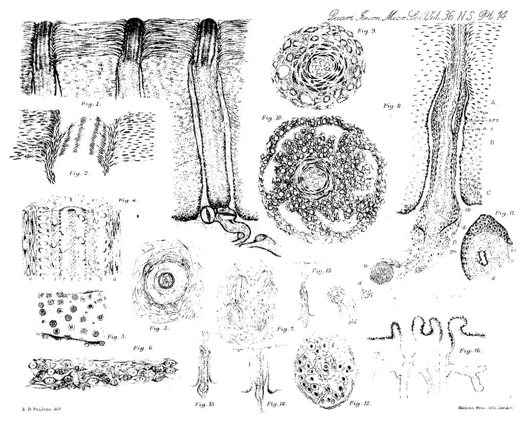
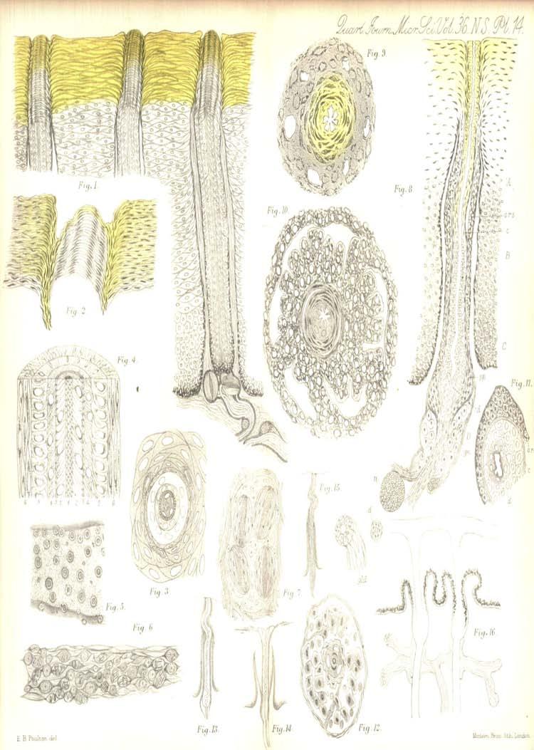
58


59 H B Poulton del
The Structure and Development of the Hairs of Monotremes and Marsupials- Part I. Monotremes.
 HAIRS OF MONOTREMBS AND MARSUPIALS. 549 The Structure and Development of the Hairs of Monotremes and Marsupials- Part I. Monotremes. By Baldwin Spencer, M.A., Professor of Biology in the University of
Hair can be defined as a slender, thread like outgrowth from a follicle in the skin of mammals. Composed mainly of keratin, it has three
 1 2 3 Hair can be defined as a slender, thread like outgrowth from a follicle in the skin of mammals. Composed mainly of keratin, it has three morphological regions the cuticle, medulla, and cortex. 4
Medical Forensics Notes
 Medical Forensics Notes The Biology of Hair Hair is composed of the protein keratin, which is also the primary component of finger and toe nails. The Biology of Hair Hair is produced from a structure called
SAMPLE COPY SAMPLE COPY SAMPLE COPY
 The Integumentary and Skeletal Systems EXPERIMENT 3.1: A CLOSER LOOK AT THE SKIN Supplies: Microscope Prepared slide: human skin (not the one with follicles or hairs) Purpose: To examine the dermis and
EXPERIMENTS ON ELECTRICAL RESISTANCE OF THE HUMAN EPIDERMIS. TAIZAN SUCHI* Institute of Physiology, University of Nagoya
 EXPERIMENTS ON ELECTRICAL RESISTANCE OF THE HUMAN EPIDERMIS TAIZAN SUCHI* Institute of Physiology, University of Nagoya In deeper layers of the epidermis, there are fine intercellular clefts separating
Unit 3 Hair as Evidence
 Unit 3 Hair as Evidence A. Hair as evidence a. Human hair is one of the most frequently pieces of evidence at the scene of a violent crime. Unfortunately, hair is not the best type of physical evidence
HAIR SCIENCE AND BIOLOGY
 HAIR SCIENCE AND BIOLOGY Your hair is composed of keratin, a strong fibrous protein, and is built from cells similar to those of your skin. The average number of hairs on the human scalp is 120,000, although
Trace Evidence: Hair. Forensic Science
 Trace Evidence: Hair Forensic Science Hair is A slender threadlike outgrowth from the follicles of the skin of mammals Found all over our bodies Head Eyebrows and Eyelashes Beard and Mustache Underarm
Integument. Sweat glands. Oil glands. Hair Nails. Sudoriferous glands. Sebaceous glands
 The Hypodermis Aka. Subcutaneous or superficial fascia Composed of Adipose Not really a part of the integument, but it is important in stabilizing the position of the skin in relation to underlying tissue
Trace Evidence: Hair. Forensic Science
 Trace Evidence: Hair Forensic Science Hair Hair is A slender threadlike outgrowth from the follicles of the skin of mammals Found all over our bodies Head Face Chest Limbs (arms and legs) Pubic region
Coat Colour. Lakeland Terrier
 A Discussion of Coat Colour Genetics in the Lakeland Terrier Chapter 2 Dog Hair By Ron Punter Page Representation of section of an active hair follicle Topcoat hair The surface Epidermis is confluent with
Accessory Structures of the Skin *
 OpenStax-CNX module: m46062 1 Accessory Structures of the Skin * OpenStax This work is produced by OpenStax-CNX and licensed under the Creative Commons Attribution License 4.0 By the end of this section,
found identity rule out corroborate
 Hair as Evidence Human hair is one of the most frequently found pieces of evidence at the scene of a violent crime. Unfortunately, hair is not the best type of physical evidence for establishing identity.
Fig 1A-1a Pre Germ Stage. Fig 1A-1b Germ Stage. Fig 1A-1c Hair Peg Stage
 1 HAIR ANATOMY AND HISTOLOGY Ronald Shapiro Md, Paul Rose MD, Michael Morgan MD, Hair Transplantation 4 th Edition, Revised and Expanded, Unger & Shapiro,2004, Chapter 1A: 25-33 EMBRYOLOGY Hair follicles
Chapter 3 The Study of Hair By the end of this chapter you will be able to:
 Chapter 3 The Study of Hair By the end of this chapter you will be able to: identify the various parts of a hair describe variations in the structure of the medulla, cortex, and cuticle distinguish between
A New Method for Staining Connective Tissue Fibres, with a Note on Liang's Method for Nerve-fibres. By G. OWEN
 421 A New Method for Staining Connective Tissue Fibres, with a Note on Liang's Method for Nerve-fibres By G. OWEN (From the Department of Zoology, The University, Glasgow) With two plates (figs, i and
FORENSIC SCIENCE. Trace Evidence
 FORENSIC SCIENCE Trace Evidence 1 Introduction Trace Evidence--any small pieces of material, man-made or naturally occurring. Trace evidence (in the absence of DNA) is considered class evidence. As early
Trace Evidence: Hair. Forensic Science
 Trace Evidence: Hair Forensic Science Hair Hair is A slender threadlike outgrowth from the follicles of the skin of mammals Found all over our bodies Head Face Chest Limbs (arms and legs) Pubic region
junction, from their deep location we can tell they are responsible of transmission of deep pressure. 2 P a g e
 7 2 1 P a g e Sensory receptors: Sensory receptors are divided into 2 types. I. Sensory receptors which are surrounded by a capsule are called encapsulated sensory receptors, Examples: 1- Meissner s corpuscles,
: An organ - two or more tissues functioning
 2 INTEGUMENTARY SYSTEM - CHAPTER 5 : An organ - two or more tissues functioning sq. m, 9-11 lbs, 7% of body weight mm thick Two layers & A.Epidermis protective layer w/o B.Dermis tissue fibrous & passes
Objectives. You will understand that: Hair
 Hair 1 Objectives You will understand that: Hair is class evidence. Hair can be used to back up circumstantial evidence. Hair absorbs and adsorbs substances both from within the body and from the external
Hair Microscopy The comparison microscope is integral to trace evidence examinations. Two matching hairs identified with the comparison microscope
 Hairs, which are composed primarily of the protein keratin, can be defined as slender outgrowths of the skin of mammals. Each species of animal possesses hair with characteristic length, color, shape,
Part 10: Chapter 17 Pleated Buttoning
 Part 10: Chapter 17 Pleated Buttoning OUR last chapter covered the upholstering of one of the commonest forms of chair frames. The same chair may be upholstered with deeper buttoning, but instead of indenting
Trace evidence is a term for small, often microscopic material. This evidence can be a significant part of an investigation. It includes an endless
 Trace Evidence Trace evidence is a term for small, often microscopic material. This evidence can be a significant part of an investigation. It includes an endless variety of materials but the trace evidence
Trace Evidence: Hair. Forensic Science
 Trace Evidence: Hair Forensic Science Human & Animal Hair 3 Main differences Pigment Medulla Scale Structure Human Hair remains constant in its color and pigmentation throughout the length of the shaft
Hair. Oleg_Mit/Shutterstock.com
 Hair Oleg_Mit/Shutterstock.com 1 Objectives You will understand that: Hair is class evidence. Hair can be used to back up circumstantial evidence. Hair absorbs and adsorbs substances both from within the
Forensic Value of Hair
 Forensic Value of Hair Hair is class evidence morphorlogy (how it looks), index (how thick the medula is), color Mitochondrial DNA from shaft. All mitochondria comes from egg only, not sperm. So ONLY maternal
Objectives. You will understand that: Hair
 Hair 1 Objectives You will understand that: Hair is class evidence. Hair can be used to back up circumstantial evidence. Hair absorbs and adsorbs substances both from within the body and from the external
Objectives. You will understand that: Hair
 Hair 1 Objectives You will understand that: Hair is class evidence. Hair can be used to back up circumstantial evidence. Hair absorbs and adsorbs substances both from within the body and from the external
Title. Author(s)KONDO, Keiji; Vanek, Milan; Rasmussen, Palle V.; Blo. CitationJournal of the Graduate School of Agriculture, Hokka. Issue Date
KONDO, Keiji; Vanek, Milan; Rasmussen, Palle V.; Blo. CitationJournal of the Graduate School of Agriculture, Hokka. Issue Date") Title The beauty of mink pelage observea with SEM Author(s)KONDO, Keiji; Vanek, Milan; Rasmussen, Palle V.; Blo CitationJournal of the Graduate School of Agriculture, Hokka Issue Date 2001-03 Doc URL http://hdl.handle.net/2115/13157
Title The beauty of mink pelage observea with SEM Author(s)KONDO, Keiji; Vanek, Milan; Rasmussen, Palle V.; Blo CitationJournal of the Graduate School of Agriculture, Hokka Issue Date 2001-03 Doc URL http://hdl.handle.net/2115/13157
DEMARCATION OF THE STONE AGES.
 20 HAMPSHIRE FLINTS. DEMARCATION OF THE STONE AGES. BY W, DALE, F.S.A., F.G.S. (Read before the Anthropological Section of -the British Association for the advancement of Science, at Birmingham, September
Copyright 2013 Crosscutting Concepts, LLC. All Rights Reserved.
 Trace Evidence Trace evidence results from the transfer of material from one place to another. Examples include: fibers glass fragments paint hair Trace Evidence Locard s principle: Every contact leaves
HAIRS. Morphology of Hair dermis 5/5/2017. Chapter 8 HAIR, FIBERS, AND PAINT. cortex medulla Sebaceous gland
 Chapter 8 HAIR, FIBERS, AND PAINT HAIRS 1 2 Introduction Hair is encountered as physical evidence in a wide variety of crimes. Although it is not yet possible to individualize a human hair to any single
Hair Restoration Gel
 Hair Restoration Gel CLINICAL STUDY Cosmetic hair tonics have been peddled for the better part of the last century, mostly in the form of inert tonics and pigmented creams that promised to restore hair
Unit-3-A Anatomy & Physiology of Skin
 In The Name of God (A PROJECT OF NEW LIFE HEALTH CARE SOCIETY, KARACHI) Unit-3-A Anatomy & Physiology of Skin Shahzad Bashir RN, BScN, DCHN, MScN (Std. DUHS) Instructor New Life College of Nursing Updated
Hair. Chapter 5: For three days after death, hair and fingernails continue to grow but phone calls taper off.
 Chapter 5: Hair For three days after death, hair and fingernails continue to grow but phone calls taper off. Johnny Carson Comedian and television host http://kids.niehs.nih.gov/illusion/illus ions.htm
Microscopic Examination of Trace Evidence
 Microscopic Examination of Trace Evidence When a forensic scientist receives hair and/or fiber evidence from a crime scene they must determine the following things: Is it a hair or is it a fiber? If a
AFFINITY - A novel system for direct Intrafollicular infusion of liquid solutions containing active ingredients.
 AFFINITY - A novel system for direct Intrafollicular infusion of liquid solutions containing active ingredients. Marina Landau, 1 M.D., Michael Tavger, PhD 1. Dermatology, Wolfson Medical Center, Holon,
Biology of Hair. Hair is composed of the protein, which is also the primary component of finger and toe.
 Prof. J. Dodd Forensic Science http://media.popularmechanics.com/images/pmx0706forensicshairsmall.jpg Biology of Hair Hair is composed of the protein, which is also the primary component of finger and
Forensics 1: Unit 3: Trace Evidence: Hair
 Forensics 1: Unit 3: Trace Evidence: Hair -Encountered as physical evidence in a wide variety of crimes. -Not yet possible to individualize a human hair to a single head or body. -When properly collected
4L1 *fr4t17? JKi3ic,4. The Function and. Structure of Hair
 excited at the moment something s happening like people do. t sits there and wafts to be --Herbert Leon MacDoneli, The Evidence NeverLies inner medulla, the cortex, and the outer cuticle. A hair has three
SERIATION: Ordering Archaeological Evidence by Stylistic Differences
 SERIATION: Ordering Archaeological Evidence by Stylistic Differences Seriation During the early stages of archaeological research in a given region, archaeologists often encounter objects or assemblages
Regardless of the animal species, the skin is made up of three layers: the epidermis, the dermis, the subcutaneous tissue.
 Hides and skins, the raw material 29 Epidermis GRAIN Dermis FLESH Subcutaneous tissues > Cut of a mammal hide HIDE / SKIN STRUCTURE It is composed of: an outer side called the grain side, including the
Study Guide-Forensic Science Chapter 5- Hair Name:
 1. Is hair class or individual evidence? Class 2. Does hair only absorb substances from inside the body? _No, Inside and out_ 3. From hair, one can determine the following: _Human or animal Race Origin
-hairs grows out of a follicle (has cells with DNA for analysis) - hair extends from here (in the follicle) has cells with DNA
 - hair extends from here (in the follicle) has cells with DNA") Name _ period Unit 4: Hair and Fibers Anatomy and Use in Forensic Science Objectives You will understand that: Hair is. Hair can be used to back up. Hair absorbs and adsorbs substances both from within
Name _ period Unit 4: Hair and Fibers Anatomy and Use in Forensic Science Objectives You will understand that: Hair is. Hair can be used to back up. Hair absorbs and adsorbs substances both from within
MacDonald of Glenaladale
 Background MacDonald of Glenaladale The MacDonald of Glenaladale is one of a small group of tartans where an extant specimen survives that can accurately be dated to the mid-c18th. For many years confusion
A COIN OF OFFA FOUND IN A VIKING-AGE BURIAL AT VOSS, NORWAY. Bergen Museum.
 A COIN OF OFFA FOUND IN A VIKING-AGE BURIAL AT VOSS, NORWAY. BY HAAKON SCHETELIG, Doct. Phil., Curator of the Bergen Museum. Communicated by G. A. AUDEN, M.A., M.D., F.S.A. URING my excavations at Voss
TRAINING LAB HAIR AS EVIDENCE: PART 1 HUMAN HAIR NAME
 TRAINING LAB HAIR AS EVIDENCE: PART 1 HUMAN HAIR NAME Background: You loose about 50 to 100 hairs a day from the approximately 100,000 total hairs present on your head. Don t worry, however, because there
TRAINING LAB HAIR AS EVIDENCE: PART 2 ANIMAL HAIR NAME
 TRAINING LAB HAIR AS EVIDENCE: PART 2 ANIMAL HAIR NAME Background: Animal hair can be just as important in helping to solve a crime as human hair. Example: a criminal breaks in to a house and steals some
Skin Care Tips and Facts
 Skin Tips and Facts * Your skin is your body's largest organ! The of the average woman weighs kilograms, while that of the average weighs five. * What's the secret to? Not surprisingly, the state of our
Forensic Science
 http://media.popularmechanics.com/images/pmx0706forensicshairsmall.jpg Forensic Science 1 Central Focus Students will understand how forensic scientists use observational, physical, chemical, and biological
A HOARD OF EARLY IRON AGE GOLD TORCS FROM IPSWICH
 A HOARD OF EARLY IRON AGE GOLD TORCS FROM IPSWICH ByJ. W. BRAILSFORD, M.A., F.S.A. On 26 October 1968 five gold torcs (Plates XX, XXI, XXII) of the Early Iron Age were found at Belstead Hills Estate, Ipswich
Anatomy of Skin and its Defense, Breakdown, and Fortification
 Anatomy of Skin and its Defense, Breakdown, and Fortification Copyright 2011 All rights reserved. The content of this presentation may not be copied, replaced, Healthy Skin Human skin is a remarkable organ,
THE PERMANENCE OF SCARRING, VISIBILITY AND COSMETIC DEFECT
 THE PERMANENCE OF SCARRING, VISIBILITY AND COSMETIC DEFECT The 13 th edition of the Judicial College Guidelines indicate a number of factors to be taken into consideration in the valuation of facial injuries
FORMATION OF NOVEL COMPOSITE FIBRES EXHIBITING THERMOCHROMIC BEHAVIOUR
 FORMATION OF NOVEL COMPOSITE FIBRES EXHIBITING THERMOCHROMIC BEHAVIOUR L. van der Werff 1,2,3 *, I. L. Kyratzis 1, A. Robinson 2, R. Cranston 1, G. Peeters 1 1 CSIRO Materials Science and Engineering,
Morphology and Structure of Hair
 Hairs & Fibers Morphology and Structure of Hair Morphology of Hair An appendage of the skin that grows out of an organ known as the hair follicle. Extends from its root or bulb embedded in the follicle,
Hair loss, alopecia areata, cicatricial alopecia. By Kai Chi Chan P-year Medical Student SGUL-UNIC at Sheba Hospital
 Hair loss, alopecia areata, cicatricial alopecia By Kai Chi Chan P-year Medical Student SGUL-UNIC at Sheba Hospital No need to pull your hair out about it! Summary: Hair Structure Hair growth cycle Male
A Highland Revival Drawstring Plaid
 Introduction A Highland Revival Drawstring Plaid The late 18th and early 19th centuries were a period of great variation and change in the development of Highland Dress. Covering much of the reign of Geo
LESSON ASSIGNMENT. Anatomy and Physiology of the Integumentary System. After completing this lesson, you should be able to:
 LESSON ASSIGNMENT LESSON 1 Anatomy and Physiology of the Integumentary System. LESSON ASSIGNMENT Paragraphs 1-1 through 1-5. LESSON OBJECTIVES After completing this lesson, you should be able to: 1-1.
The cuticle of cysticerci of Taenia saginata, T. hydatigena, and T. pisiformis By E. H. SIDDIQUI. Summary
 141 The cuticle of cysticerci of Taenia saginata, T. hydatigena, and T. pisiformis By E. H. SIDDIQUI (From the Sub-Department of Parasitology, Department of Zoology, University of Edinburgh, West Mains
DETECTION OF RADIATION EFFECTS ON HAIR ROOTS OF THE HUMAN SCALP*
 DETECTION O RADIATION EECTS ON HAIR ROOTS O THE HUMAN SCALP* EUGENE J. VAN SCOTT, M.D. AND RICHARD P. REINERT5ON, M.D. More than sixty years ago (1896), shortly after Roentgen discovered the X-ray, Daniel
THE outermost layers of the arthropod cuticle, collectively termed the
 4 1 An Examination of the Cuticles of two Scorpions, Pandinus imperator and Scorpiops hardwickii By J. KENNAUGH {From the Department of Zoology, the University, Manchester) SUMMARY The cuticles of Pandinus
T so far, by any other ruins in southwestern New Mexico. However, as
 TWO MIMBRES RIVER RUINS By EDITHA L. WATSON HE ruins along the Mimbres river offer material for study unequaled, T so far, by any other ruins in southwestern New Mexico. However, as these sites are being
HANT3 FIELD CLUB AND ARCH^OLOGICAL SOCIETY, PLATE 4
 HANT3 FIELD CLUB AND ARCH^OLOGICAL SOCIETY, 1898. PLATE 4 VUU*. ilurti.14 HALF SIZE. BRONZE PALSTAVES, FOUND AT PEAR TREE GREEN. n BRONZE IMPLEMENTS FROM THE. NEIGHBOURHOOD OF SOUTHAMPTON, BY W. DALE,
OBSERVATIONS ON THE FLUORESCENT MATERIAL IN HAIRS
 OBSERVATIONS ON THE FLUORESCENT MATERIAL IN HAIRS INFECTED BY MICROSPORON IN TINEA CAPITIS* ZACHARY FELSHER, M.D., B.S. The greenish fluorescence of children's hair infected by M. audouni and M. lanosum
Microanatomical observations of hair in domestic animals: A comparative study
 2018; 6(5): 1925-1929 E-ISSN: 2320-7078 P-ISSN: 2349-6800 JEZS 2018; 6(5): 1925-1929 2018 JEZS Received: 18-07-2018 Accepted: 19-08-2018 S Senthilkumar PG Scholar, Dept. of Veterinary Anatomy, Madras veterinary
United States Patent (19) 11 Patent Number: 4,526,488 Krull 45) Date of Patent: Jul. 2, 1985
 11 Patent Number: 4,526,488 Krull 45) Date of Patent: Jul. 2, 1985") United States Patent (19) 11 Patent Number: 4,526,488 Krull 45) Date of Patent: Jul. 2, 1985 54 INK RIBBON CASSETTE PROVIDED WITH 56) References Cited AN EMPREGNATION DEVICE U.S. PATENT DOCUMENTS s 2,76,539
United States Patent (19) 11 Patent Number: 4,526,488 Krull 45) Date of Patent: Jul. 2, 1985 54 INK RIBBON CASSETTE PROVIDED WITH 56) References Cited AN EMPREGNATION DEVICE U.S. PATENT DOCUMENTS s 2,76,539
SELYE and McKeown (1935) and Baker (1948) have noted the presence of
 and Baker (1948) have noted the presence of") A Pigment in the Rat's Uterus By ]. G. WARBRICK {From the Department of Anatomy, University of Glasgow, Glasgow, W. 2) With one plate (fig. i) SUMP4ARY 1. A yellowish-brown pigment was found at the old
A Pigment in the Rat's Uterus By ]. G. WARBRICK {From the Department of Anatomy, University of Glasgow, Glasgow, W. 2) With one plate (fig. i) SUMP4ARY 1. A yellowish-brown pigment was found at the old
T ment formation as observed in fetal and adult guinea pigs with the
 GENETIC ASPECTS OF PIGMENT PRODUCTION IN THE GUINEA PIG MARY T. HARMAN AND ANNETTE ALSOP CASEz Kansas State College, Manhattan, Kansas Received March 14, 1941 INTRODUCTION AND REVIEW OF LITERATURE HE purpose
Hair as Evidence Hair is helpful as evidence for 4 reasons 1) Wide range of classifications 2) Persistent (gets stuck in clothing) 3) Resists
 Wide range of classifications 2) Persistent (gets stuck in clothing) 3) Resists") Hair Human hair is one of the most frequently found pieces of evidence at the scene of a violent crime. It can provide a link between the criminal and the crime Hair as Evidence Hair is helpful as evidence
Hair Human hair is one of the most frequently found pieces of evidence at the scene of a violent crime. It can provide a link between the criminal and the crime Hair as Evidence Hair is helpful as evidence
Anatomical Errors - Comparing the Manoppello to the Shroud By Matthias Henrich
 Anatomical Errors - Comparing the Manoppello to the Shroud By Matthias Henrich The eyebrows run the wrong direction. The portrayal in the Manoppello-picture is therefore physignomically-anatomically wrong.
Advanced Skin Rejuvenation Wrinkle Enhancement and Skin Resurfacing Procedures
 Advanced Skin Rejuvenation Wrinkle Enhancement and Skin Resurfacing Procedures Note: Prior to reading this section, you should have read Parts I and II of this book, in particular, the section beginning
Healthy Shine Lilac. For renewed balance and shine
 Healthy Shine Lilac For renewed balance and shine NAOLYS ACTIVE CELLS Healthy Shine Lilac For renewed balance and shine A STORY The lilac Syringa vulgaris, Oleaceae Fragrant flowers, a precious remnant
(12) United States Patent (10) Patent No.: US 6,308,717 B1
 United States Patent (10) Patent No.: US 6,308,717 B1") USOO63O8717B1 (12) United States Patent (10) Patent No.: US 6,308,717 B1 Vrtaric (45) Date of Patent: Oct. 30, 2001 (54) HAIR BRUSH WITH MOVABLE BRISTLES 5,657,775 8/1997 Chou... 132/125 5,715,847 * 2/1998
USOO63O8717B1 (12) United States Patent (10) Patent No.: US 6,308,717 B1 Vrtaric (45) Date of Patent: Oct. 30, 2001 (54) HAIR BRUSH WITH MOVABLE BRISTLES 5,657,775 8/1997 Chou... 132/125 5,715,847 * 2/1998
Improvement of Grease Leakage Prevention for Ball Bearings Due to Geometrical Change of Ribbon Cages
 NTN TECHNICAL REVIEW No.78 2010 Technical Paper Improvement of Grease Leakage Prevention for Ball Bearings Due to Geometrical Change of Ribbon Cages Norihide SATO Tomoya SAKAGUCHI Grease leakage from sealed
Forensic Science. Presentation developed by T. Trimpe 2006
 Forensic Science http://media.popularmechanics.com/images/pmx0706forensicshairsmall.jpg Presentation developed by T. Trimpe 2006 http://sciencespot.net/ Biology of Hair Hair is composed of the protein
You can usually divide the hair into three different parts: the cuticle, or the cover cells, which is the outer hard layer of the hair.
 Super Wash anatomy Hair consists mostly of a protein called a-keratin. Keratin or horn substance, is a sulfur-containing protein and belongs to a group of fibrous proteins that form the basis of hair and
Pertemuan XXI, XXIII, XXV : PENGAKU (Stiffener)
") Pertemuan XXI, XXIII, XXV : PENGAKU (Stiffener) Mata Kuliah : Struktur Baja Kode MK : TKS 4019 Pengampu : Achfas Zacoeb Introduction Stiffeners are secondary plates or sections which are attached to beam
Pertemuan XXI, XXIII, XXV : PENGAKU (Stiffener) Mata Kuliah : Struktur Baja Kode MK : TKS 4019 Pengampu : Achfas Zacoeb Introduction Stiffeners are secondary plates or sections which are attached to beam
AN INVESTIGATION OF LINTING AND FLUFFING OF OFFSET NEWSPRINT. ;, l' : a Progress Report MEMBERS OF GROUP PROJECT Report Three.
 ;, l' : Institute of Paper Science and Technology. ' i,'',, AN INVESTIGATION OF LINTING AND FLUFFING OF OFFSET NEWSPRINT, Project 2979 : Report Three a Progress Report : r ''. ' ' " to MEMBERS OF GROUP
Copyright 2017 Naturalislabs Pte Ltd. All rights reserved. Published by Eric Kelly.
 UltraFX10.com 1 Copyright 2017 Naturalislabs Pte Ltd. All rights reserved. Published by Eric Kelly. No part of this publication may be reproduced, stored in a retrieval system, or transmitted in any form
SpiraBrush CX. SBX-9000 Gynecological Biopsy Device. Physician and Clinician Primer: Resource Guide and Standard Lab Tissue Removal Procedure
 SpiraBrush CX SBX-9000 Gynecological Biopsy Device Physician and Clinician Primer: Resource Guide and Standard Lab Tissue Removal Procedure Ver: September 2015 056-0012 rev. A LLC Histologics LLC 20409
Design Decisions. Copyright 2013 SAP
 Design Decisions Copyright 2013 SAP ELEMENTS OF DESIGN FORM should be in proportion to the shape of the head and face, and the length and width of neck and shoulder SPACE is the area the style occupies;
EXPERIENCE during the war with the common vesicants, mustard gas. through the skin. After penetration they produce skin reactions
 612.793.5 s O 4i.7et 1..-.s FACTORS INFLUENCING THE PENETRATION OF THE SKIN BY CHEMICAL AGENTS. By H. CULLIUMBINE, M.D., Ch.B., M.Sc. (Chemical Defence Experimental Station, Porton, Wilts.) (Received for
How To Measure In Vivo UVA and UVB Blocking Sunscreens and Cosmetics on Human Skin
 How To Measure In Vivo UVA and UVB Blocking Sunscreens and Cosmetics on Human Skin Jeffrey L. Taylor, Ph.D. Jillian F. Dlugos HUMAN HEALTH ENVIRONMENTAL HEALTH 2015 PerkinElmer Skin Related Spectral Regions
Excavations at Shikarpur, Gujarat
 Excavations at Shikarpur, Gujarat 2008-2009 The Department of Archaeology and Ancient History, the M. S. University of Baroda continued excavations at Shikarpur in the second field season in 2008-09. In
Hiliiil. R!llii i. ilijii. ill;! liiii
 Ji i Hiliiil R!llii i ill;! ilijii liiii i li ALBERT R. LIBRARY MANN New York State Colleges OF Agriculture and Home Economics AT Cornell University EVERETT FRANKLIN PHILLIPS BEEKEEPING LIBRARY PROCEEDINGS
Bacterial smear and Staining
 Practical Microbiology 18-22/11/2018 University of Sulaimani college of Pharmacy Year2 Lab. 4: Bacterial smear and Staining Before staining and observing a microbe under a microscope, a smear must be prepared.
Patterns and Necklines
 South Dakota State University Open PRAIRIE: Open Public Research Access Institutional Repository and Information Exchange Cooperative Extension Circulars: 1917-1950 SDSU Extension 11-1930 Patterns and
Trace Evidence: Hair. Forensic Science
 Trace Evidence: Hair Forensic Science Copyright and Terms of Service Copyright Texas Education Agency, 2011. These materials are copyrighted and trademarked as the property of the Texas Education Agency
International Journal of Science, Environment and Technology, Vol. 7, No 5, 2018,
 International Journal of Science, Environment and Technology, Vol. 7, No 5, 2018, 1726 1730 ISSN 2278-3687 (O) 2277-663X (P) Review Article HISTOLOGICAL STUDY OF HAIR FOLLICLES OF CATTLE BREEDS OF MAHARASHTRA
1 of 8 11/3/14 2:01 PM
 Home About Us Laboratory Services Forensic Science Communications Back Issues July 2000 Hairs, Fibers, Crime, and Evidence, Part 1, by Deedrick... Part 1: Hair Evidence July 2000 - Volume 2 - Number 3
Basics. Scalp has highest density, but declines with age from 1135/cm 2 at birth to /cm 2 in adults for a total of 100,000 follicles.
 Basics Hair has little remaining physiologic importance, but great psychological importance. Five million total hair follicles in the adult human, without significant racial or sexual differences Scalp
The Iron Handle and Bronze Bands from Read's Cavern: A Re-interpretation
 46 THE IRON HANDLE AND BRONZE BANDS FROM READ'S CAVERN The Iron Handle and Bronze Bands from Read's Cavern: A Re-interpretation By JOHN X. W. P. CORCORAN. M.A. Since the publication of the writer's study
Microscopic Evidence HAIR EVIDENCE FIBER ANALYSIS PATHOLOGY INTRO
 Microscopic Evidence HAIR EVIDENCE FIBER ANALYSIS PATHOLOGY INTRO Microscopic Hair Analysis The first step in microscopic hair evaluation is to determine whether it is animal or human. Hair (and nails)
Chapter 14 Men s Haircutting and Styling
 Chapter 14 Men s Haircutting and Styling MULTIPLE CHOICE 1. During the, the barber must determine what the client is asking for in the way of a haircut or style. a. strand test c. client consultation b.
Abstract. Greer, Southwestern Wyoming Page San Diego
 Abstract The Lucerne (48SW83) and Henry s Fork (48SW88) petroglyphs near the southern border of western Wyoming, west of Flaming Gorge Reservoir of the Green River, display characteristics of both Fremont
RESULTS AND INTERPRETATION
 CHAPTER 6 RESULTS AND INTERPRETATION 6.1 INTRODUCTION Chapter 6 deals with the factor analysis results and the interpretation of the factors identified for the product category lipstick and the three advertisements
ASMI COMPLAINTS PANEL FINAL DETERMINATION Meeting held 10 November, 2009
 ASMI COMPLAINTS PANEL FINAL DETERMINATION Meeting held 10 November, 2009 Hamilton Laboratories ( HL ) v. Johnson & Johnson Pacific ( JJP ) Neutrogena Ultra Sheer Dry-Touch Sunscreen Lotion 1. HL complains
NOTES ON THE ANCIENT ART OF CENTRAL AMERICA
 NOTES ON THE ANCIENT ART OF CENTRAL AMERICA Mi BY GEORGE GRANT MACCURDY HILE I was attending the centenary celebration of The Academy of Natural Sciences of Philadelphia, Miss H. Newel1 Wardle of the Academy's
Cinderella Beauty Protocol with SCENAR Devices. by Irina Kossovskaia, MD, P.HD., D.N.M.
 Cinderella Beauty Protocol with SCENAR Devices by Irina Kossovskaia, MD, P.HD., D.N.M. The Aging Skin Aging is an issue of mind over matter. If you don t mind, it doesn t matter. Mark Twain Inside every