Staining Criteria Handbook
|
|
|
- Julie Gardner
- 5 years ago
- Views:
Transcription


1 Staining Criteria Handbook General Pathology (Routine Histopathology) Neuropathology Edition 4 November 2015
2 Index Page Haematoxylin and Eosin Assessment Criteria 3 Special Stains A & B Assessment Criteria 6 List of Assessment Criteria 26 Haematoxylin and Eosin Special Stains Assessment Criteria Definitions 30 Haematoxylin and Eosin Special Stains Appendix 40 Haematoxylin and Eosin Model Description Scoring System Scoring based on criteria UK NEQAS CPT Stain Repertoire 2
3 Haematoxylin and Eosin Assessment Criteria 3
4 Haematoxylin and Eosin Assessment Criteria Pre Microtomy Microtomy Staining Insufficient cellular features for assessment Crush artefacts Foam inset artefact Red blood cell lysis Poor chromatin detail Cracking Nuclear bubbling Nuclear meltdown Incorrect orientation suspected Incorrect trimming suspected Chatter / vibration Displacement Folds / creases Knife back debris Knife marks Lifting Position on slide Section too thick Section too thin Section thickness variable Squames / floaters / fibres Trimming artefact Water bath bubbles Haematoxylin Intensity too strong Haematoxylin Intensity too weak Haematoxylin colour not blue Haematoxylin background staining Eosin Intensity too strong Eosin Intensity too weak Eosin colour Eosin not selective Uneven staining Stain deposit present Post Staining Air bubbles Air drying artefact Excessive mountant Mountant shrinkage Residual wax Section wiped / section off slide Tissue exposed Water present Contaminant on slide 4
5 Description of Staining Results Nuclei must be stained purple blue with haematoxylin. The intensity must be strong enough allow clear demonstration of nuclear detail at a medium power, but not too strong to cause a loss of the chromatin granularity or excessive cytoplasmic or connective tissue staining. Where the haematoxylin has been differentiated out, minimal cytoplasmic or connective tissue background staining with haematoxylin must remain. This background if present must not reduce the effectiveness of the nuclear demonstration or affect the colour and selectiveness of the eosin. The eosin should be selective enough to demonstrate different cellular components such as collagen, cytoplasm, red blood cells, cellular granules, amyloid etc. The intensity must be appropriate to the section thickness and the haematoxylin intensity. Where the eosin is too weak it will fail to allow selective demonstration of different components at low power. If the eosin intensity is too strong the colour and detail of the nuclear stain will be obscured and selectivity will be reduced. For extended description including pre Microtomy, Microtomy and post staining please see model description in appendix. 5
6 Special Stains A & B Assessment Criteria 6
7 Alcian Blue/PAS Assessment Criteria Primary Stain Alcian Blue Intensity too strong Alcian Blue Intensity too weak Alcian Blue Colour PAS Intensity too strong PAS Intensity too weak PAS Coloration Background Counterstain Nuclear Stain Intensity too strong Nuclear Stain Intensity too weak Nuclear Stain colour Post staining Air bubbles Air drying artefact Excessive mountant Mountant shrinkage Residual wax Section wiped / section off slide Tissue Damage Tissue exposed Water present Contaminant on slide Description of Staining Results Acid mucopolysaccharides should be coloured bright blue. Neutral mucopolysaccharides should be bright magenta. Mixed mucins should be purple. There should be clear distinction between acid, neutral and mixed mucins. Background staining should be negligible. A counterstain (typically haematoxylin) is considered optional. If present, it should be light and even and should not mask or alter the primary stains. It should assist location. Section quality and presentation should not impair the result. 7
8 Amyloid (method for) Assessment Criteria Primary Stain - Bright field Congo red (stain) colour Intensity too strong Intensity too weak Background Primary Stain - No green birefringence on cross polarisation Cross polarised light Birefringence intensity too weak Birefringence intensity too strong Birefringence not selective Counterstain Nuclear stain intensity too strong Nuclear stain intensity too weak Nuclear stain colour Post staining Description of Staining Results Air bubbles Air drying artefact Excessive mountant Mountant shrinkage Residual wax Section wiped / section off slide Tissue Damage Tissue exposed Water present Contaminant on slide Amyloid: There is no amyloid specific dye. However, when performed correctly the alkaline alcoholic Congo red method is highly selective for binding to amyloid, when sections are prepared at the appropriate thickness (~5 10 µm) and staining is performed stringently. Use of other dyes may produce sub optimal or erroneous results. Brightfield: Amyloid should stain pink to red (congophilic). Stain intensity is seldom homogeneous and may differ in different tissues reflecting the deposition, abundance and underlying organisation of the amyloid fibrils. Collagen and elastin may be weakly coloured. Background staining must be negligible. Haematoxylin counterstain should be light, even and not mask or alter the primary stain; it should assist location. Cross Polarised Light: Amyloid stained pink / red in brightfield microscopy must also exhibit a bright green colour in cross polarised light (green birefringence). Denser deposits of amyloid may exhibit yellow green or bright yellow birefringence. The colour and/or intensity of the primary stain must not mask the green birefringence of amyloid in cross polarised light. Collagen and elastin usually give pale yellow to vivid white polarisation colours. 8
9 Axonal Swelling (method for) Assessment Criteria Primary Stain Intensity too strong Intensity too weak Stain colour Axonal Swelling demonstration good Axonal Swelling demonstration poor Low power visibility Background Counterstain Nuclear Intensity too strong Nuclear Intensity too weak Nuclear Stain colour Post staining Air bubbles Air drying artefact Excessive mountant Mountant shrinkage Residual wax Section wiped / section off slide Tissue Damage Tissue exposed Water present Contaminant on slide Description of Staining Results Axonal swelling may be demonstrated by silver impregnation methods such as Bielschowsky or using antibodies such as amyloid precursor protein (APP). Swellings should be clearly stained, visible at low power, and discernible from other structures. Immunohistochemical methods require a light nuclear counterstain. Section quality and presentation should not impair the result. 9
10 Copper Associated Protein (CAP) Assessment Criteria Primary Stain Intensity too strong Intensity too weak Stain colour Background Counterstain Intensity too strong Intensity too weak Stain colour Post staining Air bubbles Air drying artefact Excessive mountant Mountant shrinkage Residual wax Section wiped / section off slide Tissue Damage Tissue exposed Water present Contaminant on slide Description of Staining Results All copper associated protein should be stained and clearly identifiable. HepBSA and elastin, if stained, should be readily distinguishable from CAP. Background should be negligible. If a counterstain is used it should not mask or modify the primary stain. It should assist location. Section quality and presentation should not impair the result. 10
11 Diastase/PAS Assessment Criteria Primary Stain Intensity too strong Intensity too weak Stain colour Background Residual Glycogen Counterstain Nuclear Stain Intensity too strong Nuclear Stain Intensity too weak Nuclear Stain colour Post staining Air bubbles Air drying artefact Excessive mountant Mountant shrinkage Residual wax Section wiped / section off slide Tissue Damage Tissue exposed Water present Contaminant on slide Description of Staining Results The Schiff reaction is a histochemical method for aldehyde groups. Periodic acid oxidation creates these groups on a variety of tissue structures notably glycogen, mucins, basement membrane and fungi. This modification uses the enzyme diastase (or amylase) to digest out any glycogen and is normally used in conjunction with a straight PAS (not required for EQA unless requested). Complete removal of glycogen and precise, complete demonstration of intestinal mucopolysaccharide. Membrane staining should not be confused with background, which should be minimal. Counterstain (typically haematoxylin) should provide good colour contrast and assist location. Section quality and presentation must not impair the result. 11
12 Elastin van Gieson Assessment Criteria Primary Stain Intensity too strong Intensity too weak Stain Colour No contrast Fine Fibres Background Counterstain Collagen Stain Intensity too strong Collagen Stain Intensity too weak Collagen Stain colour Cytoplasm Stain Intensity too strong Cytoplasm Stain Intensity too weak Cytoplasm Stain colour Post staining Air bubbles Air drying artefact Excessive mountant Mountant shrinkage Residual wax Section wiped / section off slide Tissue Damage Tissue exposed Water present Contaminant on slide Description of Staining Results Elastin stains fall into two main groups, Haematoxylin based (e.g. Verhoeff) and hydrophobic dye mixes (e.g. Miller s and Weigert s) Precise staining of all coarse and fine elastin fibres. The counterstain is a simple trichrome and there should be good colour separation between muscle (yellow) and collagen (red). Picro Sirius red is a suitable alternative, popular in the USA. The counterstain should provide good colour contrast and assist location. Section quality and presentation must not impair the result. 12
13 Glial Fibres (method for) Assessment Criteria Primary Stain Intensity too strong Intensity too weak Stain colour Glial Fibre demonstration good Glial Fibre demonstration poor Low power visibility Background Counterstain Intensity too strong Intensity too weak Stain colour Post staining Air bubbles Air drying artefact Excessive mountant Mountant shrinkage Residual wax Section wiped / section off slide Tissue Damage Tissue exposed Water present Contaminant on slide Description of Staining Results Glial fibres may be demonstrated using tinctorial methods such as PTAH or using antibodies such as GFAP. Astrocytic fibres should be clearly stained against a pale or clear background. With PTAH, cell nuclei and myelin will also stain this requiring no further counterstaining. Immunohistochemical methods require a light nuclear counterstain. Section quality and presentation must not impair the result. 13
14 Gram Stain Assessment Criteria Primary Stain Gram Positive Intensity too strong Gram Positive Intensity too weak Gram Positive Colour Background Low power visibility Counterstain Gram Negative Intensity too strong Gram Negative Intensity too weak Gram Negative Colour Low power visibility Post staining Air bubbles Air drying artefact Excessive mountant Mountant shrinkage Residual wax Section wiped / section off slide Tissue Damage Tissue exposed Water present Contaminant on slide Description of Staining Results Crystal Violet staining should render all Gram positive organisms blue/black without over or under differentiation. There should be no violet background. The counterstain serves to demonstrate all Gram negative organisms in a contrasting colour and to assist location. Most variations are based on the counterstain used and vary widely. Section quality and presentation must not impair the result. 14
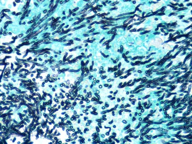
15 Grocott Assessment Criteria Primary Stain Organism Demonstration too strong Organism Demonstration too weak Organism Colour Background Non Specific Silver Precipitate Counterstain Intensity too strong Intensity too weak Stain colour Post staining Air bubbles Air drying artefact Excessive mountant Mountant shrinkage Residual wax Section wiped / section off slide Tissue Damage Tissue exposed Water present Contaminant on slide Description of Staining Results There should be dark grey/black staining of all fungal spores and/or hyphae or pneumocysts. Where appropriate, internal structures of the organisms should be visible. Impregnation of other structures, particularly collagen, should be negligible. There should be no non specific silver precipitation. The counterstain should provide good colour contrast and assist location, but must not mask any delicately impregnated fungi. Section quality and presentation must not impair the result. 15
16 Haematoxylin / Van Gieson Assessment Criteria Primary Stain Cytoplasm Stain Intensity too strong Cytoplasm Stain Intensity too weak Cytoplasm Stain colour Collagen Stain Intensity too strong Collagen Stain Intensity too weak Collagen Stain Colour Background Counterstain Nuclear Stain Intensity too strong Nuclear Stain Intensity too weak Nuclear Stain Colour Post staining Air bubbles Air drying artefact Excessive mountant Mountant shrinkage Residual wax Section wiped / section off slide Tissue Damage Tissue exposed Water present Contaminant on slide Description of Staining Results HVG is perhaps the simplest of trichrome methods which are designed to give colour separation between smooth muscle and collagen. The emphasis is on collagen, all of which should be stained bright red. Muscle, erythrocytes and general cytoplasm (including hepatocytes) should be yellow. Nuclear staining should be dark blue or black but must not influence the cytoplasmic colouration by being under differentiated. Section quality and presentation should not impair the result. 16
17 Martius Scarlet Blue Assessment Criteria Primary Stain Fibrin Demonstration too strong Fibrin Demonstration too weak Muscle Stain Intensity too strong Muscle Stain Intensity too weak Muscle Stain Colour Collagen Stain Intensity too strong Collagen Stain Intensity too weak Collagen Stain Colour Red blood cell colour Background Counterstain Nuclear Stain Intensity too strong Nuclear Stain Intensity too weak Nuclear Stain Colour Post staining Air bubbles Air drying artefact Excessive mountant Mountant shrinkage Residual wax Section wiped / section off slide Tissue Damage Tissue exposed Water present Contaminant on slide Description of Staining Results MSB is a specialised trichrome method designed to demonstrate fibrin of various ages in colours from orange to red. Early fibrin can also stain yellow and old fibrin blue. All fibrin should be brightly coloured and distinguishable from other structures. As with other trichromes, there should be good colour separation between the dyes best seen in red blood cells (yellow), muscle (red) and collagen (blue). Nuclear staining should be dark blue or black. Section quality and presentation should not impair the result. 17
18 Masson Fontana (for melanin) Assessment Criteria Primary Stain Counterstain Melanin Demonstration too strong Melanin Demonstration too weak Background Non Specific Silver Precipitate Incomplete Toning Intensity too strong Intensity too weak Stain colour Post staining Air bubbles Air drying artefact Excessive mountant Mountant shrinkage Residual wax Section wiped / section off slide Tissue Damage Tissue exposed Water present Contaminant on slide Description of Staining Results All melanin should be coloured black by silver impregnation. The reaction product should be toned to distinguish it from unreacted melanin. There should be no background staining arising from over impregnation and no non specific silver precipitation. The counterstain should provide good colour contrast and assist location. Section quality and presentation should not impair the result. 18
19 Myelin (method for) Assessment Criteria Primary Stain Intensity too strong Intensity too weak Stain colour Myelin demonstration good Myelin demonstration poor Low power visibility Background Counterstain Intensity too strong Intensity too weak Stain colour Post staining Air bubbles Air drying artefact Excessive mountant Mountant shrinkage Residual wax Section wiped / section off slide Tissue Damage Tissue exposed Water present Contaminant on slide Description of Staining Results Axonal myelin sheaths may be demonstrated by tinctorial methods such as Luxol fast blue (LFB) or using antibodies such as myelin basic protein. All myelin should be clearly stained against a pale/clear background. Fine fibres should be discernible. LFB should be counterstained, traditionally with cresyl fast violet. Section quality and presentation should not impair the result. 19
20 Neurofibrillary Tangles Assessment Criteria Primary Stain Intensity too strong Intensity too weak Stain colour Tangle demonstration good Tangle demonstration poor Low power visibility Background Counterstain Intensity too strong Intensity too weak Stain colour Post staining Air bubbles Air drying artefact Excessive mountant Mountant shrinkage Residual wax Section wiped / section off slide Tissue Damage Tissue exposed Water present Contaminant on slide Description of Staining Results Neurofibrillary Tangles may be demonstrated quite specifically using silver impregnation methods such as Gallyas or using antibodies such as Tau or Ubiquitin All tangles should be clearly stained, visible at low power, and discernible from other structures. Immunohistochemical methods require a light nuclear counterstain. Section quality and presentation should not impair the result. 20
21 Nissl Substance Assessment Criteria Primary Stain Intensity too strong Intensity too weak Stain colour Nissl demonstration good Nissl demonstration poor Low power visibility Background Counterstain Intensity too strong Intensity too weak Stain colour Post staining Air bubbles Air drying artefact Excessive mountant Mountant shrinkage Residual wax Section wiped / section off slide Tissue Damage Tissue exposed Water present Contaminant on slide Description of Staining Results All nissl granules in neuronal cell bodies should be clearly stained purple / blue. Cell nuclei should also be demonstrated. There should be no negligible background and no stain precipitate. This method may also be employed as a counterstain for other techniques. Section quality and presentation should not impair the result. 21
22 Perls Prussian Blue Assessment Criteria Primary Stain Intensity too strong Intensity too weak Stain Colour Background Unreacted iron Counterstain Intensity too strong Intensity too weak Stain colour Post staining Air bubbles Air drying artefact Excessive mountant Mountant shrinkage Residual wax Section wiped / section off slide Tissue Damage Tissue exposed Water present Contaminant on slide Description of Staining Results A histochemical reaction for ferric iron adapted for the demonstration of haemosiderin Prussian Blue colouration of haemosiderin should not be modified by counterstain or underlying, unreacted pigment. The counterstain should provide good colour contrast and assist location. Section quality and presentation should not impair the result. 22
23 Reticulin (silver method for) Assessment Criteria Primary Stain Counterstain Post staining Reticulin Demonstration too strong Reticulin Demonstration too weak Background Non Specific Silver Precipitate Incomplete Toning Intensity too strong Intensity too weak Stain colour Air bubbles Air drying artefact Excessive mountant Mountant shrinkage Residual wax Section wiped / section off slide Tissue Damage Tissue exposed Water present Contaminant on slide Description of Staining Results This method is primarily a low power scanning method so contrast is all important. There are many methods, the most popular in the UK being Gordon and Sweet. In Europe and the US Gomori is more popular. The latter impregnates nuclei which, when done well, renders counterstaining unnecessary. The main variation in method is toning. Without it, collagen would remain golden brown and cell cytoplasm a pale yellow. Again, counterstaining is unnecessary if the rest of the method is done well. Reticulin should be black, under impregnation will result in incomplete staining, over impregnation yields knobbly fibres. Fine reticulin fibres should be clearly seen at high power. There should be no non specific silver deposits and staining of cytoplasmic structures is undesirable. Counterstaining, if used, should provide good contrast and assist location. Gomori and its variants may render toned collagen rose-pink. Section quality and presentation should not impair the result. 23
24 Senile Plaques (method for) Assessment Criteria Primary Stain Intensity too strong Intensity too weak Stain colour Plaque demonstration good Plaque demonstration poor Low power visibility Background Non Specific Silver Precipitate Incomplete Toning Counterstain Intensity too strong Intensity too weak Stain colour Post staining Air bubbles Air drying artefact Excessive mountant Mountant shrinkage Residual wax Section wiped / section off slide Tissue Damage Tissue exposed Water present Contaminant on slide Description of Staining Results Plaques may be demonstrated quite specifically using silver impregnation methods such as Haga or by virtue of their amyloid component using antibodies such as β amyloid A4. All plaques should be clearly stained against a pale or clear background and be visible at low power. The dystrophic neuritis of neuritic plaques may also be demonstrated. Silver methods generally create a yellow background which requires no further counterstaining. Immunohistochemical methods require a light nuclear counterstain Section quality and presentation should not impair the result. 24
25 Trichrome (not HVG) Assessment Criteria Primary Stain Muscle Stain Intensity too strong Muscle Stain Intensity too weak Muscle Stain Colour Collagen Stain Intensity too strong Collagen Stain Intensity too weak Collagen Stain Colour Red blood cell colour Background Counterstain Nuclear Stain Intensity too strong Nuclear Stain Intensity too weak Nuclear Stain Colour Post staining Air bubbles Air drying artefact Excessive mountant Mountant shrinkage Residual wax Section wiped / section off slide Tissue Damage Tissue exposed Water present Contaminant on slide Description of Staining Results Trichrome methods are designed to give colour separation between smooth muscle and collagen. There are a variety of methods; Masson s being the most popular in the UK. All muscle should be stained red, collagen blue or green and nuclei blue/black. The collagen (milling) dye may be green or blue. Nuclear staining should be dark blue/black. Section quality and presentation should not impair the result. 25
26 Ziehl Neelsen Assessment Criteria Primary Stain Bacilli stain Intensity too strong Bacilli stain Intensity too weak Bacilli Colour Background Low power visibility Counterstain Intensity too strong Intensity too weak Stain colour Post staining Air bubbles Air drying artefact Excessive mountant Mountant shrinkage Residual wax Section wiped / section off slide Tissue Damage Tissue exposed Water present Contaminant on slide Description of Staining Results A stain used for Mycobacterium tuberculosis and closely related organisms All mycobacteria, including isolated bacilli, should be sharply stained magenta/red by carbol fuchsin with minimal background staining. The counterstain should be light, even and predominantly nuclear. It should provide good colour contrast assist location. There should be no dye precipitation and the tissue should not be damaged by heating. Section quality and presentation should not impair the result. 26
27 List of Assessment Criteria Haematoxylin and Eosin Special Stains 27
28 Haematoxylin and Eosin Assessment Criteria Pre Microtomy Microtomy Staining Insufficient cellular features for assessment Crush artefacts Foam inset artefact Red blood cell lysis Poor chromatin detail Cracking Nuclear bubbling Nuclear meltdown Incorrect orientation suspected Incorrect trimming suspected Chatter / vibration Displacement Folds / creases Knife back debris Knife marks Lifting Position on slide Section too thick Section too thin Section thickness variable Squames / floaters / fibres Trimming artefact Water bath bubbles Haematoxylin Intensity too strong Haematoxylin Intensity too weak Haematoxylin colour not purple blue Haematoxylin background staining Eosin Intensity too strong Eosin Intensity too weak Eosin colour Eosin not selective Uneven staining Stain deposit present Post Staining Air bubbles Air drying artefact Excessive mountant Mountant shrinkage Residual wax Section wiped / section off slide Tissue exposed Water present Contaminant on slide 28
29 Special Stains Assessment Criteria Primary Stain Alcian Blue Colour Alcian Blue Intensity too strong Alcian Blue Intensity too weak Amyloid Bright field Background Amyloid Bright field Deposit / Precipitate present Amyloid Bright field Intensity too strong Amyloid Bright field Intensity too weak Amyloid Bright field Stain colour Axonal Swelling demonstration good Axonal Swelling demonstration poor Bacilli Colour Bacilli stain Intensity too strong Bacilli stain Intensity too weak Background Birefringence intensity too weak Birefringence colour Birefringence not selective Collagen Stain Colour Collagen Stain Intensity too strong Collagen Stain Intensity too weak Cytoplasm Stain Colour Cytoplasm Stain Intensity too strong Cytoplasm Stain Intensity too weak Fibrin Demonstration too strong Fibrin Demonstration too weak Fine Fibres Glial Fibre demonstration poor Glial Fibre demonstration good Gram Positive Colour Gram Positive Intensity too strong Gram Positive Intensity too weak Incomplete Toning Intensity too strong Intensity too weak 29
30 Primary Stain continued Low power visibility Melanin Demonstration too strong Melanin Demonstration too weak Muscle Stain Colour Muscle Stain Intensity too strong Muscle Stain Intensity too weak Myelin demonstration poor Myelin demonstration good Nissl demonstration good Nissl demonstration poor No green birefringence on cross polarisation No Contrast Non Specific Silver Precipitate Organism Colour Organism Demonstration too strong Organism Demonstration too weak PAS Coloration PAS Intensity too strong PAS Intensity too weak Plaque demonstration good Plaque demonstration poor Red blood cell colour Residual Glycogen Reticulin Demonstration too strong Reticulin Demonstration too weak Stain colour Tangle demonstration good Tangle demonstration poor Unreacted iron 30
31 Counterstain Collagen Stain Colour Collagen Stain Intensity too strong Collagen Stain Intensity too weak Cytoplasm Stain Colour Cytoplasm Stain Intensity too strong Cytoplasm Stain Intensity too weak Gram Negative Colour Gram Negative Intensity too strong Gram Negative Intensity too weak Intensity too strong Intensity too weak Low power visibility Nuclear Stain Colour Nuclear Stain Intensity too strong Nuclear Stain Intensity too weak Stain colour Post Staining Air bubbles Air drying artefact Excessive mountant Mountant shrinkage Residual wax Section wiped / section off slide Tissue exposed Water present Contaminant on slide 31
32 Assessment Criteria Definitions Haematoxylin and Eosin Special Stains 32
33 Haematoxylin and Eosin Criteria Definitions Pre Microtomy The material supplied must be paraffin wax processed and have sufficient variety of cellular features, in particular nuclei to allow assessment. Insufficient cellular features for assessment - The material supplied does not have sufficient variety of cellular features, in particular nuclei to allow assessment Artefacts, such as crush effects and foam insert impressions should not be introduced during the laboratory handling and preparation stages. Crush artefacts - distortion of nuclei to give a thin elongated appearance because of crushing prior to fixation Foam inset artefact - triangular holes on the outer surface of biopsies produced by the surface of foam cassette inserts The tissue must show no evidence of being inadequately fixed or having had delayed fixation and there must be a clear demonstration of the chromatin detail within the nuclei at medium and low power and little cell lysis. Red blood cell lysis -loss of structure and shape and fusion of red cells Poor chromatin detail - insufficient chromatin clumping, granularity or perinuclear chromatin There must be no evidence of poor paraffin processing. Epithelial cells groups and connective tissue components must not appear to be separated by cracking. There should be no disruption of the nuclear membrane resulting in pale, fused nuclear staining (nuclear meltdown). The appearance of bubble like artefacts over the nuclei will be noted but no marks will be deducted. Cracking - excessive separation of epithelial cells, lymphoid cells or connective tissue Nuclear bubbling - the appearance of a vacuole or bubble shape over and within nuclei Nuclear meltdown - loss of a distinct nuclear membranes and pale staining in nuclei If the biopsy is suspected of being orientated in a way that fails to demonstrate the cell types or layers appropriately this will be noted but no deduction will be made from the score. Similarly if it is felt that the tissue has not been trimmed to full face, or has been trimmed past full face this will be noted but no deduction will be made. Incorrect orientation - suspected lack of all the cell layers expected in a tissue type Incorrect trimming - suspected lack of areas of epithelium or other which could be expected 33
34 Microtomy The section must be of a thickness appropriate to the tissue type. The section is too thick if it is difficult to focus on a single layer of nuclei, or too thin if staining intensity is compromised. It must be free from variations in thickness, creases, folds, scores and contaminants. No accumulation or fragments of tissue should be seen over or between sections. The section should be flat, with no lifted areas. There should be no holes, tearing or damage either as a result of trimming or sectioning or cover-slipping. The section must be positioned on the slide so that microscopy is not compromised. Chatter / vibration - very closely placed variation in thickness causing stripes of alternate staining intensity at right angles to direction of sectioning Displacement - cells that become detached and move over other areas of section Folds / creases - creases and folds in the section giving doubles layers that lift from the slide and stain intensely Knife back debris - accumulation of cells that are smeared on the edge of the knife and deposited between alternate sections Knife marks - scores and scratches at parallel to the direction of sectioning Lifting - areas of section that lift and do not lie flat Position on slide - section positioned appropriately on slide for full visualisation Section too thick - unable to focus on a single cell layer Section too thin - loss of stain intensity and contrast because of thinness of section Section thickness variable - varying thickness in different areas of the section, recognised by varying dye intensity Squames / floaters / fibres -contamination by material that is not in the block, usually above the section Trimming artefact - tears and rough edged holes, often with lifted edges, within the section Water bath bubbles circular areas of lifting, often cracked and intensely stained 34
35 Staining Nuclei must be stained purple/ blue with haematoxylin. The intensity must be strong enough allow clear demonstration of nuclear detail at a medium power, but not too strong to cause a loss of the chromatin granularity or excessive cytoplasmic or connective tissue staining. Where the haematoxylin has been differentiated out, minimal cytoplasmic or connective tissue background staining with haematoxylin must remain. This background if present must not reduce the effectiveness of the nuclear demonstration or affect the colour and selectiveness of the eosin. Haematoxylin Intensity too strong - a loss of the chromatin granularity or excessive cytoplasmic or connective tissue staining Haematoxylin Intensity too weak - intensity must be strong enough to allow clear demonstration of nuclear detail at a medium power Haematoxylin colour not purple - blue Nuclei must be stained purple/ blue with haematoxylin Haematoxylin background staining - where the haematoxylin has been differentiated out, minimal cytoplasmic or connective tissue background staining with haematoxylin must remain. If present must not reduce the effectiveness of the nuclear demonstration or affect the colour and selectiveness of the eosin The eosin should be selective enough to demonstrate different cellular components such as collagen, cytoplasm, red blood cells, cellular granules, amyloid etc. The intensity must be appropriate to the section thickness and the haematoxylin intensity. Where the eosin is too weak it will fail to allow selective demonstration of different components at low power. If the eosin intensity is too strong the colour and detail of the nuclear stain will be obscured and selectivity will be reduced. Eosin Intensity too strong - the intensity must be appropriate to the section thickness and the haematoxylin intensity. If the eosin intensity is too strong the colour and detail of the nuclear stain will be obscured and selectivity will be reduced Eosin Intensity too weak - the intensity must be appropriate to the section thickness and the haematoxylin intensity. Where the eosin is too weak it will fail to allow selective demonstration of different components at low power Eosin colour - the cytoplasm is not stained the appropriate colour based on the method employed and the expected staining results. Old dyes. Mixed incorrectly. Not evenly applied to slide. Uneven washing. Dehydrated incorrectly. Eosin not selective - the eosin should be selective enough to demonstrate different cellular components such as collagen, cytoplasm, red blood cells, cellular granules, amyloid etc All staining should be even across the section and there should be no dye deposits or precipitate. Uneven staining - all staining should be even across the section Stain deposit present -dye deposits or precipitate 35
36 Post Staining Coverslipping must be free from air bubbles, air drying artefact or drying back. There should be no excessive mountant, the sections must be totally covered and there must be no evidence of incomplete dewaxing or dehydration. The difficulties of processing and sectioning bony tissue will be taken into account when assessing. Air bubbles - air bubbles within the mountant Air drying artefact - retractile areas and tissue damage Excessive mountant - mountant outside the coverslip Mountant shrinkage - areas of mountant have dried back from the coverslip edges Residual wax - retractile areas of undissolved wax are visible Section wiped / section off slide - section scratched by hand or off edge of slide Tissue exposed - the section is not covered by the coverslip Water present - droplets of water under the coverslip Contaminant on slide - Contamination by non biological or biological material, excluding dye / reagent deposits, usually above the tissue section between the section and the coverslip. For example, squames / floaters / fibres/ pencil or ink deposits 36
37 Special Stains Criteria Definitions Primary Stain Alcian Blue Colour - the acid mucin is not bright blue, and the mixed mucins are not purple. Old dyes. Mixed incorrectly. Not evenly applied to slide. Uneven washing. Dehydrated incorrectly Alcian Blue Intensity too strong - stain intensity is so strong that it is obscuring/ interfering with the nuclear stain or staining areas that should be another colour. Other detail may be missing or selectivity could be reduced. Alcian Blue Intensity too weak - where the stain is too weak it will fail to allow the clear, selective demonstration of different components at low power the intensity must be appropriate to the section thickness. Amyloid Bright field Background - Background staining present, reducing contrast of the nuclei or obscuring detail or affecting colour of amyloid deposits. Caused by staining too long, not differentiating enough or non-selective staining, or breakdown products in old stain. Amyloid Bright field Deposit / Precipitate present - Random irregular or crystalline deposits above and around amyloid deposits. Old or unfiltered dye. Amyloid Bright field Intensity too strong - Stain intensity is so strong that it obscures / interferes with the nuclear counterstain, or staining areas that should be another colour. Other detail may be missing or selectivity could be reduced. Amyloid Bright field Intensity too weak - Stain intensity is so weak that it is failing to demonstrate amyloid deposits clearly. Amyloid Bright field Stain colour Amyloid is not stained the appropriate colour based on the method employed and the expected staining results. Old dye. Mixed incorrectly. Not evenly applied to slide. Uneven washing. Dehydrated incorrectly. Axonal Swelling demonstration good swellings should be clearly stained, visible and discernible from other structures. Axonal Swelling demonstration poor - ineffective demonstration prevents successful visualisation. Bacilli Colour - the bacilli are not stained magenta / red. Old dyes. Mixed incorrectly. Not evenly applied to slide. Uneven washing. Dehydrated incorrectly. Bacilli stain Intensity too strong - stain intensity is so strong that it is obscuring/ interfering with the clear, selective demonstration of bacilli or staining areas that should be another colour. Other detail may be missing or selectivity could be reduced. Bacilli stain Intensity too weak - where the stain is too weak it will fail to allow the clear, selective demonstration of different components at low power the intensity must be appropriate to the section thickness. 37
38 Primary Stain continued Background - background staining present, reducing contrast of the nuclei or obscuring detail or affecting colour of another feature. Caused by staining too long, not differentiating enough or nonselective breakdown products in old stains. Birefringence intensity too weak Birefringence intensity is so weak in cross polarised light that it is failing to demonstrate amyloid deposits clearly. Birefringence colour Birefringence is not the appropriate colour based on the method employed and the expected results in cross polarised light. Incorrect / non-stringent method. Old dye. Mixed incorrectly. Not evenly applied to slide. Uneven washing. Dehydrated incorrectly. Birefringence not selective Birefringence not selective of different tissue components in cross polarised light. Incorrect / non-stringent method. Old dye. Mixed incorrectly. Not evenly applied to slide. Uneven washing. Dehydrated incorrectly. Collagen Stain Colour - the collagen is not stained the appropriate colour based on the method employed and the expected staining results. Old dyes. Mixed incorrectly. Not evenly applied to slide. Uneven washing. Dehydrated incorrectly. Collagen Stain Intensity too strong - stain intensity is so strong that it is obscuring/ interfering with demonstration of collagen and different components or staining areas that should be another colour. Other detail may be missing or selectivity could be reduced. Collagen Stain Intensity too weak- where the stain is too weak it will fail to allow the clear, selective demonstration of collagen and different components at low power the intensity must be appropriate to the section thickness. Cytoplasm Stain Colour - the cytoplasm is not stained the appropriate colour based on the method employed and the expected staining results. Old dyes. Mixed incorrectly. Not evenly applied to slide. Uneven washing. Dehydrated incorrectly. Cytoplasm Stain Intensity too strong - stain intensity is so strong that it is obscuring/ interfering with demonstration of different cytoplasmic components. Other detail may be missing or selectivity could be reduced. Cytoplasm Stain Intensity too weak- where the stain is too weak it will fail to allow the clear, selective demonstration of different cytoplasmic components at low power. The intensity must be appropriate to the section thickness. Deposit / Precipitate present- random irregular or crystalline deposits above and around the cells. Old or unfiltered dyes Fibrin Demonstration too strong - stain intensity is so strong that it is obscuring/ interfering with demonstration of fibrin and different components or staining areas that should be another colour. Other detail may be missing or selectivity could be reduced. Fibrin Demonstration too weak- where the stain is too weak it will fail to allow the clear, selective demonstration of fibrin and different components at low power the intensity must be appropriate to the section thickness. 38
39 Primary Stain continued Fine Fibres effective demonstration allows fine fibres to be visualised successfully. Glial Fibre demonstration good fibres should be clearly stained against a pale or clear background, and visible and discernible from other structures. Glial Fibre demonstration poor - ineffective demonstration prevents successful visualisation. Gram Positive Colour - the gram positive organisms are not stained blue / black. Old dyes. Mixed incorrectly. Not evenly applied to slide. Uneven washing. Dehydrated incorrectly Gram Positive Intensity too strong - stain intensity is so strong that it is obscuring/ interfering with the clear, selective demonstration of bacilli or staining areas that should be another colour. Other detail may be missing or selectivity could be reduced. Gram Positive Intensity too weak- where the stain is too weak it will fail to allow the clear, selective demonstration of bacilli at low power the intensity must be appropriate to the section thickness. Incomplete Toning colouration impairs clear visualisation / demonstration. Intensity too strong - stain intensity is so strong that it is obscuring/ interfering with the nuclear stain or staining areas that should be another colour. Other detail may be missing or selectivity could be reduced. Intensity too weak - stain intensity is so weak that it is failing to demonstrate tissue components clearly Low power visibility - where it is effective and precise to allow the clear, selective demonstration of different components at low power. The intensity must be appropriate to the section thickness. Melanin Demonstration too strong - stain intensity is so strong that it is obscuring/ interfering with the clear, selective demonstration of melanin or staining areas that should be another colour. Other detail may be missing or selectivity could be reduced. Melanin Demonstration too weak- where the stain is too weak it will fail to allow the clear, selective demonstration of melanin at low power the intensity must be appropriate to the section thickness. Muscle Stain Colour - the muscle is not stained the appropriate colour based on the method employed and the expected staining results. Old dyes. Mixed incorrectly. Not evenly applied to slide. Uneven washing. Dehydrated incorrectly. Muscle Stain Intensity too strong - stain intensity is so strong that it is obscuring/ interfering with the clear, selective demonstration of muscle or staining areas that should be another colour. Other detail may be missing or selectivity could be reduced. Muscle Stain Intensity too weak- where the stain is too weak it will fail to allow the clear, selective demonstration of muscle at low power the intensity must be appropriate to the section thickness. 39
40 Primary Stain continued Myelin Demonstration good myelin should be clearly stained against a pale or clear background, and discernible from other structures. Myelin Demonstration poor - ineffective demonstration prevents successful visualisation. Nissl demonstration good myelin should be clearly stained against a pale or clear background, and discernible from other structures. Nissl demonstration poor - ineffective demonstration prevents successful visualisation. No green birefringence on cross polarisation - Amyloid deposits that are stained pink/red in brightfield microscopy must also exhibit green birefringence (strong) in cross polarised light No contrast colouration impairs clear visualisation / demonstration of target cells, tissue component or organism. Non Specific Silver Precipitate - deposit / precipitate present as deposits above and around the cells. Old or unfiltered dyes. - of different cell types or tissue components. Old dyes. Mixed incorrectly. Not evenly applied to slide. Uneven washing. Dehydrated incorrectly Organism Colour - the organisms according to the staining results of the method employed. Old dyes. Mixed incorrectly. Not evenly applied to slide. Uneven washing. Dehydrated incorrectly Organism Demonstration too strong - stain intensity is so strong that it is obscuring/ interfering with the clear, selective demonstration of organisms or staining areas that should be another colour. Other detail may be missing or selectivity could be reduced. Organism Demonstration too weak- where the stain is too weak it will fail to allow the clear, selective demonstration of organisms at low power the intensity must be appropriate to the section thickness. PAS Coloration - the neutral mucins are not bright magenta, and the mixed mucins are not purple. Old dyes. Mixed incorrectly. Not evenly applied to slide. Uneven washing. Dehydrated incorrectly PAS Intensity too strong - stain intensity is so strong that it is obscuring/ interfering with the nuclear stain or staining areas that should be another colour. Other detail may be missing or selectivity could be reduced. PAS Intensity too weak- where the stain is too weak it will fail to allow the clear, selective demonstration of different components at low power the intensity must be appropriate to the section thickness. Plaque Demonstration good plaques should be clearly stained against a pale or clear background, and discernible from other structures. Plaque Demonstration poor - ineffective demonstration prevents successful visualisation. 40
41 Primary Stain continued Red blood cell colour red blue cells must be stained according to the staining results of the method employed. Residual Glycogen residual glycogen present in tissue components after digestion, where glycogen should no longer be demonstrated. Reticulin Demonstration too strong - stain intensity is so strong that it is obscuring/ interfering with the clear, selective demonstration of thick and / or fine reticulin fibres or staining areas that should be another colour. Other detail may be missing or selectivity could be reduced. Reticulin Demonstration too weak- where the stain is too weak it will fail to allow the clear, selective demonstration of different thick and / or fine at low power the intensity must be appropriate to the section thickness. Stain Colour is not stained the appropriate colour based on the method employed and the expected staining results. Old dyes. Mixed incorrectly. Not evenly applied to slide. Uneven washing. Dehydrated incorrectly. Tangles Demonstration good neurofibrillary tangles should be clearly stained against a pale or clear background, and discernible from other structures. Tangles Demonstration poor - ineffective demonstration prevents successful visualisation. Uneven staining - varying intensity of staining in different parts of the slide preparation. Dyes applied but not spread evenly. Uneven dehydration. Unreacted iron a significant amount of haemosiderin has not reacted with the Perls reagent and remains brown in colour. Counterstain Collagen Stain Colour - the collagen is not stained the appropriate colour based on the method employed and the expected staining results. Old dyes. Mixed incorrectly. Not evenly applied to slide. Uneven washing. Dehydrated incorrectly. Collagen Stain Intensity too strong - stain intensity is so strong that it is obscuring/ interfering with demonstration of collagen and different components or staining areas that should be another colour. Other detail may be missing or selectivity could be reduced. Collagen Stain Intensity too weak- if the stain is too weak it will fail to allow the clear, selective demonstration of collagen and different components at low power the intensity must be appropriate to the section thickness. 41
42 Counterstain continued Cytoplasm Stain Colour - the cytoplasm is not stained the appropriate colour based on the method employed and the expected staining results. Old dyes. Mixed incorrectly. Not evenly applied to slide. Uneven washing. Dehydrated incorrectly. Cytoplasm Stain Intensity too strong - stain intensity is so strong that it is obscuring/ interfering with demonstration of different cytoplasmic components. Other detail may be missing or selectivity could be reduced. Cytoplasm Stain Intensity too weak- if the stain is too weak it will fail to allow the clear, selective demonstration of different cytoplasmic components at low power. The intensity must be appropriate to the section thickness. Deposit / Precipitate present - - random irregular or crystalline deposits above and around the cells. Old or unfiltered dyes. Gram Negative Colour - the gram negative organisms are not stained according to the colour of the counterstain employed. Old dyes. Mixed incorrectly. Not evenly applied to slide. Uneven washing. Dehydrated incorrectly Gram Negative Intensity too strong - stain intensity is so strong that it is obscuring/ interfering with the clear, selective demonstration of bacilli or staining areas that should be another colour. Other detail may be missing or selectivity could be reduced. Gram Negative Intensity too weak- where the stain is too weak it will fail to allow the clear, selective demonstration of bacilli at low power the intensity must be appropriate to the section thickness. Intensity too strong - stain intensity is so strong that it is obscuring/ interfering with the nuclear stain or staining areas that should be another colour. Other detail may be missing Intensity too weak - stain intensity is so weak that it is failing to demonstrate tissue components clearly Low power visibility - where it is effective and precise to allow the clear, selective demonstration of different components at low power. The intensity must be appropriate to the section thickness. - of different cell types or tissue components. Old dyes. Mixed incorrectly. Not evenly applied to slide. Uneven washing. Dehydrated incorrectly Nuclear Stain Colour nuclei are not stained the appropriate colour based on the method employed and the expected staining results. Old dyes. Mixed incorrectly. Not evenly applied to slide. Uneven washing. Dehydrated incorrectly. Nuclear Stain Intensity too strong - Obscuring chromatin detail or staining non-nuclear features. Usually caused by under differentiation/ or staining for too long. If so there may also be background staining of cytoplasm 42
Pelagia Research Library. Staining reactions of microwave processed tissues compared with conventional paraffin wax processed tissues
 Available online at www.pelagiaresearchlibrary.com European Journal of Experimental Biology, 2011, 1 (1): 57-62 Staining reactions of microwave processed tissues compared with conventional paraffin wax
: In order to study tissues with a microscope they must be preserved (fixed)- fixation Following fixation, blocks of tissue must be cut into thin
- fixation Following fixation, blocks of tissue must be cut into thin") : In order to study tissues with a microscope they must be preserved (fixed)- fixation Following fixation, blocks of tissue must be cut into thin sections.-microtomy Other techniques involve dehydration
: In order to study tissues with a microscope they must be preserved (fixed)- fixation Following fixation, blocks of tissue must be cut into thin sections.-microtomy Other techniques involve dehydration
SELYE and McKeown (1935) and Baker (1948) have noted the presence of
 and Baker (1948) have noted the presence of") A Pigment in the Rat's Uterus By ]. G. WARBRICK {From the Department of Anatomy, University of Glasgow, Glasgow, W. 2) With one plate (fig. i) SUMP4ARY 1. A yellowish-brown pigment was found at the old
A Pigment in the Rat's Uterus By ]. G. WARBRICK {From the Department of Anatomy, University of Glasgow, Glasgow, W. 2) With one plate (fig. i) SUMP4ARY 1. A yellowish-brown pigment was found at the old
SPECIAL STAINS IN HISTOPATHOLOGY
 SPECIAL STAINS IN HISTOPATHOLOGY VAN GIESON MOVAT S PENTACHROME STAIN DR RAZANA MOHD ALI SPECIAL STAINS IN HISTOLOGY STAINS FOR MICROORGANISM CONNECTIVE TISSUE STAINS STAINS FOR PIGMENTS AND MINERAL INTRODUCTION
Lab Six:- Medical Microbiology Prepared by: Luma J. Witwit. Staining
 Staining Even with the microscope, bacteria are difficult to see unless they are treated in a way that increases contrast between the organisms and their background. The most common method to increase
Staining of the clinical material or the bacteria from colonies on laboratory media provide a direct visualization of the morphology of the organisms
 COMMON STAINING PROCEDURES Staining of the clinical material or the bacteria from colonies on laboratory media provide a direct visualization of the morphology of the organisms as well as their reactions
Single Source For Your Histology Reagents. Protecting Every Life Story In Your Lab.
 A Division of General Data Healthcare Histology Innovation for a NEW Generation Ready-To-Use Reagents, Dyes & Stains Routine Product Stains Name & Special Here Stains Single Source For Your Histology Reagents.
ABOUT US. Mission. Keeping prices competitive: We take pride in making sure our products arrive at your lab with the fairest price possible.
 STAIN KITS ABOUT US Mission Keeping it real: Our job is to help you get your job done! Everything we do is designed with your needs out front. Our goal is to make sure your goals are met. Keeping the lines
Basic Microbiology and Immunology Practical Course
 Basic Microbiology and Immunology Practical Course 2 Lab # 2: Colouring the microorganisms Rules that must be followed to maintain an aseptic zone 3 For most bacterial cultures, you will use a sterile
BIOL 251 BASIC MICROBIOLOGY
 BIOL 251 BASIC MICROBIOLOGY CHARACTERISATION OF BACTERIA CHARACTERISATION OF BACTERIA CHARACTERISATION OF BACTERIA MICROSCOPIC To be able to examine microbes microscopically, they need to be stained
Bacterial smear and Staining
 Practical Microbiology 18-22/11/2018 University of Sulaimani college of Pharmacy Year2 Lab. 4: Bacterial smear and Staining Before staining and observing a microbe under a microscope, a smear must be prepared.
LAB 3 CHARACTERIZING YOUR UNKNOWN BACTERIA AND USING MORE COMPLEX STAINS. Part I: Isolating Your Unknown Bacteria and Describing Colony Morphology
 LAB 3 CHARACTERIZING YOUR UNKNOWN BACTERIA AND USING MORE COMPLEX STAINS Objectives In this lab you will learn how to: - describe bacteria on the basis of colony and cell morphology - isolate bacterial
Prisma & Film Staining Workshop. Application Specialist Mea Pelkonen
 Prisma & Film Staining Workshop Application Specialist Mea Pelkonen Tissue-Tek Prisma Tissue-Tek Prisma Always program the Prisma in the following order: 1. Edit solution names Check if desired solution
GIVD TO GMDN CORRELATION TABLE
 GIVD TO GMDN CORRELATION TABLE GIVD code: 00 00 00 00 Not for IVD use 0 32931 Bath, flotation, tissue Not for IVD use 0 32938 Forlin, neutral buffered EDMA code: 13 01 03 01 Histo/Cyto stains 1891 17080
Single Source For Your Histology Reagents. Protecting Every Life Story In Your Lab.
 A Division of General Data Healthcare Histology Innovation for a NEW Generation Reagents Histology Product Reagents Name Product Here Catalog Single Source For Your Histology Reagents. Protecting Every
COMMON STAINING TECHNIQUE
 2 COMMON STAINING TECHNIQUE 2.1 INTRODUCTION Staining is technique used in microscopy to enhance contrast in the microscopic image. Stains and dyes are frequently used in biological tissues for viewing,
Laboratory Exercise # 8: Other Staining Techniques
 Laboratory Exercise # 8: Other Staining Techniques Purpose: The purpose of this laboratory exercise is to acquaint the student with staining techniques other than the Gram stain that are routinely used
ORTON and Post (1932) and Cutler (1935) investigated the use of diethylene
 and Cutler (1935) investigated the use of diethylene") 593 A Modified Ester Wax for Embedding Tissues By W. CHESTERMAN AND E. H. LEACH {From the University Laboratory of Physiology, Oxford) SUMMARY I. A modification of Steedman's ester wax embedding method
593 A Modified Ester Wax for Embedding Tissues By W. CHESTERMAN AND E. H. LEACH {From the University Laboratory of Physiology, Oxford) SUMMARY I. A modification of Steedman's ester wax embedding method
for Stool Examination Issued by: LABORATORY MANAGER Original Date: March 13, 2000 Approved by: Laboratory Director Hematoxylin Stain
 Section: Page 28 Policy # MI\PAR\05\06\v01 Page 1 of 5 Subject Title: Laboratory Procedures for Stool Examination Issued by: LABORATORY MANAGER Original Date: March 13, 2000 Approved by: Laboratory Director
Steps of microbial smear preparation :
 Lab 4 STAINING Practical Microbiology Microbial smear : It is a very small amount of microbial growth ( broth or solid ) spreaded on a clean slide and drying by air. Fixation : The process of passing the
Special Stain Catalog
 Beratung und Vertrieb In Deutschland bei dianova GmbH Warburgstr. 45 20354 Hamburg Germany Fon + 49 (0) 40 45 067 0 Fax + 49 (0) 40 450 67 490 info@dianova.de bestellung@dianova.de www.dianova.de Special
Exercise 6-C STAINING OF MICROORGANISMS ACID-FAST STAIN
 Exercise 6-C STAINING OF MICROORGANISMS ACID-FAST STAIN Introduction The acid-fast stain is a differential stain that separates bacteria on the basis of the lipid content of their cell walls. Bacteria
A New Method for Staining Connective Tissue Fibres, with a Note on Liang's Method for Nerve-fibres. By G. OWEN
 421 A New Method for Staining Connective Tissue Fibres, with a Note on Liang's Method for Nerve-fibres By G. OWEN (From the Department of Zoology, The University, Glasgow) With two plates (figs, i and
Exercise 6-A STAINING OF MICROORGANISMS DIRECT VS INDIRECT STAINING
 Exercise 6-A STAINING OF MICROORGANISMS DIRECT VS INDIRECT STAINING Introduction The morphological features of individual microorganisms may be examined either by observing living, unstained materials,
Stains and Solutions Used in Hematology and Cytology
 Stains and Solutions Used in Hematology and Cytology A APPENDIX Acid-Fast Stain Commercially prepared acid-fast stains are available 1. Ziehl Neelsen carbolfuchsin: Dissolve 3.0 g basic fuchsin in 100
International Journal of Science, Environment and Technology, Vol. 7, No 5, 2018,
 International Journal of Science, Environment and Technology, Vol. 7, No 5, 2018, 1726 1730 ISSN 2278-3687 (O) 2277-663X (P) Review Article HISTOLOGICAL STUDY OF HAIR FOLLICLES OF CATTLE BREEDS OF MAHARASHTRA
Exercise 6-D STAINING OF MICROORGANISMS ENDOSPORE STAINS, CAPSULE STAINS & FLAGELLA
 Exercise 6-D STAINING OF MICROORGANISMS ENDOSPORE STAINS, CAPSULE STAINS & FLAGELLA Introduction Endospore stains, capsule stains, and flagellar stains are staining techniques that allow for the differentiation
EXPERIMENT. Bacterial Morphology and Staining Techniques
 EXPERIMENT Bacterial Morphology and Staining Techniques Hands-On Labs, Inc. Version 42-0240-00-02 Review the safety materials and wear goggles when working with chemicals. Read the entire exercise before
Laboratory technique and preparations
 Laboratory technique and preparations Bio 381 written by : Hind Alzaylaee Alshareef_ Maryam Alzayn Alshareef 9/17/2012 graduated cylinder Funnel Flask beaker Dropping bottle Watch glass Petri dish Reagent
Lab 6.1: Animal histology & microtechnique
 Purpose The purpose of this lab is to introduce cytology students to basic methods of tissue processing, staining, and histology. Successful completion of this lab should produce high quality microscope
Jap. J. Leprosy 49, (1984)
") Jap. J. Leprosy 49, 183-189 (1984) The Carbol Fuchsin Staining of Mycobacteria in Leprosy Institutes of Southeast Asia and Japan, Especially the Effect of "Basic Fuchsin" used in the Carbol Fuchsin Formula
Jap. J. Leprosy 49, 183-189 (1984) The Carbol Fuchsin Staining of Mycobacteria in Leprosy Institutes of Southeast Asia and Japan, Especially the Effect of "Basic Fuchsin" used in the Carbol Fuchsin Formula
ab Elastic (Connective Tissue Stain)
") Version 2 Last updated 25 June 2018 ab150667 Elastic (Connective Tissue Stain) For the histological staining of Elastin in tissue sections. This product is for research use only and is not intended for
Version 2 Last updated 25 June 2018 ab150667 Elastic (Connective Tissue Stain) For the histological staining of Elastin in tissue sections. This product is for research use only and is not intended for
ab Trichrome Stain (Connective Tissue Stain)
") Version 3 Last updated 12 February 2019 ab150686 Trichrome Stain (Connective Tissue Stain) For the histological visualization of collagenous connective tissue fibers in tissue sections. This product is
Version 3 Last updated 12 February 2019 ab150686 Trichrome Stain (Connective Tissue Stain) For the histological visualization of collagenous connective tissue fibers in tissue sections. This product is
DO DIFFERENT WOUND DRESSINGS PROMOTE WOUND HEALING?
 DO DIFFERENT WOUND DRESSINGS PROMOTE WOUND HEALING? A MUGANZA MD, FCS (SA), FRCSI Head, Burns Unit, Chris Hani Baragwanath Academic Hospital and University of Witwatersrand Wound healing is a complex and
cytokine activated technology
 cytokine activated technology + CONTENTS + INTRODUCTION + TECHNOLOGY + 4-1N-1 FACIAL SERUM + EYELASH CONDITIONING SERUM + EYEBROW CONDITIONING SERUM + CLINICAL FACIAL SERUM + HAIR FOLLICLE CONDITIONING
ROUTINE TECHNIC FOR SURGICAL SPECIMENS. Fixation, Dehydration and Embedding
 A TRICHROME STAINING METHOD FOR ROUTINE USE SERGIO A. BENCOSME, M.D. Department of Pathology, University of Ottawa, and the Ottawa General Hospital, Ottawa, Ontario, Canada Despite the added information
MODULE UNDERSTANDING SKIN
 MODULE UNDERSTANDING SKIN representative training programme DID YOU KNOW YOUR SKIN IS SIMILAR TO A CAKE? Like your skin, a cake is made up of layers Let s use a two layer cake as an example: two layers
ab Gram Stain Kit (Microorganism Stain)
") Version 2 Last updated 27 June 2018 ab150672 Gram Stain Kit (Microorganism Stain) For the histological differentiation of Gram-Positive and Gram- Negative bacteria. This product is for research use only
Version 2 Last updated 27 June 2018 ab150672 Gram Stain Kit (Microorganism Stain) For the histological differentiation of Gram-Positive and Gram- Negative bacteria. This product is for research use only
Phenion FT Skin Model Histological processing Paraffin sections
 Phenion FT Skin Model Histological processing Paraffin sections Objective This Standard Operation Procedure is recommended to fix and embed Phenion FT Skin Models in order to prepare paraffin sections.
Parasitology PARASITOLOGY FIXATIVES, REAGENTS & STAINS FIXATIVES, REAGENTS & STAINS. Hymenolepis nana Paragonimus species Diphyllobothrium latum
 FIXATIVES, REAGENTS & STAINS Hymenolepis nana Paragonimus species Diphyllobothrium latum FIXATIVES, REAGENTS & STAINS Clonorchis Hymenolepis diminuta Trichuris trichiura Taenia species Hookworm Fasciola
fully a good result. However, it was not until addition of
 A METHOD OF STAINING BACTERIAL FLAGELLA AND CAPSULES TOGETHER WITH A STUDY OF THE ORIGIN OF FLAGELLA EINAR LEIFSON From the Department of Pathology and Bacterioogy, Johns Hopkins University, Baltimore
What is Life? Project PART 1: Looking at Cells Lab
 What is Life? Project PART 1: Looking at Cells Lab Directions: Complete the drawings and answer the questions in the space provided. For each drawing: Title the drawing of the specimen (e.g. Cork Cells)
WHAT IS GEL ELECTROPHORESIS?
 Getting Started With Gel Electrophoresis a world of learning Presented by Peter J Ball, Southern Biological. For further information, please contact the author by phone (03) 9877-4597 or by email peterjball@southernbiological.com.
SHAW ACADEMY NOTES. Diploma in Beauty
 SHAW ACADEMY NOTES Diploma in Beauty Lesson 8 Makeup Finishing Touches with Tips & Tricks Different Types of Makeup Categories Day Makeup - created with the use of neutral colours to enhance the person
smart cleaning proper hearing
 smart cleaning proper hearing Information brochure on cleaning and care of hearing aids Why is cleaning so important? The complex electronic systems of a hearing aid are subjected to many strains and stresses
Trace Evidence: Hair. Forensic Science
 Trace Evidence: Hair Forensic Science Hair Hair is A slender threadlike outgrowth from the follicles of the skin of mammals Found all over our bodies Head Face Chest Limbs (arms and legs) Pubic region
ANALYSIS OF FINGERPRINTS, LIPSTICK 2 ND HAIR
 ANALYSIS OF FINGERPRINTS, LIPSTICK 2 ND HAIR LAB FORENSICS.3 From Sourcebook, National Science Foundation, 1997 INTRODUCTION PART A. OBTAINING A FINGERPRINT Black ink stamp pad Tissue paper 4 x 4 cm Card
UNIT 7 BASIC TECHNIQUES OF SLIDE PREPARATION
 UNIT 7 BASIC TECHNIQUES OF SLIDE PREPARATION Structure 7.1 Introduction Objectives 7.2 Cleaning, Care and Storage of Slides Washing up Used Slides Cleaning Routine for New Slides Storage of Prepared Slides
DERMACO PRO VX. Thermotherapy. Why Thermotherapy?
 DERMACO PRO VX Thermotherapy Why Thermotherapy? Improves Blood Circulation Softens Surface Skin Tissue Aids the Body in releasing Toxins Naturally through opening the pores Thermotherapy is the first procedure
Chapter 18 Haircoloring and Lightening
 Chapter 18 Haircoloring and Lightening MULTIPLE CHOICE 1. Which hair characteristic is an indication of the strength of the cortex, including cross-bonds and melanin molecules? a. Texture. c. Porosity.
are limited to those of acidic function. The periodic acid-schiff (PAS) technique demonstrates
 technique demonstrates") THE DEMONSTRATION OF ACID SUBSTANCES IN NORMAL SKIN BY ALCIAN BLUE* ROBERT W. GOLTZ, M.D., RAMON M. FUSARO, M.D. AND JAMES JARVIS, B.S. This report arose out of the application of the Alcian blue staining
THE DEMONSTRATION OF ACID SUBSTANCES IN NORMAL SKIN BY ALCIAN BLUE* ROBERT W. GOLTZ, M.D., RAMON M. FUSARO, M.D. AND JAMES JARVIS, B.S. This report arose out of the application of the Alcian blue staining
SKANSN6 (SQA Unit Code - F9KV 04) Enhance and maintain nails using UV gel
 Enhance and maintain nails using UV gel") Overview This unit is providing services to enhance, maintain, repair and remove nails using UV gel, on the hands and feed. It covers consulting with the client to establish their requirements and recognising
Overview This unit is providing services to enhance, maintain, repair and remove nails using UV gel, on the hands and feed. It covers consulting with the client to establish their requirements and recognising
Session 4. Basic Science. Trainer requirements to teach this lesson. Trainer notes. For this session you will need the following:
 Basic Science Trainer requirements to teach this lesson For this session you will need the following: Handout.4.1a Handout.4.1b Handout.4.1c (2 pages) Activity.4.1d Handout.4.2 Handout.4.3 (2 pages) Activity.4.3
SPECIALONECOLOR - BLEACH&COLOR USAGE & DIRECTIONS
 SPECIALONECOLOR - BLEACH&COLOR USAGE & DIRECTIONS USAGE & DIRECTIONS SPECIALONECOLOR SEMI-PERMANENT MASK With Lotus Flower & Baobab - Ammonia Free Add vibrancy to natural or colored hair Enhance existing
Demystifying Skin Care for Massage Therapists Chapter 3
 1 Skin Concerns - FACE Demystifying Skin Care for Massage Therapists Chapter 3 Created by Nina Howard, Founder and Master Trainer Adapted and Edited by Kathryn Myers, CEO Bellanina Institute BELLANINA
7 Common Mistakes People Make
 7 Common Mistakes People Make When It Comes To Skincare And Well Being By Another Level Medispa www.anotherlevelmedispa.com OUR BIG CONCEPT We have created this with you in mind. Helping our Clients make
Medical Forensics Notes
 Medical Forensics Notes The Biology of Hair Hair is composed of the protein keratin, which is also the primary component of finger and toe nails. The Biology of Hair Hair is produced from a structure called
Trace Evidence: Hair. Forensic Science
 Trace Evidence: Hair Forensic Science Hair is A slender threadlike outgrowth from the follicles of the skin of mammals Found all over our bodies Head Eyebrows and Eyelashes Beard and Mustache Underarm
MOLLUSCS PROCESSING FOR DIAGNOSIS BY HISTOLOGY
 European Union Reference Laboratory for Molluscs Diseases MOLLUSCS PROCESSING FOR DIAGNOSIS BY HISTOLOGY SUMMARY 1. SCOPE...2 2. REFERENCES...2 3. GENERAL INFORMATION...2 4. EQUIPMENT AND ENVIRONMENT...2
T e c h n i ca l M a n ua l co lo r
 TechnicalManualcolor TechnicalManualcolor USER S GUIDE INDEX Presentation of colorations...4 Dilution ratios...6 Method...8 Colorè...14 How to apply Coloré Oil and Colorcream... 16 Remix...22 Remix special
TAKING ON THE CHALLENGE OF DEVELOPING 100% PLANT-BASED HAIR DYES
 Textes et photos mis librement à disposition des médias pour diffusion journalistique TAKING ON THE CHALLENGE OF DEVELOPING 100% PLANT-BASED HAIR DYES I told myself many times, We ll never do it, recalls
ABSTRACT. The other materials supporting the line, the technical sheets for each product and the De-Coloration price lists are available on demand.
 ABSTRACT 1 Introducing de-coloration. The de-coloration service in the salon, definitions, tools, the action of the products 2 De-Coloration di Elgon: plus della linea e d'uso dei prodotti The other materials
1 of 8 11/3/14 2:01 PM
 Home About Us Laboratory Services Forensic Science Communications Back Issues July 2000 Hairs, Fibers, Crime, and Evidence, Part 1, by Deedrick... Part 1: Hair Evidence July 2000 - Volume 2 - Number 3
SKANSN7 (SQA Unit Code - F9KY 04) Enhance and maintain nails using liquid and powder
 Enhance and maintain nails using liquid and powder") Overview This unit is providing services to enhance, maintain, repair and remove nails on the hands and feet, using liquid and powder. It covers consulting with the client to establish their requirements
Overview This unit is providing services to enhance, maintain, repair and remove nails on the hands and feet, using liquid and powder. It covers consulting with the client to establish their requirements
The Q-switched neodymium:yag laser and tattoos: a microscopic analysis of laser tattoo interactions
 British Journal of Dermatology 1997; 137: 405 410. The Q-switched neodymium:yag laser and tattoos: a microscopic analysis of laser tattoo interactions J.E.FERGUSON, S.M.ANDREW,* C.J.P.JONES* AND P.J.AUGUST
STUDENT LABORATORY PACKET
 L5 Elodea-Onion-Cheek-Cell_Size Page 1 of 7 STUDENT LABORATORY PACKET Student s Full Name Lab #5: Elodea, Onion, Cheek Cells-Cell Size Lab Instructor Date Points Microscope # OBJECTIVES: a. to examine
SKANSN6 (2) N6 Enhance and maintain nails using UV gel
 N6 Enhance and maintain nails using UV gel") Overview 1. [OVERVIEW] SKANSN6 (2) 1 Performance criteria [PERFORMANCE] You must be able to: SKANSN6 (2) 2 Knowledge and understanding You need to know and understand: To perform this unit successfully,
Overview 1. [OVERVIEW] SKANSN6 (2) 1 Performance criteria [PERFORMANCE] You must be able to: SKANSN6 (2) 2 Knowledge and understanding You need to know and understand: To perform this unit successfully,
Student Performance Guide. Student Performance Guide. Student Performance Guide. Student Performance Guide. LESSON 3-3 Bleeding Time
 LESSON 3-3 Bleeding Time Student Performance Guide LESSON 3-4 Prothrombin Time Student Performance Guide LESSON 3-5 Activated Partial Thromboplastin Time Student Performance Guide LESSON 3-6 Rapid Tests
The cuticle of cysticerci of Taenia saginata, T. hydatigena, and T. pisiformis By E. H. SIDDIQUI. Summary
 141 The cuticle of cysticerci of Taenia saginata, T. hydatigena, and T. pisiformis By E. H. SIDDIQUI (From the Sub-Department of Parasitology, Department of Zoology, University of Edinburgh, West Mains
Patient Information Leaflet. Dermal Filler
 Patient Information Leaflet Dermal Filler When considering treatment with dermal fillers we want you to have a safe treatment. Some risks are unavoidable and out of your control. The following information
UPHOLSTERY CLEANING - COMMON QUESTIONS & ANSWERS
 UPHOLSTERY CLEANING - COMMON QUESTIONS & ANSWERS COLOUR CHANGE I ve cleaned a sofa in the usual way and now there are pink patches in various places. What can I do? The dyes in the fabric have reacted
Microscopic Examination of Trace Evidence
 Microscopic Examination of Trace Evidence When a forensic scientist receives hair and/or fiber evidence from a crime scene they must determine the following things: Is it a hair or is it a fiber? If a
A complete bleaching system lightening up to 7 tones.
 a complete range of products, designed to respect hair s specific needs and create cutting-edge treatments that are in line with the latest fashion trends. A complete bleaching system lightening up to
POWERGLIDE TITANIUM FEATURES
 Model 7448TU POWERGLIDE TITANIUM The BaByliss for Men PowerGlide Titanium offers precision honed titanium cutting blades and mains operation for continuous high power to deliver an ultra smooth hair cut
SHAW ACADEMY NOTES. Diploma in
 SHAW ACADEMY NOTES Diploma in Diploma in Personal Beauty Lesson 6 Makeup Perfecting your Eyes & Brows Different Types of Makeup Categories Day Makeup - created with the use of neutral colours to enhance
Acid Or Alkali? Testing With Cabbage
 Acid Or Alkali? Testing With Cabbage Topic Using vegetables as an acid/base indicator Introduction Forensic scientists need to discover if someone has tampered with liquids (e.g., cosmetics, cleaning products,
EC Altering Women's Ready Made Dresses
 University of Nebraska - Lincoln DigitalCommons@University of Nebraska - Lincoln Historical Materials from University of Nebraska- Lincoln Extension Extension 1972 EC72-427 Altering Women's Ready Made
EC Altering Women's Ready-Made Dresses
 University of Nebraska - Lincoln DigitalCommons@University of Nebraska - Lincoln Historical Materials from University of Nebraska- Lincoln Extension Extension 1961 EC61-427 Altering Women's Ready-Made
SOLE: The inner sole is where your foot rests when wearing the boot. The outer sole is the bottom of the boot.
 Understanding basic boot terminology SOLE: The inner sole is where your foot rests when wearing the boot. The outer sole is the bottom of the boot. HEEL: Heels are usually leather, formed and glued together,
Hair Clipper. User's manual
 Hair Clipper User's manual EN SHP 3301BL / SHP 3302RD SHP 4301BL / SHP 4302RD Prior to using this appliance, please read the user's manual thoroughly, even in cases, when one has already familiarised themselves
Determining the Effects of Age of Stain on Stain Removal Annabel Winterberg, Skye Murray October 3rd Introduction
 Determining the Effects of Age of Stain on Stain Removal Annabel Winterberg, Skye Murray October 3rd 2014 Introduction The purpose of this experiment was to determine the effect of the age of a stain on
Chapter 3 The Study of Hair By the end of this chapter you will be able to:
 Chapter 3 The Study of Hair By the end of this chapter you will be able to: identify the various parts of a hair describe variations in the structure of the medulla, cortex, and cuticle distinguish between
Hair Microscopy The comparison microscope is integral to trace evidence examinations. Two matching hairs identified with the comparison microscope
 Hairs, which are composed primarily of the protein keratin, can be defined as slender outgrowths of the skin of mammals. Each species of animal possesses hair with characteristic length, color, shape,
Instructions for use of FARGOTEX furniture fabrics from the collection Magic Home Collection
 Instructions for use of FARGOTEX furniture fabrics from the collection Magic Home Collection I. FLOCK CHARACTERISTICS 1. Flock an article having a covering attached to the backing. 2. A characteristic
were made by the National Physical Laboratory, were collected into EDTA-K2 anticoagulant (1-5 films were made shortly after blood collection.
 J. clin. Path., 97, 8, 8-8 An evaluation of some commercial Romanowsky stains P. N. MARSHALL, S. A. BENTLEY, AND S. M. LEWIS From the Department ofhaematology, Royal Postgraduate Medical School, Hammersmith
: An organ - two or more tissues functioning
 2 INTEGUMENTARY SYSTEM - CHAPTER 5 : An organ - two or more tissues functioning sq. m, 9-11 lbs, 7% of body weight mm thick Two layers & A.Epidermis protective layer w/o B.Dermis tissue fibrous & passes
EnglisH introduction general description (Fig. 1) Preparing for use Charging
 Preparing for use Charging") User manual HP6581 1 4 English Introduction Congratulations on your purchase and welcome to Philips! To fully benefit from the support that Philips offers, register your product at www.philips.com/welcome.
User manual HP6581 1 4 English Introduction Congratulations on your purchase and welcome to Philips! To fully benefit from the support that Philips offers, register your product at www.philips.com/welcome.
Instrument Cleaning. Dental Clinics
 1 CLEANING FOR INFECTION CONTROL Dental Clinics 2 This presentation is designed to reflect the recommendations given in the following Australian Standards and Guidelines. Throughout this discussion we
CHEMICAL SKIN PEELING
 CLIENT REF DATE OF TREATMENT The CIBTAC / SALLY DURANT Level 4 Qualifications in Advanced Skin Studies and Aesthetic Practice COMPETENCY ASSESSMENT CHEMICAL SKIN PEELING Student Name Candidate Number LEVEL
Enhance and maintain nails using liquid and powder
 Enhance and maintain nails using liquid and powder This unit is about providing services to enhance, maintain, repair and remove nails on the hands and feet, using liquid and powder. It covers consulting
-hairs grows out of a follicle (has cells with DNA for analysis) - hair extends from here (in the follicle) has cells with DNA
 - hair extends from here (in the follicle) has cells with DNA") Name _ period Unit 4: Hair and Fibers Anatomy and Use in Forensic Science Objectives You will understand that: Hair is. Hair can be used to back up. Hair absorbs and adsorbs substances both from within
Name _ period Unit 4: Hair and Fibers Anatomy and Use in Forensic Science Objectives You will understand that: Hair is. Hair can be used to back up. Hair absorbs and adsorbs substances both from within
names 1 inch + Black Vis-à-Vis Black Sharpie
 Types of Covalent Compounds: Polar and Nonpolar If you ever had a piece of paper get wet, you ve noticed that the ink making up the lines of the paper or the ink from your carefully collected notes travel
Glycoprotein Staining Kit
 193PR 02 G-Biosciences 1-800-628-7730 1-314-991-6034 technical@gbiosciences.com A Geno Technology, Inc. (USA) brand name Glycoprotein Staining Kit With Rapidstain for Enhanced Glycoprotein & Non Glycoproteins
Unit 3 Hair as Evidence
 Unit 3 Hair as Evidence A. Hair as evidence a. Human hair is one of the most frequently pieces of evidence at the scene of a violent crime. Unfortunately, hair is not the best type of physical evidence
Name(s) School Region Sub-region GARDE MANGER RUBRIC
 School Region Sub-region GARDE MANGER RUBRIC") GARDE MANGER RUBRIC Instructions: Check the indicators demonstrated by the student. Circle the score that best describes the level of the performance based on the indicators for each element. Write positive,
GARDE MANGER RUBRIC Instructions: Check the indicators demonstrated by the student. Circle the score that best describes the level of the performance based on the indicators for each element. Write positive,
VINTAGE ART Fluorescent Porcelain Stains
 VINTAGE ART Fluorescent Porcelain Stains Introduction VINTAGE Art fluorescent porcelain stains are designed to realize the internal and external modification of shades for all existing high fusing PFM
TRAINING LAB HAIR AS EVIDENCE: PART 1 HUMAN HAIR NAME
 TRAINING LAB HAIR AS EVIDENCE: PART 1 HUMAN HAIR NAME Background: You loose about 50 to 100 hairs a day from the approximately 100,000 total hairs present on your head. Don t worry, however, because there
Standard Operating Procedures
 Standard Operating Procedures (V1_4/7/16) Safe Working Practices for Leica Laser Micro Dissection Microscope Table of Contents I. General Information II. Facility Orientation and Training III. Startup
A complete bleaching system lightening up to 9 tones. For all techniques, all kinds of results, all types of hair structure.
 A complete bleaching system lightening up to 9 tones. For all techniques, all kinds of results, all types of hair structure. nova ultra performing bleaching powder 1000g plus scalp art all-purpose bleaching
AN INVESTIGATION OF LINTING AND FLUFFING OF OFFSET NEWSPRINT. ;, l' : a Progress Report MEMBERS OF GROUP PROJECT Report Three.
 ;, l' : Institute of Paper Science and Technology. ' i,'',, AN INVESTIGATION OF LINTING AND FLUFFING OF OFFSET NEWSPRINT, Project 2979 : Report Three a Progress Report : r ''. ' ' " to MEMBERS OF GROUP