EDITORIAL 79 Support Your Local Researcher. FEATURE ARTICLE 80 Separating Natural and Synthetic Rubies on the
|
|
|
- Amie Arnold
- 6 years ago
- Views:
Transcription
1


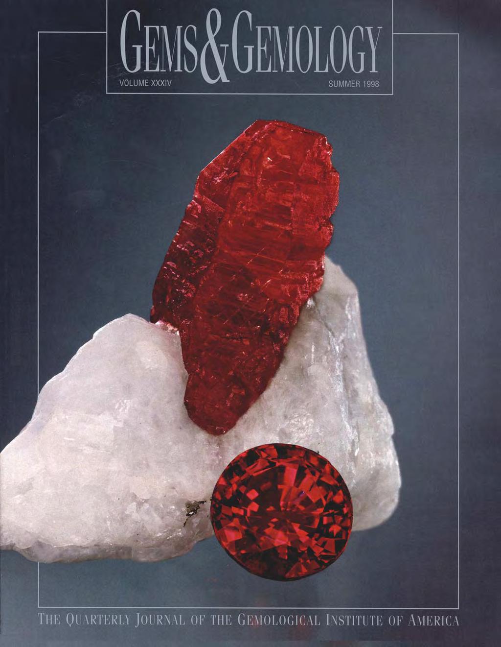
2 VOLUME 34 NO. 2 SUMMER 1998 T A B L E O F C O N T E N T S EDITORIAL 79 Support Your Local Researcher Alice S. Keller pg. 97 pg. 109 FEATURE ARTICLE 80 Separating Natural and Synthetic Rubies on the Basis of Trace-Element Chemistry Sam Muhlmeister, Emmanuel Fritsch, James E. Shigley, Bertrand Devouard, and Brendan M. Laurs NOTES AND NEW TECHNIQUES 102 Raman Investigations on Two Historical Objects from Basel Cathedral: The Reliquary Cross and Dorothy Monstrance Henry A. Hänni, Benno Schubiger, Lore Kiefert, and Sabine Häberli 114 Topaz, Aquamarine, and Other Beryls from Klein Spitzkoppe, Namibia Bruce Cairncross, Ian C. Campbell, and Jan Marten Huizenga REGULAR FEATURES 127 Gem Trade Lab Notes 134 Gem News 147 Thank You, Donors 148 Book Reviews 150 Gemological Abstracts pg. 115 pg. 145 ABOUT THE COVER: One of the greatest challenges for the contemporary gemologist is the separation of natural and synthetic rubies, especially when there are no characteristic internal features. A further consideration in many markets is identifying the deposit or country of origin of the ruby. The lead article in this issue reports the results of a comprehensive study on the trace-element chemistry (as determined by EDXRF analysis) of synthetic rubies from various manufacturers and natural rubies from several different deposits. The natural ruby crystal shown here on a calcite marble matrix is from Jegdalek, Afghanistan, and measures 17 x 10 x 9 mm. The accompanying 1.29 ct faceted ruby is from Mogok, Myanmar. Both are courtesy of the collection of Michael M. Scott. Photo Harold & Erica Van Pelt Photographers, Los Angeles, California. Color separations for Gems & Gemology are by Pacific Color, Carlsbad, California. Printing is by Fry Communications, Inc., Mechanicsburg, Pennsylvania Gemological Institute of America All rights reserved. ISSN X


3 SUPPORT YOUR LOCAL RESEARCHER Last February, many leading gemologists attended the very successful First World Emerald Congress in Bogotá, Colombia. Of particular concern to the participants was the topic of emerald treatments that is, the use of different fillers to improve the apparent clarity of the stone. All were aware of the negative publicity that emerald filling has received in recent months, and of the fact that some fillers are less effective or less durable than others. At the Congress, representatives from many of the major gemological laboratories sat together on a single panel to discuss this problem. A constant refrain from the audience, which included dealers as well as retailers, was: When were the GIA Gem Trade Laboratory, the Gübelin Gemmological Laboratory, SSEF, AGTA, and even Gems & Gemology, among others, going to resolve the treatment issue? We, however, cannot address such problems without help from you in the trade. A meaningful gemological study whether done in the U.S., Europe, Asia, or South America often requires large numbers of samples, hundreds of hours of testing, and the participation of several experts to analyze the results. For example, the trace-element-chemistry project reported by Sam Muhlmeister and colleagues in this issue used 283 natural and synthetic rubies, from 14 localities and 12 synthetics manufacturers. As Dr. Mary Johnson reported at the Emerald Congress, the emerald treatment study that GIA is currently spearheading required almost 300 emerald samples, all natural, from a number of known localities. Several fillers are being tested by various means to determine both their effectiveness and their durability under normal conditions of wear and care. Often the greatest delay in conducting such a study is in acquiring the samples. Unlike universities or government research institutions, the research conducted by most gemological laboratories is not supported by outside funding. You are the experts on buying and selling gems, so most of you know what it costs to purchase 300 one-half to one carat emeralds or rubies. In addition, even though for many projects the samples do not have to be large or expensive, it is critical that they be representative of the particular locality, manufacturing process, or treatment under investigation information that often can be provided only by members of the trade. As a result, researchers may have to work for years to obtain the stones for a single research project. For the ruby study, some samples were donated, many were loaned, and others were purchased. Our hats are off to all of the people who participated (more than 30 individuals and companies for that study alone). Still, some dealers leave request letters unanswered or make promises they cannot keep, consuming precious time. Then, when the pressure is on from consumers and the media, these same dealers want to know what s taking so long. As the gem and jewelry industry is faced with ever-morepressing issues regarding the disclosure of treatments and synthetics, comprehensive research will become even more important. Knowledge gained from the scientific study of gem materials and their treatments is necessary to maintain confidence in the industry by our colleagues and consumers alike. The use of broad, representative sets of samples, accompanied by reliable information about the sources (or treatments) of the stones, is critical to many of these research projects. So, please, think about it. And the next time you re asked, send some stones, provide information, share your experience and... support your local researcher. Alice S. Keller EDITOR Editorial GEMS & GEMOLOGY Summer
4 SEPARATING NATURAL AND SYNTHETIC RUBIES ON THE BASIS OF TRACE-ELEMENT CHEMISTRY By Sam Muhlmeister, Emmanuel Fritsch, James E. Shigley, Bertrand Devouard, and Brendan M. Laurs Natural and synthetic gem rubies can be separated on the basis of their trace-element chemistry as determined by energy-dispersive X-ray fluorescence (EDXRF) spectrometry. This method is especially important for rubies that do not have diagnostic inclusions or growth features, since such stones are difficult to identify using traditional gem testing methods. The results of this study indicate that the presence of nickel, molybdenum, lanthanum, tungsten, platinum, lead, or bismuth proves synthetic origin, but these elements were not detectable in most of the synthetic rubies tested. Alternatively, the concentrations of titanium, vanadium, iron, and gallium considered together, as a trace-element signature provide a means for separating nearly all synthetic from natural rubies. EDXRF can also help identify the geologic environment in which a ruby formed, and thus imply a geographic origin. ABOUT THE AUTHORS Mr. Muhlmeister (smeister@gia.edu) is research associate, and Dr. Shigley is director, at GIA Research in Carlsbad, California. Dr. Fritsch (fritsch@cnrs-imn.fr) is professor of physics at the University of Nantes, France. Dr. Devouard is assistant professor at Clermont-Ferrand University, France. Mr. Laurs, a geologist and gemologist, is senior editor of Gems & Gemology, Gemological Institute of America, Carlsbad. Please see acknowledgments at end of article. Gems & Gemology, Vol. 34, No. 2, pp Gemological Institute of America Correct gem identification is crucial to the gem and jewelry trade. However, accurate information on a gem s origin rarely accompanies a stone from the mine, or follows a synthetic through the trade after it leaves its place of manufacture. Today, natural and synthetic rubies from a variety of sources are seen routinely (figure 1). Usually, careful visual observation and measurement of gemological properties are sufficient to make important distinctions (Schmetzer, 1986a; Hughes, 1997). In some cases, however, traditional gemological methods are not adequate; this is particularly true of rubies that are free of internal characteristics or that contain inclusions and growth features that are ambiguous as to their origin (Hänni, 1993; Smith and Bosshart, 1993; Smith, 1996). The consequences of a misidentification can be in the tens of thousands, and even hundreds of thousands, of dollars. Ruby is a gem variety of corundum (Al 2 ) that is colored red by trivalent chromium (Cr 3+ ). Besides Cr, most rubies contain other elements in trace amounts that were incorporated during their growth, whether in nature or in the laboratory. For the purpose of this article, we consider trace elements to be those elements other than aluminum, oxygen, and chromium. These trace elements (such as vanadium [V] and iron [Fe]) substitute for Al 3+ in the corundum crystal structure, or they may be present as various mineral inclusions (such as zirconium [Zr] in zircon) or as constituents in fractures. The particular assemblage of trace elements (i.e., which ones are present and their concentrations) provides a distinctive chemical signature for many gem materials. Since the trade places little emphasis on establishing the manufacturer of synthetic products, this article will focus on how trace-element chemistry, as determined by EDXRF, can be used for the basic identification of natural versus synthetic rubies. It will also explore how EDXRF can 80 Separating Rubies GEMS & GEMOLOGY Summer 1998

5 Figure 1. These six natural and synthetic rubies are typical of material that might be submitted to a gemological laboratory for identification. From top to bottom and left to right: 1.29 ct Kashan flux-grown synthetic ruby, 1.10 ct natural ruby, 0.93 ct Czochralski-pulled synthetic ruby, 0.95 ct Ramaura synthetic ruby, 1.05 ct natural ruby from Tanzania, and 0.57 ct Swarovski flame-fusion synthetic ruby with flux-induced fingerprints. The 1.10 ct natural ruby was reported to have come from Mogok, but trace-element chemistry indicated that the stone was from a basalt-hosted deposit (such as Thailand); microscopy also indicated a basaltic origin. Photo GIA and Tino Hammid. help determine the geologic origin of a natural ruby, which is useful for identifying the country of origin. BACKGROUND Previous research has indicated the potential of trace-element chemistry for separating natural from synthetic rubies (Hänni and Stern, 1982; Stern and Hänni, 1982; Kuhlmann, 1983; Schrader and Henn, 1986; Tang et al., 1989; Muhlmeister and Devouard, 1991; Yu and Mok, 1993; Acharya et al., 1997), and also for differentiating rubies from different localities (Harder, 1969; Kuhlmann, 1983; Tang et al., 1988, 1989; Delé-Dubois et al., 1993; Osipowicz et al., 1995; Sanchez et al., 1997). The two most common analytical techniques for determining ruby chemistry are electron microprobe and EDXRF. Other methods include wavelength dispersive X-ray fluorescence spectrometry, neutron activation analysis, proton-induced X-ray emission (PIXE) analysis, and optical emission spectroscopy. Most of these methods involve instrumentation that is not readily available to gemological laboratories, or use techniques that may damage the sample. EDXRF, however, is nondestructive and has become standard equipment in many gemological laboratories. Qualitative EDXRF analyses have shown that synthetic rubies contain relatively few trace elements, and that the presence of molybdenum (Mo) indicates a flux origin; natural rubies, on the other hand, show a greater number of trace elements, and Myanmar rubies show relatively low Fe and high V (Muhlmeister and Devouard, 1991; Yu and Mok, 1993). Analyses by electron microprobe (Delé- Dubois et al., 1993) and PIXE (Tang et al., 1988, 1989; Sun, 1992; Osipowicz et al., 1995) have revealed the same trends for Myanmar rubies, and have indicated that Thai stones show high Fe and low V; these studies have also shown significant variations in trace elements in rubies from the same deposit. PIXE analyses of rubies from several deposits have detected numerous trace elements, including silicon (Si), sulfur (S), chlorine, potassium Separating Rubies GEMS & GEMOLOGY Summer
 are from Mong Hsu, Myanmar, which first became known only in the early 1990s. Photo Tino Hammid.")
6 Figure 2. The discovery of new gem localities presents constant challenges for the gemologist. These two rubies (2.59 ct total weight) are from Mong Hsu, Myanmar, which first became known only in the early 1990s. Photo Tino Hammid. (K), calcium (Ca), titanium (Ti), V, Cr, manganese (Mn), Fe, and gallium (Ga); in contrast, with the exception of Cr, synthetic stones from Chatham, Inamori, and Seiko showed no significant trace elements, Kashan synthetic rubies showed some Ti, and Knischka and Ramaura synthetics contained Fe (Tang et al., 1989). Using optical emission spectroscopy, Kuhlmann (1983) reported the following trace elements (besides Cr) at levels >0.001 wt.% in these synthetics: Chatham and Verneuil Fe, Si, Mo, beryllium (Be); Knischka Fe, Si, Be, copper (Cu); Kashan Si, Ti, Fe, Cu, Mn, Be. In natural rubies, the following (besides Cr) were detected at levels >0.001 wt.%: Mogok, Myanmar V, Fe, Ti, Si, tin (Sn), Mn, Be; Jegdalek, Afghanistan Si, Fe, Be; Umba Valley, Tanzania Fe, Si, Ti, Cr, V, Mn; Morogoro, Tanzania Ti, Si, Cr, Fe, V; Tsavo Park, Kenya Cr, Fe, Si, Ti, V; Rajasthan, India Fe, Si, Ti, Sn, Cu, V, Mn, Be. In recent years, a number of new synthetic ruby products have entered the market, including those grown by the flux method (Smith and Bosshart, 1993; Henn and Bank, 1993; Henn, 1994; Hänni et al., 1994); by the hydrothermal method (Peretti and Smith, 1993); and by the Czochralski technique with natural ruby as the feed material, marketed as recrystallized (Kammerling et al., 1995b,c; Nassau, 1995). Other synthetic materials that have caused identification problems for the contemporary gemologist include flame-fusion synthetic rubies with flux-induced fingerprints (Koivula, 1983; Schmetzer and Schupp, 1994; Kammerling et al., 1995a) and Czochralski-pulled synthetic rubies that are inclusion-free (Kammerling and Koivula, 1994). Natural rubies can also present identification problems. Heat treatment can significantly alter the appearance of the inclusions in natural ruby (Gübelin and Koivula, 1986; Themelis, 1992; Hughes, 1997). Fluid inclusions commonly rupture when heated, creating fractures that can be subsequently filled by the fluid or partially repaired on cooling (Gübelin and Koivula, 1986). These secondary fingerprints common in heat-treated rubies from Mong Hsu, Myanmar, for example are similar to the white, high-relief fingerprint-like inclusions commonly seen in flux synthetics (Laughter, 1993; Smith and Surdez, 1994; Peretti et al., 1995). In addition, the locality of a natural ruby can have a significant impact on its market value (figure 2). Although the importance of country of origin in helping establish a natural ruby s value is a topic of ongoing debate (Liddicoat, 1990; Hughes, 1990a), it is nonetheless a significant consideration in some trading circles, especially for larger stones. GEOLOGIC ORIGINS OF GEM-QUALITY RUBIES Rubies are mined from both primary deposits (the host rock where the ruby formed) and secondary deposits (i.e., alluvial stream transported, or eluvial weathered in place) [Simonet, 1997]. Because of its durability and relatively high specific gravity, ruby is readily concentrated into secondary deposits; the processes of weathering and alluvial transport tend to destroy all but the best material, so these deposits can be quite valuable. In general, primary deposits are economically important only if the ruby is concentrated in distinct mineable areas, such as layers within marble. The conditions under which ruby forms are important to gemologists, because the chemical composition, inclusions, and growth features visible in fashioned stones are influenced by the composition, pressure, and temperature of the ruby-forming environment (see, e.g., Peretti et al., 1996). The trace elements in a ruby are incorporated into the crystal structure, or are present as mineral inclusions (table 1) or as constituents in fractures. Therefore, the geologic environment influences the assemblage of trace elements present. Corundum (Al 2 ) crystallizes only in silica-deficient environments because, in the presence of Si, the Al is used to form more common minerals such as kyanite, 82 Separating Rubies GEMS & GEMOLOGY Summer 1998
7 TABLE 1. Geology and mineralogy of gem-quality ruby deposits. Deposit type Geology Typical associated Typical mineral Localities a minerals inclusions Basalt-hosted Xenocrysts in Sapphire, clino- Pyrrhotite (com- Australia Barrington (Sutherland, 1996a,b; Suthalkali basalt (al- pyroxene, zircon, monly altered to erland and Coenraads, 1996; Webb, 1997; luvial and eluvial Fe-rich spinel, gar- goethite), apatite; Sutherland et al., 1998) deposits) net; sometimes sometimes spinel, Cambodia Pailin (Jobbins and Berrangé, 1981; sapphirine almandine garnet Sutherland et al., 1998) Thailand Chanthaburi, at Bo Rai Bo Waen (Charaljavanaphet, 1951; Gübelin, 1971; Keller, 1982; Vichit, 1987; Coenraads et al., 1995) Vietnam southern, at Dak Nong (Kane et al., 1991; Poirot, 1997) Marble-hosted Calcite or dolo- Calcite, dolomite, Calcite, dolomite, Afghanistan Jegdalek (Hughes, 1994; Bowersox mite marble, com- spinel, pargasite, rutile, apatite, py- and Chamberlin, 1995) monly interlayered phlogopite, rutile, rite, phlogopite, China Aliao Mountains, Yunnan Province (Keller with schists and Cr-muscovite, boehmite; some- and Fuquan, 1986; ICA Gembureau, 1991) gneisses; may or chlorite, tremolite, times spinel, zir- India southern (Viswanatha, 1982) may not be cut tourmaline, apatite, con, margarite, Myanmar Mogok (Iyer, 1953; Keller, 1983; Gübelin by granite intru- sphene, anorthite, graphite, pyrrho- and Koivula, 1986; Kammerling et al., 1994) sions (primary margarite, pyrrhot- tite Myanmar Mong Hsu (Peretti and Mouawad, 1994; and alluvial de- ite, pyrite, ilmenite, Smith and Surdez, 1994; Peretti et al., 1995) posits) graphite, fluorite Nepal Taplejung district (Harding and Scarratt, 1986; Smith et al., 1997) Pakistan Hunza (Bank and Okrusch, 1976; Okrusch et al., 1976; Gübelin, 1982; Hunstiger, 1990b) Pakistan Kashmir ( Kashmir Yields Ruby, Tourmaline, 1992; Kane, 1997) Russia Ural Mountains (Kissin, 1994) Tanzania Morogoro (Hänni and Schmetzer, 1991) Tajikistan eastern Pamir Mtns. (Henn et al., 1990b; Smith, 1998) Vietnam northern, at Luc Yen (Henn and Bank, 1990; Henn, 1991; Kane et al., 1991; Delé-Dubois et al., 1993; Poirot, 1997) Metasomatic Desilicated peg- Plagioclase, ver- Rutile, boehmite; India southern (Viswanatha, 1982; Menon et al., matite cutting miculite, phlogo- sometimes apatite, 1994) ultramafic rock pite, muscovite, zircon, spinel, ver- Kenya Mangari (Pohl et al., 1977; Pohl and Horor marble (pri- tourmaline (highly miculite, musco- kel, 1980; Bridges, 1982; Hunstiger, 1990b; mary, eluvial, variable) vite, pyrrhotite, Levitski and Sims, 1997) and alluvial de- graphite (highly Tanzania Umba (Solesbury, 1967; Zwaan, 1974; posits) variable) Hänni, 1987) Vietnam central, at Quy Chau (Kane et al., 1991; Poirot, 1997) Metasomatic Desilicated alu- Plagioclase, amphi- Rutile, plagioclase, India southern (Viswanatha, 1982; Hunstiger, 1990a) minous schist/ bole, epidote, tour- apatite, zircon, Kenya Mangari (Pohl et al., 1977; Pohl and Horgneiss adjacent maline, micas, silli- graphite, sillimanite, kel, 1980; Hunstiger, 1990b; Key and Ochieng, to ultramafic rock manite, kyanite amphibole, ilmen- 1991; Levitski and Sims, 1997) (primary, eluvial, (highly variable) ite (highly variable) Malawi Chimwadzulu Hill (Rutland, 1969; Henn and alluvial de- et al., 1990a) posits) Sri Lanka Ratnapura, Elahera (Dahanayake and Ranasinghe, 1981, 1985; Munasinghe and Dissanayake, 1981; Dahanayake, 1985; Rupashinge and Dissanayake, 1985; Gunawardene and Rupasinghe, 1986) a Although there are known deposit types in the countries listed, there may also be deposits (e.g., alluvial) that have not yet been characterized. Consequently, we do not know the specific geologic environment for all of the rubies obtained for this study. feldspars, and micas. The scarcity of gem corundum especially ruby results from this requirement for Si-depleted conditions in the presence of the appropriate chromophore(s) (i.e., Cr for ruby and Fe and Ti for blue sapphire), under the appropriate temperature and pressure conditions. The mechanisms by which gem corundum forms are still debated by geologists (see, e.g., Levinson and Cook, Separating Rubies GEMS & GEMOLOGY Summer
: basalt-hosted, marble-hosted, and metasomatic.")
8 Figure 3. This specimen (4.6 cm high) from Jegdalek, Afghanistan, shows a ruby embedded in calcite marble, along with traces of associated minerals. Courtesy of H. Obodda; photo Jeffrey Scovil. 1994). With the exception of a few occurrences, gem-quality ruby has been found in three types of primary deposits (again, see table 1): basalt-hosted, marble-hosted, and metasomatic. (The latter two form by different metamorphic processes, as explained below.) Basalt-Hosted Deposits. Some of the world s largest ruby deposits, past and present, are secondary deposits associated with alkali basalts (e.g., Cambodia and Thailand). However, the formation conditions of this type of corundum are the least understood. The occurrence of corundum in basalt is similar to that of diamond in kimberlite: The ruby and sapphire crystals (xenocrysts) are transported in molten rock from lower levels of the earth s crust (or upper mantle) to the surface. During transport from depths of km (Levinson and Cook, 1994), the corundum is partially resorbed, as shown by the rounded edges and surface etch patterns typically seen on basalt-hosted corundum (Coenraads, 1992). From evidence revealed by trace elements, mineral inclusions, fluid inclusions, and associated mineral assemblages observed in rare corundumbearing assemblages (as xenoliths), geologists have suggested that both metamorphic- and igneousformed rubies may be present in a given basalt-hosted deposit (see, e.g., Sutherland and Coenraads, 1996; Sutherland et al., 1998). The metamorphic rubies in such deposits may be derived from regional metamorphism of aluminous rocks (such as shales, laterites, and bauxites) that were subducted to great depths (Levinson and Cook, 1994). Two models have been proposed for the igneous origin of gem corundum: (1) from magma mixing at midcrustal levels (Guo et al., 1996a,b), or (2) from the pegmatite-like crystallization of silica-poor magma in the deep crust or upper mantle (Coenraads et al., 1995). Regardless of the specific origin, the mineral inclusions and associated minerals suggest that the corundum formed in an environment containing Fe, S, and geochemically incompatible elements (Coenraads, 1992). This geochemical (or trace-element) signature is consistent with the relatively enriched Fe content that is characteristically shown by basalt-hosted rubies (see Results section, below). Marble-Hosted Deposits. Some of the world s finest rubies form in marble-hosted deposits, such as those in Myanmar. These deposits are commonly thought to have formed as a result of the regional metamorphism of limestone by heat and pressure (see, e.g., Okrusch et al., 1976). At some localities, the close association of granitic intrusions with the ruby-bearing marble has led some researchers to consider them metasomatic (e.g., Mogok; Iyer, 1953); that is, chemical interaction between the marble and fluids associated with the intrusions caused ruby mineralization. Therefore, marble-hosted ruby deposits may form from regional metamorphism or contact metasomatism, or from a combination of the two (Konovalenko, 1990). Depending on the composition of the original limestone, ruby-bearing marbles may be composed of calcite (CaC ) and/or dolomite [CaMg(C ) 2 ]. Ruby and associated minerals, such as spinel and micas (again, see table 1), form in layers that are irregularly distributed within the marble (figure 3). These ruby-bearing assemblages are thought to represent impure horizons within the original limestone, where Al-rich clays or sediments (such as bauxite) were deposited (see, e.g., Platen, 1988; Okrusch et al., 1976). The composition of the asso- 84 Separating Rubies GEMS & GEMOLOGY Summer 1998
 are prized for their pure red color (i.e., absence of brown modifying hues), which is supposedly due to their lack of iron; this is consistent with the low iron content of their marble host rocks.")
9 ciated minerals (table 1) indicates that these impure layers contain traces of Si, S, K, Ti, V, Cr, and, in general, are low in Fe. Rubies from certain marbletype deposits (such as Mogok) are prized for their pure red color (i.e., absence of brown modifying hues), which is supposedly due to their lack of iron; this is consistent with the low iron content of their marble host rocks. Metasomatic Deposits. Metasomatism is a metamorphic process whereby chemical components are exchanged in the presence of fluids. One important mechanism is desilication, in which Si is mobilized (i.e., removed from the rock), leaving Al behind to form corundum. Metasomatic deposits of gem ruby can be divided into two groups: (1) desilicated pegmatites intruding silica-poor rocks such as serpentinite (as, e.g., at Umba, Tanzania: Solesbury, 1967) or marble (as at Quy Chau, Vietnam; Poirot, 1997); and (2) desilicated schists and gneisses that have been altered by metasomatic fluids in the presence of ultramafic (low Si; high Mg, Fe) rock (e.g., Malawi: Rutland, 1969; again, see table 1). As stated above, the ruby deposits at Mogok may also be metasomatic; if so, they would constitute a third type of metasomatic deposit marble that has been altered by pegmatite-derived fluids. The trace-element chemistry of rubies formed in metasomatic deposits is variable because of the different rock types and the particular local geochemical conditions under which these rubies formed (see, e.g., Kuhlmann, 1983). For example, rubies from different metasomatic deposits at Mangari, Kenya, show different characteristics: At the John Saul mine, pure red ruby coloration is common; at the Penny Lane deposit, the ruby is darker (Bridges, 1982; Levitski and Sims, 1997). These color variations most likely result from the variable composition of the host rocks; that is, at John Saul, the host rocks contain lower concentrations of Fe than at Penny Lane. Other metasomatic-type ruby localities with variable host rocks are in India (see, e.g., Viswanatha, 1982) and Sri Lanka (see, e.g., Dahanayake and Ranasinghe, 1985; Rupasinghe and Dissanayake, 1985). MANUFACTURE OF SYNTHETIC RUBY Synthetic rubies were introduced to the gem market in Originally sold as natural rubies from a fictitious mine near Geneva, and hence named Geneva ruby, they were later proved to be synthetic (Nassau and Crowningshield, 1969; Nassau, Figure 4. Synthetic rubies were first manufactured more than 100 years ago. Today, several types are available in the gem marketplace. Here, a Chatham flux-grown synthetic ruby is set in 14k gold. Ring courtesy of Chatham Created Gems, photo Tino Hammid. 1980, 1995). The producer of these early synthetic rubies was never disclosed. The first scientific paper describing ruby synthesis using the flame-fusion process was published by Auguste Verneuil in 1904, two years after his first success. The flame-fusion process used today to grow rubies is largely unchanged from the one Verneuil introduced at the turn of the century (Nassau and Crowningshield, 1969; Nassau, 1980; Hughes, 1990b). Although flame-fusion material is still the most common synthetic corundum used in jewelry, other techniques are commercially available (figure 4; table 2). Ruby manufacturing methods can be divided into two general categories: melt and solution. Melt-grown synthetic rubies including flamefusion, Czochralski, and floating zone are produced by melting and crystallizing aluminum oxide powder to which traces of Cr and (possibly) various trace elements have been added. With the Czochralski and floating-zone methods, crystallization typically takes place in an iridium crucible; with flame-fusion, crystallization occurs on a rotating boule, without a container. The chemistry of melt-grown synthetic rubies is usually relatively pure that is, they contain detectable amounts of relatively few elements in comparison to other synthetic rubies, because the melt generally contains few additives. Separating Rubies GEMS & GEMOLOGY Summer
10 Solution-grown synthetic rubies are crystallized from a solution in which aluminum and trace elements are dissolved. There are two types of growth solutions: flux and hydrothermal. A variety of chemical fluxes are used (again, see table 2), and the solutions are contained within a metal crucible, usually platinum. Hydrothermal synthetic rubies are grown from a water-rich solution enclosed in a pressurized autoclave. These synthetic rubies may contain trace elements originating from the flux or the hydrothermal solution, and possibly from the crucible or autoclave. MATERIALS AND METHODS Materials. For this study, we examined 121 natural and 162 synthetic rubies, most of which were faceted. These samples were chosen to represent the known commercially available sources of gem-quality material. The stones ranged from 0.14 to ct, with most samples between 0.50 and 3.00 ct. The majority were transparent, but some were translucent; many had eye-visible inclusions. The samples were provided by individuals who had reliable information on the country of origin, although in some cases the specific deposit was not known. Note that some corundums with Lechleitner synthetic ruby overgrowth were included in this study, although they are not true synthetic rubies (Schmetzer, 1986b; Schmetzer and Bank, 1987). Red diffusion-treated corundum and recrystallized synthetic ruby were also analyzed. At least seven representative samples were obtained for most localities and manufacturers, although the actual number varied from one to 31 for each type. Due to the limited number of samples from some deposits or manufacturers, and the possible future compositional variations of these rubies, the results of this study should only be considered an indication of the chemistry from these sources. Also, the trace-element data in this study are valid only for rubies, and do not necessarily apply to corundum of other colors (including pink). The fol- TABLE 2. Manufacturing methods and possible sources of trace elements in synthetic rubies. Manufacturing method/ Growth material References manufacturer a Melt Feed Czochralski-pulled Alumina and Cr 2 Rubin and Van Uitert, 1966; Nassau, 1980 Flame-fusion Alumina and Cr 2 Verneuil, 1904; Nassau, 1980; Yaverbaum, 1980 Floating zone Alumina and Cr 2 Nassau, 1980; Sloan and McGhie, 1988 Induced fingerprint (Flame-fusion synthetic rubies with Koivula, 1983; Schmetzer and Schupp, 1994; Kammerling et al., induced fingerprints by any of 1995a various fluxes) Solution Flux Fluxes Chatham Li 2 O-Mo -PbF 2 and/or PbO Schmetzer, 1986b Douros PbF 2 or PbO 4 Smith and Bosshart, 1993; Hänni et al., 1994 Kashan Na 3 AlF 6 Henn and Schrader, 1985; Schmetzer, 1986b; Weldon, 1994 Knischka Li 2 O-W -PbF 2, PbO, Knischka and Gübelin, 1980; Schmetzer, 1986b, 1987; Galia, Na 2 W 2 O 7, and Ta 2 O ; Brown and Kelly, 1989 Lechleitner Flux overgrowth (using Li 2 O-Mo - Schmetzer, 1986b; Schmetzer and Bank, 1987 PbF 2 and/or PbO flux) on natural corundum Ramaura Bi 2 -PbF 2, also rare-earth Kane, 1983; Schmetzer, 1986b dopant b added to flux as well as La 2 O c 3 Solution Hydrothermal Solution Tairus (Russia) Alumina or aluminum hydrates Nassau, 1980; Yaverbaum, 1980; Peretti and Smith, 1993; partially dissolved in an aqueous Peretti et al., 1997; Qi and Lin, 1998 medium with Cr compounds such as Na 2 Cr 2 O 7 a The following containers are typically used during manufacture: Melt iridium crucible (except no container is used for flame-fusion), Flux platinum crucible, Hydrothermal metal autoclave containing Fe, Ni, and Cu, possibly lined with silver, gold, or platinum. b Produces a yellow fluorescence, which is usually absent in faceted material (Kane, 1983). c Promotes the growth of facetable crystals. 86 Separating Rubies GEMS & GEMOLOGY Summer 1998
11 lowing were not included in the study samples: material from localities not known to provide crystals suitable for faceting, synthetic rubies grown for experimental purposes only, and phenomenal rubies (e.g., star rubies). Although it is possible to obtain analyses of larger mounted stones with EDXRF, we did not include mounted samples. Methods. We used a TN Spectrace 5000 EDXRF spectrometer for the chemical analyses. This instrument can detect the elements sodium (atomic number 11) through uranium (atomic number 92), using an X-ray tube voltage of up to 50 kv and a current of 0.01 ma to 0.35 ma. Two sets of analytical conditions were used to maximize sensitivity: for sodium (atomic number 11) through sulfur (atomic number 16) a voltage of 15 kv, current of 0.15 ma, no filter, and a count time of 200 seconds; for chlorine (atomic number 17) through bromine (atomic number 35) a voltage of 25 kv, current of 0.25 ma, mm aluminum filter, and a count time of 200 seconds. Each sample was run once under the two separate instrument conditions, in order to generate one analysis. We used the spectra derived from these procedures to obtain semi-quantitative data for the elements Al, Ca, Ti, V, Cr, Mn, Fe, and Ga using the Fundamental Parameters (FP) method of Criss and Birks (1968; see also Jenkins, 1980). (Oxygen was assumed present in stoichiometric proportions, and Fe is reported as FeO [i.e., +2 oxidation state]). In addition, the presence of heavier elements (e.g., Zr, Ni, Cu, Mo, lanthanum [La], tungsten [W], and lead [Pb]) was noted from the spectra, but these elements were not analyzed quantitatively. The samples were analyzed for Si; however, because of peak overlap with Al, results for Si were unreliable and are not reported in this study. The following standards were used for calibration and to check the instrument s performance: colorless Czochralski-pulled synthetic corundum for Al; tsavorite garnet for Al and Ca; and almandine garnet for Al, Mn, and Fe. The compositions of the standards were quantitatively determined at the California Institute of Technology by Paul Carpenter using an electron microprobe. We used a 3 mm diameter X-ray beam collimator to limit the beam size to about 20 mm 2. (However, the actual area analyzed varied depending on the size of the sample; on most, it was less than 20 mm 2.) This area was analyzed to a depth of approximately 0.1 mm. Whenever possible, we oriented each sample to avoid prominent color zoning or conspicuous mineral inclusions. We usually analyzed the table facet; for some samples, we analyzed the pavilion. We did not include analyses if we suspected that the presence of diffraction peaks caused erroneous quantitative results. Both the accuracy and the sensitivity of the analyses are affected by the size of the area analyzed, because this determines the number of counts obtained for a given element. Table 3 illustrates the differences in detection limits for rubies of three different sizes. In general, the detection limits decreased with increasing sample size. Above approximately 1 ct, there were only minor improvements in the detection limits. The concentrations of each element in the sample are calculated from the counting statistics and the FP algorithm, and they are expressed as weight percent oxides. The number of counts determined for each element had to be normalized to 100% to compensate for the different sample sizes. Because the concentrations reported are not directly calculated from the peak counts, our analyses are considered semi-quantitative. For comparison purposes, we had five natural rubies from our study analyzed by Dr. W. B. Stern of the University of Basel using his EDXRF. Dr. Stern s results for these samples were very similar to our own. Based on multiple analyses of three of the samples used in this study, we found the repeatability of the trace-element data to be generally within 10% 20%. RESULTS Qualitative Results. Synthetic Rubies. The following elements were detected in all of the synthetic TABLE 3. Variation in EDXRF detection limits according to sample size. a Oxide 0.20 ct 0.65 ct 1.27 ct (wt.%) Al Cr CaO TiO V MnO FeO Ga a Calculated after Jenkins, Separating Rubies GEMS & GEMOLOGY Summer
12 ruby growth types (but not all samples) we examined: Ca, Ti, Cr, Mn, and Fe. Ga and V were also detected in the products of all specific manufacturers except the one Inamori sample we examined. The greatest variety of trace elements was detected in the flux-grown synthetics. In addition to the elements noted above, the Chatham flux synthetic rubies had Mo (six out of 21 samples), the Douros contained Pb (seven out of 15), and the Knischka samples showed W (five out of 14). La was seen only in Ramaura flux-grown synthetic rubies (12 out of 31), as previously reported by Schmetzer (1986b); Pt was also detected in one Ramaura synthetic ruby, Pb in four, and Bi (bismuth) in three. All of the flame-fusion synthetic rubies with flux-induced fingerprints revealed traces of Mo or Zr; otherwise, the melt-grown synthetics contained no trace elements other than those mentioned above. Traces of Ni were detected in 15 samples, and Cu in six samples, of the Tairus hydrothermal synthetic rubies (as previously reported by Peretti and Smith, 1993; Peretti et al., 1997; and Qi and Lin, 1998). One Tairus sample also contained cobalt, which we did not detect in any of the other synthetic or natural samples. Natural Rubies. Traces of Ca, Ti, V, Cr, Mn, Fe, and Ga were noted in rubies from all of the localities we analyzed. Zr was detected in seven of the eight rubies analyzed from India, all of which contained numerous zircon inclusions. Cu was detected in one sample from Mong Hsu, Myanmar. Semi-Quantitative Results. In addition to Al, the following elements were analyzed semi-quantitatively: Ca, Ti, V, Cr, Mn, Fe, and Ga. The natural rubies we examined typically contained a greater variety and higher quantity of these elements than the synthetics (tables 4 and 5; figures 5 8), which is consistent with the results reported by Kuhlmann (1983) and Tang et al. (1989). Synthetic Rubies. Compared to many natural rubies, most of the synthetic samples contained little V and Ga (table 5; figures 5 and 6). In general, the melt-grown synthetic rubies contained the lowest concentrations of the elements listed above, especially Fe (figure 7; see also Box A). Two of the 10 flame-fusion samples had relatively high Ti (nearly 0.05 wt.% TiO 2 ; figure 8); one of these also contained relatively high V (nearly 0.02 wt.% V 2 ), and the other contained the highest Cr level measured on the synthetics in this study (2.41 wt.% Cr 2 ), with the exception of the red diffusion-treated corundum (see Box B). All the other synthetic rubies had Cr contents that ranged from about 0.07 to 1.70 wt.% Cr 2, without any discernable trends. The floating-zone synthetic rubies were the only TABLE 4. Summary of natural ruby chemistry. a Basalt-hosted Marble-hosted Metasomatic Oxide Mogok, Mong Hsu, Southern Umba Valley, (wt.%) Cambodia (1) b Thailand (15) Afghanistan (15) Myanmar (19) Myanmar (11) Nepal (3) Yunnan (2) Kenya (8) Tanzania (4) Al (99.06) (98.31) (99.27) (98.69) (99.57) (98.88) (98.22) (98.11) Cr (0.419) (0.350) (0.562) (0.887) (0.260) (0.950) (0.675) (0.372) CaO bdl bdl 3.11 bdl bdl bdl bdl (0.023) (0.784) (0.029) (0.030) (0.025) (0.068) (0.068) (0.023) TiO bdl (0.020) (0.045) (0.026) (0.078) (0.080) (0.016) (0.033) (0.009) V bdl bdl (0.004) (0.009) (0.066) (0.048) (0.017) (0.020) (0.018) (0.004) MnO bdl bdl bdl bdl bdl (0.005) (bdl) (0.004) (0.006) (0.010) (0.005) (0.006) FeO bdl (0.468) (0.070) (0.028) (0.010) (0.029) (0.042) (0.020) (1.46) Ga bdl bdl bdl (0.005) (0.006) (0.012) (0.009) (0.018) (0.008) (0.033) (0.014) a Values shown are normalized wt.%. Minimum and maximum values are given, along with the average (in parentheses below each range). bdl = below detection limits (varies according to size of sample; see table 3). b Number of samples in parentheses. 88 Separating Rubies GEMS & GEMOLOGY Summer 1998

13 Figure 5. Most synthetic rubies contain little vanadium compared to natural rubies. In general, rubies from marble-hosted deposits contained the highest V content, although those from Afghanistan showed atypically low V. (Note that the Czochralski samples in these graphs are all produced by Union Carbide, as distinct from the Inamori Czochralski-pulled sample that was also analyzed.) Unknown deposit type Morogoro, Tanzania, India (8) Tanzania (5) Sri Lanka (7) misc. (7) Vietnam (16) (99.10) (98.52) (99.14) (99.08) (98.70) (0.358) (1.30) (0.327) (0.713) (1.01) bdl bdl (bdl) (0.035) (0.024) (0.052) (0.062) (0.017) (0.032) (0.083) (0.031) (0.071) bdl (0.006) (0.011) (0.027) (0.008) (0.030) bdl bdl bdl (0.002) (0.010) (0.003) (0.004) (0.011) (0.502) (0.093) (0.178) (0.098) (0.103) (0.007) (0.007) (0.011) (0.013) (0.014) synthetic samples with a relatively high average V content (0.012 wt.% V 2 ). Variable trace-element contents were measured in the flux-grown synthetic rubies. The Chatham samples contained insignificant amounts of most trace elements, except for wt.% Ga 2 in one sample. The Douros synthetics contained elevated Fe ( wt.% FeO), and the highest concentration of Ga of all the natural and synthetic rubies tested for this study ( wt.% Ga 2 ). The Kashan synthetic rubies showed very low Fe, and they had the most Ti of all the synthetics analyzed ( wt.% TiO 2 ). One Knischka sample also contained elevated Ti (0.159 wt.% TiO 2 ), and another showed high Ga (0.071 wt.% Ga 2 ). The Knischka synthetic rubies contained about the same amount of Fe as the Douros samples, with the exception of one Knischka sample that contained 1.15 wt.% FeO; this anomalous sample contained the greatest amount of Fe of all the synthetics analyzed. Ramaura synthetic rubies typically contained some Ga (up to wt.% Ga 2 ) and Fe Separating Rubies GEMS & GEMOLOGY Summer
 were exceptions to this, with more than 0.050 wt.% Ga 2. TABLE 5.")
14 Figure 6. Gallium is another key trace element for separating natural from synthetic rubies. While fewer than one-seventh of the natural stones contained less than wt.% Ga 2, more than threequarters of the synthetic samples did. However, Douros synthetic rubies (and one Knischka sample) were exceptions to this, with more than wt.% Ga 2. TABLE 5. Summary of synthetic ruby chemistry. a Melt Flux Induced finger- Oxide Czochralski: Flame- Floating Czochralski: print flame- (wt.%) Union Carbide (10) e fusion (10) zone (16) Inamori (1) b fusion (4) Chatham (21) Douros (15) Kashan (15) Al (98.86) (99.15) (99.40) (98.84) (98.98) (99.32) (99.57) Cr (1.08) (0.788) (0.521) (1.12) (0.956) (0.484) (0.291) CaO bdl bdl bdl bdl bdl bdl bdl (bdl) (0.034) (0.024) (0.018) (0.040) bdl (0.023) TiO 2 bdl bdl bdl bdl bdl bdl (0.002) (0.013) (bdl) (0.009) (0.009) (0.013) (0.096) V 2 bdl bdl bdl bdl bdl bdl bdl bdl bdl (0.003) (0.012) (0.001) (0.005) MnO bdl bdl bdl bdl bdl (0.007) (0.005) (0.003) (0.007) (0.006) (0.004) (0.003) FeO bdl (0.003) (0.007) (0.006) (0.005) (0.010) (0.085) (0.007) Ga 2 bdl bdl bdl bdl bdl bdl bdl (0.001) (0.002) bdl bdl (0.002) (0.064) (0.001) a Values are normalized wt.%. Minimum and maximum values are given, along with the average (in parentheses below each range). bdl= below detection limits (varies according to size of sample; see table 3). b Number of samples in parentheses. 90 Separating Rubies GEMS & GEMOLOGY Summer 1998

15 Figure 7. Iron is also useful for separating natural from synthetic rubies, especially when used in conjunction with vanadium. Only one of the melt-grown synthetic rubies and approximately half of the flux-grown synthetic rubies displayed an FeO content greater than 0.02 wt.%, while three-quarters of the natural stones did. Hydrothermal synthetic rubies had the highest overall Fe contents of the synthetics we analyzed. Flux (cont d) Hydrothermal Ramaura c Ramaura d (FeO < 0.075) (FeO > 0.075) Knischka (14) Lechleitner (4) (15) (15) Tairus (21) (98.97) (99.00) (99.23) (99.01) (98.51) (0.690) (0.933) (0.688) (0.822) (1.12) bdl bdl bdl bdl bdl (0.034) (0.031) (0.033) (0.039) (0.030) bdl bdl bdl (0.023) (0.026) bdl bdl (0.023) bdl bdl bdl bdl bdl bdl bdl bdl bdl bdl bdl BDL (0.006) (0.008) (0.006) (0.005) (0.008) (0.159) (0.006) (0.037) (0.112) (0.302) bdl bdl bdl bdl (0.008) bdl (0.005) (0.008) (0.001) c Includes all Burma tone samples. d Includes all Thai tone samples. ( wt.% FeO), with those Ramaura synthetic rubies marketed as Thai tone richer in Fe than those marketed as Burma tone. The Tairus hydrothermal synthetic rubies contained relatively high Fe (up to wt.% FeO) and traces of Ti (up to wt.% TiO 2 ). Natural Rubies. In general, there was greater variability in the trace-element chemistry of the natural rubies than of the synthetics, but there were also some distinct trends according to geologic occurrence. Basalt-hosted rubies were the most consistent: Samples from Thailand and Cambodia showed relatively high Fe ( wt.% FeO) and low V (up to wt.% V 2 ). Marble-hosted rubies showed the opposite trend: low Fe (up to wt.% FeO) and high V (typically wt.% V 2 ), with the exception of Afghan rubies, which showed low V contents (less than 0.02 wt.% V 2 ). The Afghan rubies contained the highest levels of Ca measured in this study (up to 3.11 wt.% CaO). Rubies from Myanmar contained the most V measured in this study (up to wt.% V 2 ). Some Separating Rubies GEMS & GEMOLOGY Summer
 ruby weigh 0.45 ct (rounds) and 0.65 ct (oval). Photo by Maha DeMaggio.")
16 BOX A: TRACE-ELEMENT CHEMISTRY OF RECRYSTALLIZED OR RECONSTRUCTED RUBY Figure A-1. The trace-element chemistry of TrueRuby most closely resembles that of melt-grown synthetic ruby. These samples of TrueGem s recrystallized (synthetic) ruby weigh 0.45 ct (rounds) and 0.65 ct (oval). Photo by Maha DeMaggio. In late 1994 and early 1995, the trade was introduced to recrystallized Czochralski-pulled synthetic ruby by TrueGem of Las Vegas, Nevada, and Argos Scientific of Temecula, California (Kammerling et al., 1995b,c). Reconstructed rubies of South African origin have also been reported (Brown, 1996). TrueGem sells its synthetic ruby product under the trademarked name TrueRuby. In its promotional material, TrueGem has claimed that its process enhances the clarity of ruby and sapphire to perfection, while retaining nature s elemental formula for the finest colored stones ever mined. In this process, natural ruby is reportedly used as feed material, rather than the alumina used by most corundum manufacturers. The final step in the growth process is Czochralski pulling. We analyzed four TrueGem synthetic rubies (see, e.g., figure A-1, table A-1). These samples showed a composition that is different from any of the natural rubies we analyzed: Cr is high, and the absence of V combined with low Fe precludes natural origin. The trace-element chemistry of this material is similar to that of the melt-grown synthetic rubies we examined, except for the enriched Cr contents. This result contradicts the manufacturer s claim that this ruby product retains nature s elemental formula. In agreement with the arguments of others (e.g., Nassau, 1995), the GIA Gem Trade Laboratory calls this material synthetic ruby on its reports (T. Moses, pers. comm., 1998). TABLE A-1. Trace-element chemistry of TrueGem recrystallized rubies. Oxide Specimen number Average (wt.%) Al Cr CaO TiO 2 bdl bdl V 2 bdl bdl bdl bdl bdl MnO FeO Ga Mong Hsu samples contained up to wt.% TiO 2 (high Ti was also reported by Smith and Surdez [1994] and Peretti et al. [1995]). The rubies from metasomatic deposits showed wide variations in composition. Samples from Umba in Tanzania had the greatest Fe measured in this study ( wt.% FeO), combined with low V and Ti. Rubies from Kenya, however, showed low Fe and the most Ga ( wt.% Ga 2 ) measured in the natural samples. DISCUSSION Chemical Variations in Synthetic and Natural Rubies. Synthetic Rubies. Verneuil s original flamefusion process required that the feed powder used be free of Fe, since Fe caused the boule to turn brown (Verneuil, 1904; Nassau, 1980). Today s melt-grown products are still essentially Fe-free, and they typically contain none or very small amounts of other elements besides Cr. Flux-grown synthetic rubies revealed the most trace elements, which is consistent with the variety of fluxes used to manufacture these synthetics (again, see table 2). Li, F, Na, Al, Mo, Ta (tantalum), W, Pb, or Bi may be present in the flux in which these rubies were grown or treated (for synthetic rubies with induced fingerprints ) [Schmetzer, 1986a,b, 1987; Schmetzer and Schupp, 1994; Kammerling et al., 1995a]. Lanthanum has been used experimentally to help grow crystals with rhombohedral rather than flat, tabular habits 92 Separating Rubies GEMS & GEMOLOGY Summer 1998

17 Figure 8. Natural rubies contain, on average, more titanium than synthetic rubies, but there was significant overlap in this study. Therefore, Ti alone is not useful for separating most natural from synthetic rubies. Among the synthetic rubies we analyzed, the Kashan samples had the highest Ti contents, while melt-grown and some flux-grown synthetics had the lowest. (Chase, 1966), which significantly increases cutting yield from the rough. Because their charges and atomic radii are different from Al 3+, some of the elements detected (e.g., Mo, La, W, Pt, Pb, and Bi) do not readily enter the corundum structure. It is interesting to note, therefore, that the samples in which these elements were detected had no flux inclusions visible with 63 magnification. In the Tairus hydrothermal samples, the Fe, Ni, and Cu that we detected were probably derived from the autoclave in which they were grown (see, e.g., Peretti and Smith, 1993). Natural Rubies. Some of the elements detected in the natural rubies (i.e., Ca and Zr) do not readily substitute for Al 3+ in the corundum crystal structure, and were probably present in mineral inclusions. The Afghan rubies, in particular, contained abundant inclusions, and these samples showed elevated amounts of Ca. Zr was detected in those Indian samples that contained abundant zircon inclusions. The other trace elements measured in the natural samples (Fe, V, Ti, and Ga) can substitute for Al 3+, and their abundance reflects the geologic environment of formation (see below). Distinguishing Natural from Synthetic Ruby. According to this study, the presence of Mo, La, W, Pt, Pb or Bi proves that a ruby is synthetic. Ni and Cu suggest synthetic origin; however, both of these elements could be present in sulfide inclusions within natural rubies, and Cu has been detected in natural rubies (Kuhlmann, 1983; Osipowicz et al., 1995; Sanchez et al., 1997). Although no single trace element proves natural origin, trace-element assemblages can provide the distinction for nearly all samples. Many of the synthetic rubies contained low amounts of Fe (<0.02 wt.% FeO), whereas most of the natural stones had more than 0.02 wt.% FeO (again, see figure 7 and table 4). Tairus hydrothermal synthetic rubies contained elevated Fe, but the common presence of Ni Separating Rubies GEMS & GEMOLOGY Summer

18 BOX B: TRACE-ELEMENT CHEMISTRY OF RED DIFFUSION-TREATED CORUNDUM In the diffusion-treatment process, faceted palecolored to colorless corundum is embedded in a powder that consists of aluminum oxide and color-causing trace elements, within an alumina crucible. Prolonged heating of the crucible to high temperatures (usually C) causes the chemicals to diffuse into a thin outer layer of treated color over the entire surface of the stone. Blue diffusion-treated corundum first appeared in the late 1970s, although it did not become widely available until 1989 (Kane et al., 1990). Red diffusion-treated corundum was introduced in the early 1990s (McClure et al., 1993; figure B-1). Although diffusion treatment initially caused a great deal of concern in the jewelry trade, it is relatively easy to identify with standard gemological techniques. When observed in immersion with diffused transmitted light, diffusion-treated corundum displays uneven or patchy coloration, color concentration along facet junctions, and a high relief when compared to natural or synthetic stones. Other identifying features include spherical voids in the diffusion layer, dense concentrations of very small white inclusions just under the surface, and anomalous refractive index readings including different readings from different facets and some R.I. values over the limit of the refractometer (McClure et al., 1993). In the event, however, that a deeper diffusion layer is achieved, perhaps making some of these identifying features less obvious, we decided to include red diffusion-treated corundum in this study to determine the effectiveness of EDXRF analysis in making this separation. Because the color of a diffusion-treated stone is present only in a thin surface layer, this layer has to be significantly darker than a similarly colored natural or synthetic ruby to achieve an equivalent depth of color. Thus, the layer of diffused color must have a high Cr concentration. To document this characteristic, we analyzed 23 samples from a number of sources using EDXRF (table B-1). The analyses revealed high to extremely high Cr contents ( wt.% Cr 2 ). In fact, the diffusiontreated sample with the least amount of Cr contained even more of this element than any of the other samples analyzed for this study; the nexthighest Cr content (2.41 wt.% Cr 2 ) was measured in a flame-fusion synthetic ruby. The enriched Cr concentrations measured in diffusion-treated corundum probably cause the anomalously high R.I. readings that are commonly measured on this material (see, e.g., McClure et al., 1993). Figure B-1. EDXRF analyses of red diffusiontreated corundums such as this 2.55 ct stone showed that this material had a higher Cr content than any of the other samples examined for this study. TABLE B-1. Trace-element chemistry of red diffusion treated corundum. Oxide (wt.%) Min. Max. Average a Std. Dev. b Al Cr CaO TiO V 2 bdl MnO bdl FeO Ga 2 bdl a Average of 23 samples. b By definition, about two-thirds of the analyses will fall within one standard deviation of the average. 94 Separating Rubies GEMS & GEMOLOGY Summer 1998

19 Figure 9. This graph of FeO content vs. V 2 content illustrates the usefulness of these elements in making the separation between natural and synthetic rubies, even though there is some overlap between these populations. Most of the synthetic samples cluster near the origin of the diagram (due to their lower Fe and V contents), while the natural samples plot further from the origin and also display greater variations in their contents of Fe or V. and Cu, combined with a lack of V and Ga, identifies them as synthetic. Relatively high concentrations of V were measured in the natural rubies. Few of the synthetic rubies we examined contained detectable amounts of V, which is consistent with the findings of Kuhlmann (1983) and Tang et al. (1989). However, a low V content is not proof of synthetic origin, since several of the natural rubies we analyzed contained relatively low V (again, see figure 5). Nevertheless, many synthetics, especially melt products, can be identified by their relative lack of both Fe and V (figure 9). In contrast to the synthetics, natural rubies contained a broad range of Fe and V contents. Therefore, with the exception of one anomalous (Knischka) sample, the data for synthetics are clustered below 0.60 wt.% FeO, and 0.02 wt.% V 2, toward the corner of the graph in figure 9. Although most synthetics contained very little Ga and in many cases, none a few samples (i.e., Ramaura, Knischka, and Chatham) showed Ga contents similar to those recorded in natural samples (again, see figure 6). Douros synthetic rubies have a consistently high Ga content, which is unique among both synthetic and natural rubies. In our analyses, a concentration above wt.% Ga 2 with some Fe, little or no Ti, and no V strongly indicates Douros as the source. When Ga is plotted together with V and Fe in a triangular diagram (figure 10), further distinctions can be made. The triangular V-Fe-Ga diagram shows a ratio of these three elements, rather than their actual concentrations. Many of the synthetics plot near the edges of the triangle. These samples are distributed fairly evenly between the V and Fe apexes (i.e., little or no Ga, variable Fe/V ratio), or extend halfway from Fe to the Ga apex (i.e., little or no V, variable Fe/Ga ratio with Fe>Ga). The natural samples, by contrast, are distributed throughout much of the triangle. Within the triangle, the considerable overlap in the region between the Fe and V apexes is due mostly to the Ga-enriched composition of the Douros synthetic rubies. Synthetic rubies generally contain less Ti than Separating Rubies GEMS & GEMOLOGY Summer

20 nificant amount of Ti and some Fe. Attempting to determine this ruby s origin based on trace-element chemistry alone would lead one to erroneously conclude that the sample was natural, yet curved striae were clearly visible with a microscope. Determining the Geologic Origin of a Natural Ruby. Using EDXRF trace-element data, we could often determine the type of geologic deposit in which a stone originated, but not its specific locality (table 4). However, optical microscopy, growth structure analysis, and laser Raman microspectroscopy studies have provided useful data on the internal features that are present in rubies from various localities (see, e.g., Gübelin, 1974; Gübelin and Koivula, 1986; Delé-Dubois et al., 1993; Peretti et al., 1995; Smith, 1996). Combining EDXRF data with these other techniques can help establish the geographic origin of a sample. Our purpose here is to demonstrate how trace-element chemistry can help determine the geologic environment in which a natural ruby formed, which is one step toward identifying its geographic source (figure 11). Figure 10. This triangular plot shows the ratio of the contents of V 2, FeO, and Ga 2 in natural and synthetic rubies. Most of the synthetics plot near the Fe apex or along the V-Fe and Fe-Ga edges, reflecting the low V and Ga contents. Natural stones plot throughout the area of the triangle, which is consistent with their more complex trace-element chemistry. do natural rubies (again, see figure 8), although there is a large overlap between the natural stones and the flux and hydrothermal synthetics. Therefore, we did not find Ti alone useful for separating natural from synthetic rubies. Kashan synthetic rubies have a high Ti content (>0.042 wt.% TiO 2 ; table 4), but low V, Fe, and Ga concentrations. This distinctive chemistry makes it easy to identify most Kashan rubies by EDXRF. We did not find Cr to be useful for separating natural from synthetic rubies. However, red diffusion-treated corundum consistently shows high to extremely high concentrations of Cr, which is diagnostic (see Box B). Even if abundant chemical evidence is available, it is important to combine chemical data with standard gemological observations when making a ruby identification. For example, one flame-fusion synthetic contained wt.% V 2, as well as a sig- Basalt-Hosted Rubies. The rubies from Thailand and Cambodia contained relatively high contents of Fe and low amounts of V. Although the formation conditions of basalt-hosted rubies are not well known, the compositions of associated minerals and mineral inclusions are consistent with the Ferich compositional trends measured in this study. In a triangular plot of V-Fe-Ga, the basalt-hosted rubies form a tight cluster at the Fe apex (i.e., high Fe, little or no Ga or V; figure 12). Marble-Hosted Rubies. A low Fe content characterized the rubies from Afghanistan, Myanmar, Nepal, and China (southern Yunnan), and is consistent with the Fe-deficient geochemistry of the host marbles. With the exception of Afghanistan, the marble-hosted rubies contained high amounts of V (table 4, figure 5). Elevated V contents are consistent with the fact that this element, like Cr, is concentrated into clays during weathering and the formation of residual bauxites (Wedepohl, 1978), which are thought to account for the aluminous layers in ruby-bearing marbles (see, e.g., Okrusch et al., 1976). Most of the marble-hosted rubies plot in a diagnostic area within figure 12, toward the V apex (i.e., V>Fe and V>Ga). Afghan rubies are an exception, however; because they contain relatively low V compared to Fe, some samples plot near the Fe apex. 96 Separating Rubies GEMS & GEMOLOGY Summer 1998
 are: a crystal from Mogok, Myanmar (Hixon ruby, 196.")
, a round brilliant from Myanmar (stone courtesy of Amba Gem Corp., New York; photo Tino Hammid), and an 8.")

.")
21 Figure 11. In some gem markets, the geographic origin is an important consideration toward its value. EDXRF analysis can help establish the geologic origin in many cases. Shown here (clockwise from the left) are: a crystal from Mogok, Myanmar (Hixon ruby, ct; specimen courtesy of the Los Angeles County Museum of Natural History, photo Harold a & Erica Van Pelt), a 2.98 ct step cut from Thailand (photo Tino Hammid), a round brilliant from Myanmar (stone courtesy of Amba Gem Corp., New York; photo Tino Hammid), and an 8.33 ct oval brilliant from Sri Lanka (photo courtesy of ICA/Bart Curren). Metasomatic Rubies. As one would expect from the variable composition of their host rocks, metasomatic rubies have widely varying trace-element chemistries. This was apparent in our samples. Rubies from Kenya plotted over a wide area of the V-Fe-Ga diagram (figure 12). Two of the analyses overlapped with the field of marble-hosted rubies, while the remainder of the samples plotted in a distinctive area closer to the Ga apex (i.e., Ga>Fe and Ga>V). Because of their enriched Fe content, the Umba rubies plotted within the field for basalt-hosted rubies. Overall, the variable composition of rubies from metasomatic deposits makes separation by trace elements alone ambiguous. However, if such data are combined with gemological observations (e.g., the presence of carbonate inclusions in marble-hosted rubies), then a more meaningful distinction between deposit types could be made. CONCLUSION Trace-element chemistry as provided by EDXRF spectrometry is effective for separating natural and synthetic rubies. It can also help determine the deposit type of a natural ruby. Because of the chemistry of the laboratory growth environment, certain trace elements are diagnostic of a synthetic product. Ni, Cu, Mo, La, W, Pt, Pb, and Bi were found only in synthetic rubies. When present, Mo, La, W, Pt, Pb, and Bi are diagnostic of flux synthetic rubies, and Ni and Cu are indicative of (Tairus) hydrothermal synthetic rubies. In those cases where these elements are not detected, the concentrations of Ti, V, Fe, and Ga provide a means for separating nearly all natural stones from synthetic rubies. Generally, the natural stones in our study contained higher concentrations of these four elements than the synthetics. However, it is important that a sample s entire trace-element signature be considered when attempting this separation. For example, the presence of significant amounts of V should not be considered proof of natural origin, since some melt-grown synthetic rubies contained V in amounts similar to those typically seen in natural stones. Furthermore, trace-element chemistry should be used in conjunction with traditional gemological methods, because a few synthetic rubies had trace-element assemblages that suggested a natural origin. Trace-element chemistry can also help determine the geologic origin of a sample, although for these distinctions, too, EDXRF should be used in conjunction with traditional methods such as microscopy. Rubies from basalt-hosted deposits typically contained little V and moderate to high Fe. The opposite trends were seen in rubies from mar- Separating Rubies GEMS & GEMOLOGY Summer

22 producers continue to improve the quality of their product or possibly change the trace-element contents. As rubies from new localities, new manufacturers, or new processes are introduced to the market, they will have to be characterized to keep this database current. It should be recognized that although trace-element chemistry can correctly separate nearly all natural and synthetic rubies, it does not substitute for careful gemological measurements and observations. Rather, chemical analysis provides a useful supplement to standard gemological techniques for ruby identification. Figure 12. Natural rubies from different deposit types are plotted on this V-Fe-Ga diagram. All of the rubies from basalt-hosted deposits plot near the Fe apex, due to their high-fe, low-v contents. Most of the marble-hosted rubies plot closer to the center of the diagram, toward the V apex; however, due to their low V contents, the marble-hosted Afghan rubies plot closer to the Fe apex. Metasomatic rubies are less systematic due to their variable growth environment: Some plotted near the Fe apex (Umba), whereas others showed a wide distribution (Kenya). ble-hosted deposits. Rubies from metasomatic deposits, however, displayed a wide variety of traceelement chemistries, so it may be difficult to establish origin for some of these stones. Combining trace-element chemistry with traditional gemological methods, such as microscopy, will strengthen the means for determining the geographic origin of rubies. Difficulties with separating natural from synthetic rubies using traditional gemological techniques will, in all likelihood, persist in the future, as Acknowledgments: The authors thank the following persons for their comments and for providing samples for analyses: Judith Osmer and Virginia Carter of J. O. Crystal Co., Long Beach, California; Angelos Douros of J.&A. Douros O.E., Piraeus, Greece; Kenneth Scarratt of the AGTA laboratory, New York; the late Robert Kammerling; and Thomas Moses, Shane McClure, Karin Hurwit, and John Koivula of the GIA Gem Trade Laboratory. The authors appreciate the helpful comments made by: Relly Knischka of Kristallzucht in Steyr, Austria; Shane Elen of GIA Research; and Dr. Mary Johnson of the GIA Gem Trade Laboratory. The authors also thank Dr. Adi Peretti of Adligenswil, Switzerland, for loaning samples from the Gübelin Gemmological Laboratory; and Dr. W. B. Stern of the Institute for Mineralogy and Petrography, Basel, Switzerland, for EDXRF analyses of some samples. Paul Carpenter of the California Institute of Technology is thanked for electron microprobe characterization of the standards used for this study. The following persons also provided samples for analysis: Musin Baba of Expo International, Dehiwala, Sri Lanka; Walter Barshai of Pinky Trading Co., Bangkok; Gordon Bleck of Ratnapura, Sri Lanka; Jay Boyle of Novastar, Fairfield, Iowa; Evan Caplan of Evan Caplan & Co., Los Angeles; Tom Chatham of Chatham Created Gems, San Francisco; Mr. and Mrs. Kei Chung of Gemstones & Fine Jewelry Co., Los Angeles; Martin Chung of PGW, Los Angeles; Don Clary of I. I. Stanley Co., Irvine, California; Terence S. Coldham of Sapphex Pty., Sydney, Australia; Gene Dente of Serengeti Co., San Diego, California; the late Vahan Djevahirdjian of Industrie De Pierres Scientifiques Hrand Djevahirdjian S.A., Monthey, Switzerland; Dorothy Ettensohn of the Los Angeles County Museum of Natural History; M. Yahiya Farook of Sapphire Gem, Colombo, Sri Lanka; Jan Goodman of Gem Age Trading Co., San Francisco; Jackie Grande of Radiance International, San Diego; Robert Kane of the Gübelin Gemmological Laboratory, Lucerne, Switzerland; Dr. Roch R. Monchamp of Goleta, California; Carlo Mora and Madeleine Florida of Tecno-Resource S.R.L., in Turin, Italy; Ralph Mueller of Ralph Mueller & Associates, Scottsdale, Arizona; Dr. Kurt Nassau of Lebanon, New Jersey; Dr. Wolfgang Porcham of 98 Separating Rubies GEMS & GEMOLOGY Summer 1998
23 D. Swarovski & Co., Wattens, Austria; Michael Randall of Gem Reflections, San Anselmo, California; H. V. Duleep Ratnavira of D&R Gems, Colombo, Sri Lanka; Dr. D. Schwarz of the Gübelin Gemmological Laboratory; Stephen H. Silver of S. H. Silver Co., Menlo Park, California; Ultragem Precious Stones, New York; and June York of the GIA GTL, Carlsbad. It would have been impossible to complete this project without their generous assistance. REFERENCES Acharya R.N., Burte P.P., Manohar S.B. (1997) Multielement analysis of natural ruby samples by neutron activation using the single comparator method. Journal of Radioanalytical and Nuclear Chemistry, Vol. 220, No. 2, pp Bank H., Okrusch M. (1976) Über Rubinvorkommen in Marmoren von Hunza (Pakistan). Zeitschrift der Deutschen Gemmologischen Gesellschaft, Vol. 25, No. 2, pp Bowersox G.W., Chamberlin B.E. (1995) Ruby (Yakut) and sapphire (Yakut Habut) deposits of Jegdalek and Gandamak. In Gemstones of Afghanistan, Geoscience Press, Tucson, AZ, pp Bridges C.R. (1982) Gemstones of East Africa. In D. M. Eash, Ed., Proceedings of the International Gemological Symposium, Gemological Institute of America, Santa Monica, CA, pp Brown G. (1996)? Reconstructed rubies of South African origin. South African Gemmologist, Vol. 10, Nos. 2 and 3, pp Brown G., Kelly S.M.B. (1989) Knischka-created rubies. Australian Gemmologist, Vol. 17, No. 5, pp Charaljavanaphet J. (1951) Gem deposits at Bo Na-Wong, Tok- Phrom, Bo-Rai in Chanthaburi and Trat Provinces and Bo Phloi in Kanchanaburi Province. Geologic Reconnaissance of the Mineral Deposits of Thailand, U.S. Geological Survey Bulletin 984, pp Chase A.B. (1966) Habit modification of corundum crystals grown from molten PbF 2 -Bi 2. Journal of the American Ceramics Society, Vol. 49, No. 5, pp Coenraads R.R. (1992) Surface features on natural rubies and sapphires derived from volcanic provinces. Journal of Gemmology, Vol. 23, No. 3, pp Coenraads R.R., Vichit P., Sutherland F.L. (1995) An unusual sapphire-zircon-magnetite xenolith from the Chanthaburi gem province, Thailand. Mineralogical Magazine, Vol. 59, pp Criss J.W., Birks L.S. (1968) Calculation methods for fluorescent X-ray spectrometry. Analytical Chemistry, Vol. 40, No. 7, pp Dahanayake K. (1985) Modes of occurrence and provenance of gemstones of Sri Lanka. Mineralium Deposita, Vol. 15, pp Dahanayake K., Ranasinghe A.P. (1981) Source rocks of gem materials: A case study from Sri Lanka. Mineralium Deposita, Vol.. 16, pp Dahanayake K., Ranasinghe A.P. (1985) Geology and mineralogy of gemming terrains of Sri Lanka. Bulletin of the Geological Society of Finland, No. 57, Part 1 2, pp Delé-Dubois M.-L., Fournier J., Peretti A. (1993) Rubis du Vietnam. Revue de Gemmologie a.f.g., No. 114, pp Galia W. (1987) Eine neue Generation synthetischer Rubine von P.O. Knischka unter Verwendung natürlicher Nährsubstanz. Zeitschrift der Deutschen Gemmologischen Gesellschaft, Vol. 36, No. 1/2, pp Gübelin E. (1971) New analytical results of the inclusions in Siam rubies. Journal of Australian Gemmology, Vol. 12, No. 7, pp Gübelin E. (1974) Internal World of Gemstones, ABC Edition, Zurich, Switzerland. Gübelin E. (1982) Die Edelsteinvorkommen Pakistans 1. Teil: Die Rubine aus dem Hunzatal. Lapis, Vol. 7, No. 5, pp Gübelin E.J., Koivula J.I. (1986) Photoatlas of Inclusions in Gemstones, ABC Edition, Zurich, Switzerland. Gunawardene M., Rupasinghe M.S. (1986) The Elahera gem field in central Sri Lanka. Gems & Gemology, Vol. 22, No. 2, pp Guo J., O Reilly S.Y., Griffin W.L. (1996a) Corundum from basaltic terrains: A mineral inclusion approach to the enigma. Contributions to Mineralogy and Petrology, Vol. 122, pp Guo J., O Reilly S.Y., Griffin W.L. (1996b) Zircon inclusions in corundum megacrysts: I. Trace element geochemistry and clues to the origin of corundum megacrysts in alkali basalts. Geochimica et Cosmochimica Acta, Vol. 60, No. 13, pp Hänni H.A. (1987) On corundums from Umba Valley, Tanzania. Journal of Gemmology, Vol. 20, No. 5, pp Hänni H.A. (1993) A new synthetic ruby from Greece poses challenges for gemologists. Rapaport Diamond Report, Vol. 16, No. 30, pp Hänni H.A., Schmetzer K. (1991) New rubies from the Morogoro area, Tanzania. Gems & Gemology, Vol. 27, No. 3, pp Hänni H.A., Schmetzer K., Bernhardt H.J. (1994) Synthetic rubies by Douros: A new challenge for gemologists. Gems & Gemology, Vol. 30, No. 2, pp Hänni H.A., Stern W.B. (1982) Über die gemmologische Bedeutung des Gallium-Nachweises in Korunden. Zeitschrift der Deutschen Gemmologischen Gesellschaft, Vol. 31, No. 4, pp Harder H. (1969) Farbgebende Spurenelemente in den natürlichen Korunden. Neues Jahrbuch für Mineralogie Abhandlungen, Vol. 110, No. 2, pp Harding R.R., Scarratt K. (1986) A description of ruby from Nepal. Journal of Gemmology, Vol. 20, No. 1, pp Henn U. (1991) Burma-type rubies from Vietnam. Australian Gemmologist, Vol. 17, No. 12, pp Henn U. (1994) A new type of synthetic ruby from Russia. Australian Gemmologist, Vol. 18, No. 11, pp Henn U., Bank H. (1990) A gemological examination of ruby from Vietnam. ICA Gazette, November, pp Henn U., Bank H. (1993) Flux-grown synthetic rubies from Russia. Journal of Gemmology, Vol. 23, No. 7, pp Henn U., Bank H., Bank F.H. (1990a) Red and orange corundum (ruby and padparadscha) from Malawi. Journal of Gemmology, Vol. 22, No. 2, pp Henn U., Bank H., Bank-Scherner M. (1990b) Rubine aus dem Pamir-Gebirge, UdSSR. Zeitschrift der Deutschen Gemmologischen Gesellschaft, Vol. 39, No. 4, pp Henn U., Schrader H. (1985) Some aspects of identification of Kashan synthetic rubies. Journal of Gemmology, Vol. 19, No. 6, pp Hughes R.W. (1990a) A question of origin. Gemological Digest, Vol. 3, No. 1, pp Hughes R.W. (1990b) Corundum, Butterworth-Heinemann, London. Hughes R.W. (1994) The rubies and spinels of Afghanistan a brief history. Journal of Gemmology, Vol. 24, No. 4, pp Separating Rubies GEMS & GEMOLOGY Summer
24 Hughes R.W. (1997) Ruby & Sapphire, RWH Publishing, Boulder, Colorado. Hunstiger C. (1990a) Darstellung und Vergleich primärer Rubinvorkommen in metamorphen Muttergesteinen: Petrographie und Phasenpetrologie; Teil II. Zeitschrift der Deutschen Gemmologischen Gesellschaft, Vol. 39, No. 1, pp Hunstiger C. (1990b) Darstellung und Vergleich primärer Rubinvorkommen in metamorphen Muttergesteinen: Petrographie und Phasenpetrologie; Teil III. Zeitschrift der Deutschen Gemmologischen Gesellschaft, Vol. 39, No. 2/3, pp ICA Gembureau (1991) Ruby discovered in China is similar to Vietnamese. ICA Gazette, August, p. 11. Iyer L.A.N. (1953) The geology and gem-stones of the Mogok Stone Tract, Burma. Memoirs of the Geological Survey of India, Vol. 82, pp Jenkins R. (1980) An Introduction to X-Ray Spectrometry, Heyden, Philadelphia. Jobbins E.A., Berrangé J.P. (1981) The Pailin ruby and sapphire gemfield, Cambodia. Journal of Gemmology, Vol. 17, No. 8, pp Kammerling R.C., Koivula J.I. (1994) Microscopic features of synthetic rubies, Part 1: Melt Products. Canadian Gemmologist, Vol. 15, No. 3, pp Kammerling R.C., Fritsch E., Koivula J.I. (1995a) Swarogem s new synthetic ruby with induced fingerprints. Jewellery News Asia, No. 127, pp Kammerling R.C., Koivula J.I., Fritsch E. (1995b) Gem news: Recrystallized synthetics. Gems & Gemology, Vol. 31, No. 1, p. 71. Kammerling R.C., Koivula J.I., Fritsch E. (1995c) Gem news: Update on recrystallized corundum. Gems & Gemology, Vol. 31, No. 2, pp Kammerling R.C., Scarratt K., Bosshart G., Jobbins E.A., Kane R.E., Gübelin E.J., Levinson A.A. (1994) Myanmar and its gems an update. Journal of Gemmology, Vol. 24, No. 1, pp Kane R.E. (1983) The Ramaura synthetic ruby. Gems & Gemology, Vol. 19, No. 3, pp Kane R.E. (1997) Kashmir ruby a preliminary report on the deposit at Nangimali, Azad Kashmir, Pakistan. Proceedings of the 26th International Gemological Congress, September 27 October 3, Idar-Oberstein, Germany, pp Kane R.E., Kammerling R.C., Koivula J.I., Shigley J.E., Fritsch E. (1990) The identification of blue diffusion-treated sapphires. Gems & Gemology, Vol. 26, No. 2, pp Kane R.E., McClure S.F., Kammerling R.C., Khoa N.D, Mora C., Repetto S., Khai N.D., Koivula J.I. (1991) Rubies and fancy sapphires from Vietnam. Gems & Gemology, Vol. 27, No. 3, pp Kashmir yields ruby, tourmaline (1992) National Jeweler, Vol. 36, No. 7, p. 17. Keller P.C. (1982) The Chanthaburi-Trat gem field, Thailand. Gems & Gemology, Vol. 18, No. 4, pp Keller P.C. (1983) The rubies of Burma: A review of the Mogok stone tract. Gems & Gemology, Vol. 19, No. 4, pp Keller P.C., Fuquan W. (1986) A survey of the gemstone resources of China. Gems & Gemology, Vol. 22, No. 1, pp Key R.M., Ochieng J.O. (1991) The growth of rubies in southeast Kenya. Journal of Gemmology, Vol. 22, No. 8, pp Kissin A.J. (1994) Ruby and sapphire from the southern Ural Mountains, Russia. Gems & Gemology, Vol. 30, No. 4, pp Knischka P.O., Gübelin E. (1980) Synthetische Rubine mit Edelsteinqualität, isometrischem Habitus und hoher Zahl unbeschädigter Kristallflächen. Zeitschrift der Deutschen Gemmologischen Gesellschaft, Vol. 29, No. 3/4, pp Koivula J.I. (1983) Induced fingerprints. Gems & Gemology, Vol. 19, No. 4, pp Konovalenko S.I. (1990) Mineral associations of corundum bearing marbles and the problem of ruby genesis. Proceedings of the 15th General Meeting of the International Mineralogical Association, Beijing, China, pp Kuhlmann H. (1983) Emissionsspektralanalyse von natürlichen und synthetischen Rubinen, Sapphiren, Smaragden und Alexandriten. Zeitschrift der Deutschen Gemmologischen Gesellschaft, Vol. 32, No. 4, pp Laughter T. (1993) How do you do? I m from Mong Hsu. JewelSiam, Vol. 4, No. 5, pp Levinson A.A., Cook F.A. (1994) Gem corundum in alkali basalt: Origin and occurrence. Gems & Gemology, Vol. 30, No. 4, pp Levitski A.G., Sims D.H.R. (1997) Surface geochemical techniques in gemstone exploration at the Rockland ruby mine, Mangare area, SE Kenya. Journal of Geochemical Exploration, Vol. 59, No. 2, pp Liddicoat R.T. Jr. (1990) The country of origin question. Gems & Gemology, Vol. 26, No. 4, p McClure S.F., Kammerling R.C., Fritsch E. (1993) Update on diffusion-treated corundum: Red and other colors. Gems & Gemology, Vol. 29, No. 1, pp Menon R.D., Santosh M., Yoshida M. (1994) Gemstone mineralization in southern Kerala, India. Journal of the Geological Society of India, Vol. 44, pp Muhlmeister S., Devouard B. (1991) Trace element chemistry of natural and synthetic rubies. Proceedings of the International Gemological Symposium 1991, A. S. Keller, Ed., Gemological Institute of America, Santa Monica, CA, pp Munasinghe T., Dissanayake C.B. (1981) The origin of gemstones of Sri Lanka. Economic Geology, Vol. 76, No. 5, pp Nassau K. (1980) Gems Made by Man, Chilton Book Co., Radnor, PA. Nassau K. (1995) Reconstituted, reconstructed, recrystallized, regrown-again! Jewelers Circular-Keystone, Vol. 166, No. 9, pp Nassau K., Crowningshield R. (1969) The nineteenth century, Verneuil, and the mystery of reconstructed ruby... Lapidary Journal, Vol. 23, No. 1, pp ; No. 2, pp , Okrusch M., Bunch T.E., Bank H. (1976) Paragenesis and petrogenesis of a corundum-bearing marble at Hunza (Kashmir). Mineralium Deposita, Vol. 11, pp Osipowicz T., Tay T.S., Orlic I., Tang S.M., Watt F. (1995) Nuclear microscopy of rubies: Trace elements and inclusions. Nuclear Instruments and Methods in Physics Research B, No. 104, pp Peretti A., Mouawad F. (1994) Fluorite inclusions in Mong Hsu ruby. JewelSiam, Vol. 5, No. 4, pp Peretti A., Mullis J., Mouawad F. (1996) The role of fluorine in the formation of colour zoning in rubies from Mong Hsu, Myanmar. Journal of Gemmology, Vol. 25, No. 1, pp Peretti A., Mullis J., Mouawad F., Guggenheim R. (1997) Inclusions in synthetic rubies and synthetic sapphires produced by hydrothermal methods (Tairus, Novosibirsk, Russia). Journal of Gemmology, Vol. 25, No. 8, pp [See abstract on pp of this issue of Gems & Gemology.] 100 Separating Rubies GEMS & GEMOLOGY Summer 1998
25 Peretti A., Schmetzer K., Bernhardt H.J., Mouawad F. (1995) Rubies from Mong Hsu. Gems & Gemology, Vol. 31, No. 1, pp Peretti H.A. [sic], Smith C.P. (1993) A new type of synthetic ruby on the market: Offered as hydrothermal rubies from Novosibirsk. Australian Gemmologist, Vol. 18, No. 5, pp Platen H.v. (1988) Zur Genese von Korund in metamorphen Bauxiten und Tonen. Zeitschrift der Deutschen Gemmologischen Gesellschaft, Vol. 37, No. 3/4, pp Pohl W., Horkel A. (1980) Notes on the geology and mineral resources of the Mtito-Andei-Taita area (southern Kenya). Mitteilungen der Oesterreichischen Geologie Gesellschaft, Vol. 73, pp Pohl W., Niedermayr G., Horkel A. (1977) Geology of the Mangari ruby mines, Kenya. Austria Mineral Exploration Project, Report No. 9, 70 pp. Poirot J.-P. (1997) Rubis et saphirs du Viêt-Nam. Revue de Gemmologie a.f.g., No. 131, June, pp Qi L., Lin S. (1998) Tairus hydrothermal synthetic ruby. China Gems, Vol. 7, No. 1, pp [in Chinese].[See abstract on p. 156 of this issue of Gems & Gemology.] Rubin J.J., Van Uitert L.G. (1966) Growth of sapphire and ruby by the Czochralski technique. Materials Research Bulletin, Vol. 1, pp Rupasinghe M.S., Dissanayake C.B. (1985) Charnockites and the genesis of gem minerals. Chemical Geology, Vol. 53, pp Rutland E.H. (1969) Corundum from Malawi. Journal of Gemmology, Vol. 11, No. 8, pp Sanchez J.L., Osipowicz T., Tang S.M., Tay T.S., Win T.T. (1997) Micro-PIXE analysis of trace element concentrations of natural rubies from different locations in Myanmar. Nuclear Instruments and Methods in Physics Research B, No. 130, pp Schmetzer K. (1986a) Rubine. E. Schweizerbart sche Verlagsbuchhandlung (Nägele u. Obermiller), Stuttgart, Germany. Schmetzer K. (1986b) Production techniques of commercially available gem rubies. Australian Gemmologist, Vol. 16, No. 3, pp Schmetzer K. (1987) Production techniques of commercially available Knischka synthetic rubies an additional note. Australian Gemmologist, Vol. 16, No. 5, pp Schmetzer K., Bank J. (1987) Synthetische Lechleitner-Rubine mit natürlichen Kernen und synthetischen Überzügen. Zeitschrift der Deutschen Gemmologischen Gesellschaft. Vol. 36, No. 1/2, pp Schmetzer K., Schupp F.-J. (1994) Flux-induced fingerprint patterns in synthetic ruby: An update. Gems & Gemology, Vol. 30, No. 1, pp Schrader H.W., Henn U. (1986) On the problems of using the gallium content as a means of distinction between natural and synthetic gemstones. Journal of Gemmology, Vol. 20, No. 2, pp Simonet C. (1997) La géologie des gisements de saphirs. Revue de Gemmologie, No. 132, pp [See abstract on p. 151 of this issue of Gems & Gemology.] Sloan G.J., McGhie A.R. (1988) Techniques of Melt Crystallization. Techniques of Chemistry, Vol. 19, John Wiley & Sons, New York. Smith C.P. (1996) Introduction to analyzing internal growth structures: Identification of the negative d plane in natural ruby. Gems & Gemology, Vol. 32, No. 3, pp Smith C.P. (1998) Rubies and pink sapphires from the Pamir mountain range in Tajikistan, former USSR. Journal of Gemmology, Vol. 26, No. 2, pp Smith C.P., Bosshart G. (1993) New flux-grown synthetic rubies from Greece. JewelSiam, Vol. 4, No. 4, pp Smith C.P., Gübelin E.J., Bassett A.M., Manandhar M.N. (1997) Rubies and fancy-color sapphires from Nepal. Gems & Gemology, Vol. 33, No. 1, pp Smith C.P., Surdez N. (1994) The Mong Hsu ruby: A new type of Burmese ruby. JewelSiam, Vol. 4, No. 6, pp Solesbury F.W. (1967) Gem corundum pegmatites in NE Tanganyika. Economic Geology, Vol. 62, pp Stern W.B., Hänni H.A. (1982) Energy dispersive X-ray spectrometry: A non-destructive tool in gemmology. Journal of Gemmology, Vol. 18, No. 4, pp Sun T.T. (1992) Analysis of Burmese and Thai rubies by Pixe [sic]. Zeitschrift der Deutschen Gemmologischen Gesellschaft, Vol. 41, No. 4, pp Sutherland F.L. (1996a) Alkaline rocks and gemstones, Australia: A review and synthesis. Australian Journal of Earth Sciences, Vol. 43, pp Sutherland F.L. (1996b) Volcanic minerals and rocks around Australia. The Volcanic Earth, UNSW Press, Sydney, Australia, Chap. 10, pp Sutherland F.L., Coenraads R.R. (1996) An unusual ruby-sapphire-sapphirine-spinel assemblage from the Tertiary Barrington volcanic province, New South Wales, Australia. Mineralogical Magazine, Vol. 60, pp Sutherland F.L., Schwarz D., Jobbins E.A., Coenraads R.R., Webb G. (1998) Distinctive gem corundum suites from discrete basalt fields: Barrington, Australia, and West Pailin, Cambodia, gemfields. Journal of Gemmology, Vol. 26, No. 2, pp Tang S.M., Tang S.H., Mok K.F., Retty A.T., Tay T.S. (1989) A study of natural and synthetic rubies by PIXE. Applied Spectroscopy, Vol. 43, No. 2, pp Tang S.M., Tang S.H., Tay T.S., Retty A.T. (1988) Analysis of Burmese and Thai rubies by PIXE. Applied Spectroscopy, Vol. 42, No. 1, pp Themelis T. (1992) The effect of heat on inclusions in corundum. The Heat Treatment of Ruby and Sapphire. Gemlab Inc., Chap. 3, pp Verneuil A. (1904) Mémoire sur la reproduction du rubis par fusion. Annales de Chimie et de Physique, Vol. 8, No. 3, pp Vichit P. (1987) Gemstones in Thailand. Journal of the Geological Society of Thailand, Vol. 9, No. 1 2, pp Viswanatha M.N. (1982) Economic potentiality of gem tracts of southern India and other aspects of gem exploration and marketing. Records of the Geological Survey of India, Vol. 114, Pt. 5, pp Webb G. (1997) Gemmological features of rubies and sapphires from the Barrington Volcano, Eastern Australia. Australian Gemmologist, Vol. 19, No. 11, pp Wedepohl K.H., Ed. (1978) Handbook of Geochemistry, Springer-Verlag, Berlin. Weldon R. (1994) Yes, that is a Kashan! Jewelers Circular Keystone, Vol. 165, No. 10, pp Yaverbaum L.H. (1980) Corundum. Synthetic Gems Production Techniques, Noyes Data Corp., Park Ridge, New Jersey, p Yu P., Mok D. (1993) Separation of natural and synthetic rubies using X-ray fluorescence analysis. Journal of the Gemmological Association of Hong Kong, Vol. 16, pp Zwaan P.C. (1974) Garnet, corundum, and other gem minerals from Umba, Tanzania. Scripta Geologica, Vol. 20, pp Separating Rubies GEMS & GEMOLOGY Summer
26 NOTES AND NEW TECHNIQUES RAMAN INVESTIGATIONS ON TWO HISTORICAL OBJECTS FROM BASEL CATHEDRAL: THE RELIQUARY CROSS AND DOROTHY MONSTRANCE By Henry A. Hänni, Benno Schubiger, Lore Kiefert, and Sabine Häberli Two ecclesiastical objects of the late Gothic period ( ) were investigated to identify the gemstones that adorn them. Both are from the treasury of Basel Cathedral (Basler Münster, Basel, Switzerland). To avoid potential damage, the identifications were conducted using only optical microscopy and Raman spectroscopy. Most of the mounted materials were found to be varieties of quartz, either as polished single pieces or as doublets with evidence of what may once have been dyed cement. Glass of various colors was also identified, as were peridot, sapphire, garnet, spinel, and turquoise. In 1996, the SSEF Swiss Gemmological Institute investigated the adornments on two ecclesiastical objects from the treasury of Basel Cathedral: the Reliquary cross and the Dorothy monstrance (fig- ABOUT THE AUTHORS Dr. Hänni FGA, is director of the SSEF Swiss Gemmological Institute, Basel, and professor of gemology at Basel University, Switzerland. Dr. Kiefert, FGA, is research scientist at the SSEF Swiss Gemmological Institute, Basel, Switzerland. Dr. Schubiger is curator of the Art History Department of the Historical Museum of Basel. Ms. Häberli, FGA, is an art historian working as assistant curator at the Art History Department of the Historical Museum of Basel. Please see acknowledgments at end of article. Gems & Gemology, Vol. 34, No. 2, pp Gemological Institute of America ures 1 and 2). These objects have been described previously with regard to their historical and cultural importance (Burckhardt, 1933; Barth, 1990), but not with regard to the materials themselves. Only rarely does a gemologist get the opportunity to examine the stones adorning historical religious items. The investigations of Meixner (1952), CISGEM (1986), Köseoglu (1987), Querré et al. (1996), Bouquillon et al. (1995), Querré et al. (1995), and Scarratt (1998) are notable exceptions. The prudence and concern of museum curators is undoubtedly a major reason why so many important historical and religious pieces of jewelry and other artwork lack precise descriptions of their materials. Another explanation is that the fancy names and historical terminology for gemstones in old inventories are often simply accepted, without taking into account 102 Notes and New Techniques GEMS & GEMOLOGY Summer 1998

 monstrance, another piece from the treasury of Basel Cathedral, is believed to have been manufactured around 1440 as well.")
27 Figure 1. The Reliquary cross from the treasury of Basel Cathedral, Basel, Switzerland, is believed to have been manufactured around Fashioned from silver with rock crystal quartz, it is adorned with a variety of stones including glass and quartz doublets, as well as natural gem materials. The 37 cm high piece is currently in the collection of the Historical Museum of Basel. Photo by P. Portner, Basel. Figure 2. The Dorothy (also known as Offenburg, in honor of the donor) monstrance, another piece from the treasury of Basel Cathedral, is believed to have been manufactured around 1440 as well. The stones in this religious artifact, which is constructed from silver with partial gilt, also include glass and doublets, as well as various natural gem materials. It is 55 cm high and currently resides in the Historical Museum of Basel. Photo by P. Portner, Basel. Notes and New Techniques GEMS & GEMOLOGY Summer
28 BOX A: RAMAN ANALYSIS Laser Raman microspectrometry has a unique combination of properties that make it useful in tackling gemological problems. These properties include a high spatial resolution that permits the study of inclusions as small as one micrometer. With a high-power objective and spatial light filtering, the system can be focused to a point inside a sample. This allows the analysis of layered compounds or inclusions inside gemstones with minimal contribution from the main bulk species. In addition, the analysis is rapid, requires no sample preparation, and, most importantly, is (with a few known exceptions) damage-free. The Raman effect is a light-scattering technique that uses a monochromatic light source (usually a visible laser). While most of the light focused on a sample is scattered and contains no useful information (so-called Rayleigh, or elastic, scattering), a small amount, typically one photon in , is re-emitted after having lost some energy. This shifted, or Stokes, radiation appears as lines in a spectrum that is characteristic for the substance under study. Most materials that the gemologist or mineralogist will encounter have a distinctive Raman spectrum, which is dependent on the material s crystal structure and atomic bonding. Even subtle changes within a single material, such as alterations in crystallinity and composition, often can be detected. However, the operator must be aware that Raman spectra can change depending on the orientation of the sample (just as color can change with pleochroism). Also, inclusions within fluorescent minerals (such as ruby) must be at or very near the surface, because of interference caused by fluorescence. The only major subset of materials that cannot be studied are metals and alloys. Using the extended scanning facility, the operator can measure a complete spectrum from 100 up to 9000 cm - 1 with a resolution of 2 cm - 1. This allows not only Raman measurements, but also simultaneous Raman and luminescence studies, to be performed between 520 and 1000 nm. The spectrum obtained can be identified by comparison with known spectra, which means that a large set of reference spectra is an important condition for the successful use of the method. A number of references are available for basic descriptions of the technique (McMillan and Hofmeister, 1988; McMillan, 1989) and its mineralogical (Griffin, 1987; Smith, 1987; Malézieux, 1990; Ostertag, 1996) and gemological applications (Dhamelincourt and Schubnel, 1977; Delé- Dubois et al., 1980; Pinet et al., 1992; Schubnel, 1992; Lasnier, 1995; Hänni et al., 1997). Geologic and gemological applications of the Raman method were also discussed at the Georaman 96 Congress (as summarized by E. Fritsch in Johnson and Koivula, 1996) and the 2nd Australian Conference on Vibrational Spectroscopy (Kiefert et al., 1996). contemporary nomenclature rules and the fact that gemstone names today agree (more or less) with modern mineralogical identifications. However, the scarcity of both analytical techniques and research organizations specialized in nondestructive methods may be the primary reason for so many vague descriptions of the materials used in such objects. The Louvre Museum in Paris is one of relatively few repositories that have a well-equipped analytical laboratory; at the Louvre, various microprobes are used to investigate a wide variety of very delicate, rare, historical objects (Querré et al., 1995). The present article reports on the examination of two historical religious objects with two nondestructive means microscopy and Raman spectroscopy available at the SSEF. Specifically, this article describes the identification of cut stones used in Christian ceremonial objects of the 15th century, during the late Gothic period. The two religious artifacts a reliquary cross (i.e., one that contains some aspect, or relic, of a holy person) and a monstrance (a transparent vessel, surrounded by metalwork) were brought to SSEF for gem identification in Such unique objects should, of course, be tested only by techniques that ensure no risk of damage. Among the available techniques, visible-range spectroscopy does not provide characteristic data for many (especially colorless) materials. Fourier-transform infrared (FTIR) spectroscopy might be appropriate, but Raman microspectrometry better accommodates large objects (figure 3) and provides a more rapid analysis (see Box A). We com- 104 Notes and New Techniques GEMS & GEMOLOGY Summer 1998

29 bined Raman analysis with observation through a microscope to arrive at the results presented below. We hope that this article will give the gemologist dealing with antique objects information about a relatively new nondestructive method of identification. We also hope that more historians will take Raman spectroscopy into account when they are considering how to describe or catalogue pieces in their inventory with respect to the gem materials they contain. HISTORICAL BACKGROUND Places of worship in many cultures are characterized by magnificent construction and rich decoration as an expression of religious devotion. Such sacred places frequently are reservoirs for gifts and votive offerings from believers, some of whom may be quite wealthy. Such gifts can result in the accumulation of a significant treasury of gold, silver, and gems. Both of the objects described here are part of the treasury of Basel Cathedral, in Basel, Switzerland. The Cathedral treasury is fascinating not only for the artistic quality and richness of its pieces, but especially for its long history (as described in Burckhardt, 1933; Barth, 1990). Through donations and, in the odd case, purchases during the 500 years from the early 11th century until the advent of the Reformation in the early 16th century, hundreds of examples of the handiwork of goldsmiths and other artisans were collected in the cathedral of the Bishopric of Basel. These incense burners, chalices, crosses, monstrances, and various other religious artifacts were the centerpieces during mass or other church ceremonies. The key piece in this collection, and also the oldest object (made in 1020), is the Golden altarpiece, which is now at the Cluny Museum in Paris. It previously belonged to Emperor Heinrich II of the Holy Roman Empire ( ). Before the Reformation, Basel Cathedral had the richest church treasury in the region that is today southern Germany and Switzerland. The treasury was spared destruction during the Reformation when the members of the cathedral chapter hid the items in a cupboard in the vestry of the cathedral in 1528, right before they fled the city. The treasures remained hidden in the vestry, removed only occasionally for inventory, until Subsequent local politics in Basel, however, finally led to the breakup of this collection. In 1833, Basel Canton divided into the half cantons of Basel- Stadt (city) and Basel-Land, and the ancient Figure 3. The Reliquary cross is shown here under the Raman microscope. To record the spectra of the different stones, the investigators had to carefully adjust the rather bulky object and direct the laser beam to the surface of the specific stone. Cathedral treasury was likewise divided. The canton of Basel-Land received two-thirds of the treasury, which it sold at auction in its capital city Liestal in Whereas the city of Basel retained its share of the treasury items, the Basel-Land pieces were dispersed in many directions. Today, those pieces that have not been destroyed or lost can be found in museums in Berlin, Paris, London, New York, St. Petersburg, Vienna, and Zürich, as well as in Basel in the church of St. Clara. A large number are now in the Historical Museum of Basel, which over the last hundred years has purchased approximately two-thirds of the existing objects from the treasury, including the portion that remained with Basel-Stadt and 11 pieces that had been sold by Basel-Land. The Reliquary cross and the Dorothy (or Offenburg) monstrance (again, see figures 1 and 2), on which we conducted our gemological investigations, came from this collection. The Reliquary cross purportedly contains relics of St. Catherine and St. Jacob, whereas the monstrance is believed to contain a relic of St. Dorothy. A precise dating of the two objects is not possible, because little information is available on the origin or history of the pieces. The first record of them is in the Cathedral treasury inventory of However, on the basis of stylistic features and what Notes and New Techniques GEMS & GEMOLOGY Summer

30 TABLE 1. Physical properties of the gem materials set in the Reliquary Cross. No. Shape/cut Color Measurements Internal features Identification (mm) A. Quatrefoil at left 1 Octagonal, faceted Violet Color zoning Amethyst 2 Octagonal, faceted Yellow Bubbles Paste/glass 3 Rectangular, cut corners Blue Bubbles Paste/glass 4 Rectangular, cut Red Tiny red particles Doublet with corners, domed table in cement layer quartz top and dyed red cement 5 Octagonal, engraved Brown Two-phase Smoky man s head inclusions quartz 6 Rectangular, cut corners Red Tiny particles in Doublet with cement layer quartz top and dyed red cement 7 Colorless piece, drilled, Colorless Drilled, with metal Quartz with metal core core B. Quatrefoil on top 1 Rectangular, domed table Blue Bubbles Paste/glass 2 Octagonal, domed table Violet Color banding Amethyst 3 Rectangular, domed table Blue Elongated bubbles Paste/glass 4 Octagonal, cut corners Violet Two-phase inclusions Amethyst 5 Rectangular, domed table Yellow Bubbles Doublet with glass top 6 Octagonal, cut corners Violet Two-phase inclusions Amethyst-quartz 7 Colorless piece, drilled, Colorless Drilled, with metal Quartz with metal core core C. Quatrefoil on right 1 Rectangular, cut corners Violet Two-phase inclusions Amethyst 2 Rectangular Red Bubbles and red Doublet with quartz top particles in cement layer 3 Rectangular, domed table Blue Bubbles Paste/glass 4 Rectangular, cut corners Yellow Bubbles Paste/glass 5 Octagonal, cut corners, Brown Two-phase inclusions Smoky quartz engraved woman s head 6 Octagonal, cut corners Violet Two-phase inclusions Amethyst 7 Colorless piece, drilled, Colorless Drilled with metal core Quartz with metal core D. Quatrefoil on bottom 1 Rectangular, cut corners, Red Bubbles and red Doublet with quartz top domed table particles in cement layer and dyed red cement 2 Octagonal, cut corners Violet Two-phase inclusions Amethyst 3 Octagonal, domed table Yellow Elongated bubbles Paste/glass 4 Octagonal, cut corners Violet Color banding; Amethyst inclusions 5 Octagonal, cut corners Brownish Two-phase inclusions Citrine yellow 6 Oval, cabochon Light Black veins Turquoise greenish blue 7 Colorless piece, drilled, Colorless Drilled, with metal core Quartz with metal core little is known of their history, both objects are believed to date from approximately 1440 (Burckhardt, 1933; Barth, 1990). Specifically, the style of the mold-made crucifix figure and the engraved foliage scrolls on the back of the cross are typical of this period. It is likely, too, that the donor of the Dorothy monstrance, Henmann Offenburg ( ), had it made to hold a relic of Saint Dorothy that he brought from Rome after being knighted there in Notes and New Techniques GEMS & GEMOLOGY Summer 1998

31 TABLE 2. Physical properties of the gem materials set in the Dorothy monstrance. No. Shape/cut Color Measurements Internal features Identification (mm) 1 Polished pebble Olive green Slight turbidity Peridot 2 Polished pebble Colorless 6.3 diameter Elongated bubbles Paste/glass 3 Octagonal lozenge, Blue Spheric bubbles Paste/glass sl. domed table 4 Octagonal, slightly Colorless Cement layer, Doublet with domed table decomposed quartz top 5 Octagonal, slightly Blue Spheric bubbles Paste/glass domed table 6 Round disc Orange None observed Chalcedony, engraved goat var. carnelian 7 Octagonal, slightly Colorless Cement layer, Doublet with domed table decomposed quartz top 8 Octagonal, slightly Blue Elongated bubbles Paste/glass domed table 9 Oval, engraved Cream and Weak banding Agate, layered onyx woman s head grey layers 10 Round cabochon Green Spherical bubbles Paste/glass 11 Oval, unpolished Black None observed Black chalcedony 12 Oval, cabochon Light blue Bubbles Paste/glass doublet 13 Octagonal, slightly Blue Bubbles Paste/glass domed table 14 Octagonal, slightly Colorless Cement layer, Doublet with quartz top domed table decomposed 15 Octagonal, slightly Blue Bubbles Paste/glass domed table 16 Rectangular, blocky Violet None observed Amethyst 17 Oval, cabochon Blue Turbid growth layers Sapphire 18 Octagonal, Red Rutile needles and Garnet cabochon mineral inclusions 19 Round, Pink 4.7 diameter Mineral inclusions Spinel cabochon MATERIALS AND METHODS The Reliquary cross and Dorothy monstrance both contain a variety of stones mounted in metal settings that are attached to the pieces (tables 1 and 2). The two pedestal-based objects are comparatively large, 37 cm and 55 cm high, respectively, and difficult to handle under a microscope. The settings of the stones as well as the fragility of the pieces themselves inhibit the use of a refractometer. To avoid any damage or contamination to those parts made of silver, all of the investigators wore cotton gloves. For the microscopic examination, we used a Leica Stereozoom Binocular Microscope with a fiber-optic light source. The microscope was equipped with a camera enabling us to take photomicrographs with magnifications from 10 to 50. A handheld OPL spectroscope was used in connection with a fiber-optic light to analyze the blue stones that looked like a cobalt glass. Because the objects were so bulky, chemical analysis was not possible. Conclusive identifications were made by taking Raman spectra of each of the stones in both objects and comparing the spectra to those in our reference data file. The spectra were obtained using a Renishaw Raman System 1000 equipped with a Peltier-cooled CCD detector, together with a 25 mw air-cooled argon ion laser (Spectra Physics; 514 nm) The laser beam was focused onto the sample, and the scattering was collected with an Olympus BH series microscope equipped with 10, 20, and 50 MSPlan objectives. Because most minerals have their characteristic Raman peaks between 100 and 1800 cm - 1, we measured spectra from 100 to 1900 cm - 1 using the extended scanning facility (again, see Box A). A standard personal computer with GRAMS/386 software was used to collect and store Notes and New Techniques GEMS & GEMOLOGY Summer


32 metrical shapes (e.g., oval or octagonal). The style of cutting usually uses a slightly domed table and one step of parallel facets on the crown. In most cases, the cutting style could be identified only on the crown side; because the stones are held in closedback settings, any fashioning of the pavilion generally was not visible. In some doublets, the pavilion was completely invisible from above because of the decomposed cement layer. For the most part, those stones that were not faceted including turquoise, sapphire, spinel, and garnet were cabochons. Figure 4. Four of the stones tested were found (by microscopy and Raman analysis) to be doublets with quartz tops and a layer of red cement. This stone, in the Reliquary cross, measures mm. the Raman spectra, as well as to analyze the data and compare the collected spectra to reference spectra stored in electronic files (Schubnel, 1992; Hänni et al., 1997 ). The metals in the two objects were not analyzed, but the white metal is believed to be silver and the yellow appears to be gold-plated silver. RESULTS Description of the Objects. With few exceptions, the faceted stones in both objects are cut in sym- Figure 5. Red pigment is seen here mixed in the cement of the doublet in figure 4. Magnified 60 The Reliquary Cross. The partially gold-plated Reliquary cross (again, see figure 1) is in the Gothic style. The design of the mold-made crucifix and the stamped evangelical medallions on the reverse of the quatrefoil (four-lobed decorative motif) that terminates each arm of the cross has many similarities to comparable crucifixes of the same era. However, the use of rock crystal in the arms of the crucifix is unusual, if not singular. The presence of gem materials on the front of the cross gives the impression that this was a cherished object. On the back, at the intersection of the arms, a rock crystal capsule contains the relics of St. Catherine and St. Jacob, which can be seen through the transparent quartz window. The Dorothy Monstrance. Crockets (in Gothic architecture, a crocket is an ornament that resembles an outward-curving leaf) frame the almondshaped vessel like tongues of flame (again, see figure 2). The base, which consists of an eight-lobed foot, a smooth shaft, and a knob that resembles bundled rods, is more soberly treated. A red niello coat of arms riveted to the door of the rear opening suggests that the monstrance was a gift from master craftsman Henmann Offenburg, of the Saffron guild of Basel. Restrained gilding on the front of this slender object creates a charming effect. The embossed figure of St. Dorothy in lavishly draped garments, hand-in-hand with the naked Christ child, appears to float within the mandorla (an almond-shaped halo of light enclosing the whole of a sacred figure). The preciousness of the object is enhanced by the fashioned stones and other ornamental materials set around the saint and by the cameo at her feet. Identification of the Ornamental Materials. Tables 1 and 2 list the results of our testing on the stones in the Reliquary cross and Dorothy monstrance, respectively. 108 Notes and New Techniques GEMS & GEMOLOGY Summer 1998
 and glass imitations in the Reliquary cross.")


33 Figure 6. This piece of fashioned blue glass in the Reliquary cross showed the air bubbles typical of glass. Magnified 40. Optical Microscopy. We identified many doublets (figure 4) and glass imitations in the Reliquary cross. Because all the gem materials are mounted in closed-back settings, in most cases we could not identify the pavilion material of the doublets. In some cases, however, gas bubbles were visible with magnification. The red doublets were found by microscopy to have quartz tops, with a cement layer that showed a red pigment mixed into the adhesive (figure 5). Probably due to their age, the cement layers in most of the doublets appeared to have dried out or shrunk, where air had entered the joining plane. The blue glasses showed air bubbles, either round or elongated (figure 6), that are typical of glass; with the spectroscope, we observed a weak cobalt spectrum. Figure 8. Raman analysis identified this mm stone in the Dorothy monstrance as peridot. Figure 7. This yellow stone, set in the Reliquary cross, contains fluid and two-phase (fluid and gas) inclusions that are typical of crystalline quartz, in this case citrine. Magnified approximately 60 Among the natural stones found, amethyst, citrine, and colorless quartz contained typical fluid and two-phase (fluid and gas) inclusions (figure 7). The turquoise cabochon (subsequently identified by Raman analysis) has a slightly greenish color that is typical of many turquoises seen today. The Dorothy monstrance also contains a number of glass imitations (table 2), but it has some interesting natural stones as well. We identified peridot (figure 8), blue sapphire, red garnet, pink spinel (figure 9), and amethyst. A carnelian finely engraved with a goat (figure 10) suggests an interesting link to Greek art (Gray, 1983). An agate is engraved with the profile of a woman s head (figure Figure 9. Blue sapphire, red garnet, and pink spinel were identified in the Dorothy monstrance. The center stone (garnet) is approximately 6 mm in longest dimension. Notes and New Techniques GEMS & GEMOLOGY Summer
. Raman Analysis.")
 that were present in the Reliquary cross and the Dorothy monstrance.")
.")
34 Figure 10. This intaglio engraved with the figure of a goat was found to be carnelian. From the Dorothy monstrance, it is approximately mm. 11). Four colorless to slightly yellow quartz doublets with heavily decomposed cement layers are particularly interesting (figure 12). Raman Analysis. For the most part, the Raman spectra simply confirmed the microscopic observations. Figure 13 shows typical spectra from samples of the quartz varieties (rock crystal, amethyst, citrine) that were present in the Reliquary cross and the Dorothy monstrance. These spectra do not differ greatly from one another, but the quartz spectrum itself is very distinct from other materials. Figure 14, in comparison, shows the Raman spectrum taken at the porous surface of a black cabochon (stone no. 11 of the Dorothy monstrance). In addition to a small peak at 461 cm -1, the spectrum shows two broad peaks at 1355 and 1605 cm -1. On the basis of the peak at 461 cm -1, the stone was identified as chalcedony. The peak at 1605 cm -1 is seen in epoxy resins, such as those used for emerald treatments. To protect the pieces, a layer of varnish was applied to them around 1970; museum personnel removed this layer with acetone prior to these analyses. Because of the porous nature of the chalcedony, it is likely that the protective varnish was not completely removed by the cleaning process, which resulted in the peak at 1605 cm -1. The broad peak at 1355 cm -1 is possibly due to carbon, present as the coloring agent. Good spectra were also obtained from peridot, garnet (figure 15), and sapphire mounted in the Figure 11. This cameo, also part of the Dorothy monstrance, was fashioned from agate. It is approximately mm. Dorothy monstrance; whereas glass gives a relatively nonspecific spectrum, with broad bands varying from sample to sample. The spectrum of the spinel in the Dorothy monstrance (figure 16) and that of Figure 12. A decomposed layer of cement is evident in this approximately mm quartz doublet from the Dorothy monstrance. 110 Notes and New Techniques GEMS & GEMOLOGY Summer 1998
.")
35 the turquoise in the Reliquary cross showed high fluorescence and small, but characteristic, peaks. In the pedestal to the Dorothy monstrance, the ends of the bundled rods are adorned with red and green enamel inlay (again, see figure 2). Although more precise characterization of this enamel is possible with Raman analysis, as recently demonstrated by Menu (1996), we did not pursue this because we were not equipped with reference data for enamel and its pigments. DISCUSSION We were surprised to find so many imitations, such as glass and doublets, in a historic piece of art with outstanding metal work. We cannot evaluate the significance of these imitations that today we call fakes. Blue (cobalt?) glass (figure 6) seems to have been a common substitute for sapphire. Red doublets (figure 4) might have been selected to mimic ruby. We believe that the near-colorless quartz and glass doublets once contained colored cement, but that the color faded over the centuries because of the organic dyes used at that time. Given that colorless glass and quartz usually exist in fairly large pieces, this seems to be the only explanation for such assemblages. In those cases where the cement layer still exhibited color, such as in the red stone A4 (figure 4) of the Reliquary cross, we recognized pigment powder. Among the natural gemstones encountered, varieties of quartz (rock crystal, amethyst, citrine, smoky quartz, and chalcedony) were the most common. In central Europe, a few occurrences of such stones have been known for centuries. The spinel and sapphire, however, show inclusions and growth zoning that suggest a Sri Lankan origin (Hänni, 1994). Although many occurrences of red garnets in Europe have been known since ancient times, there was only very limited use of this material. The turquoise and peridot probably originated from Near East deposits (Khorassan, Persia; and Zabargad, Egypt, respectively), since the pieces predate the discovery of America. Regarding the engraved carnelian and agate, we consider a Greek or Roman origin, dating from the classical age, as most probable, inasmuch as engraved gemstones from the classical Greek and Roman period were frequently recycled in the Gothic era. However, it is also possible that they were fashioned at approximately the same time as the objects themselves, because there was a resurgence of interest in engraved gems in the 15th century (Gray, 1983). Figure 13. Note the similarities in the Raman spectra of the rock crystal top of a red quartz doublet (bottom), amethyst (middle), and citrine (top) set in the Reliquary cross. The most distinctive peak for quartz is at 461 (±2) cm - 1. Additional characteristic peaks are at 124, 204, and 261 cm - 1. Other peaks are less distinct and occur in different intensities, depending on the crystallographic orientation of the gem. CONCLUSION Our investigations of the Reliquary cross and Dorothy monstrance provided a great deal of information both on the gem materials used in these two 15th century religious objects and on the viability of Raman analysis for the characterization of Notes and New Techniques GEMS & GEMOLOGY Summer


36 Figure 14. On the basis of the 461 cm - 1 Raman peak, we identified this black opaque stone in the Dorothy monstrance as chalcedony. The peak at 1605 cm - 1 is derived from residue of the varnish; whereas the broad peak at 1355 cm - 1 may be due to the presence of carbon, which is the typical coloring agent for black chalcedony. Figure 15. The Raman spectrum of a garnet set in the Dorothy monstrance shows its strongest peak at 913 cm - 1, with peaks of lower intensity at 341, 496, 862, and 1039 cm - 1. If we compare this spectrum with our reference spectra for garnet, the stone appears to be garnet with a high percentage of almandine. such unique objets d art. Although the metalwork appears to be quite fine, many of the stones in both pieces are actually colorless materials with a pigment backing, colored glass, or quartz doublets Figure 16. Spinel generally shows a high Raman fluorescence at low wavenumbers, which is overlain by its distinct peaks at 403, 308, 663, and 765 cm - 1. The intensity of the peaks depends on the color of the spinel. In this Raman spectrum of a pink spinel set in the Dorothy monstrance, only the peak at 403 cm - 1 is clearly visible. with a (presumably dyed) cement layer. Nevertheless, there are also some attractive natural gems, such as peridot, sapphire, garnet, spinel, and turquoise, as well as varieties of quartz. Raman analysis provided a manageable, relatively quick method for identifying these materials without any damage to either the gems themselves or the metal in which they were set. Acknowledgments: The authors thank Ms. Jane Hinwood, London, for the translation of the historical part. Mr. Martin Sauter of the Historical Museum of Basel assisted in handling the two items at SSEF. All photos, unless otherwise indicated, were taken at SSEF by the senior author (H.A.H.). REFERENCES Barth U. (1990) Erlesenes aus dem Basler Münsterschatz (Masterpieces from the Basel Münster Treasure). Historisches Museum Basel, Basel, Switzerland. Bouquillon A., Querré G., Poirot J-P. (1995) Pierres naturelles et matières synthétiques utilisées dans la joaillerie égyptienne. Revue Française de Gemmologie, Vol. 124, pp Burckhardt R.F. (1933) Die Kunstdenkmäler des Kantons Basel- Stadt, Vol. II: Der Basler Münsterschatz. E. Birkhäuser Verlag and Cie., Basel, pp (Dorothy monstrance), pp (Reliquary cross). CISGEM (1986) Analisi gemmologica del Tresoro del Duomo di Milano. CISGEM, Milan, Italy. Delé-Dubois M.-L., Dhamelincourt P., Schubnel H.-J. (1980) Étude par spectrométrie Raman d inclusions dans les diamants, saphirs et émeraudes. Revue de Gemmologie, No. 63, pp , and No. 64, pp Notes and New Techniques GEMS & GEMOLOGY Summer 1998
37 Dhamelincourt P., Schubnel H.-J. (1977) La microsonde moleculaire à laser et son application dans la minéralogie et la gemmologie. Revue de Gemmologie, No. 52, pp Gray F. (1983) Engraved gems: A historical perspective. Gems & Gemology, Vol. 19, No. 4, pp Griffith W.P. (1987) Advances in the Raman and infrared spectroscopy of minerals. In R.J.H. Clark and R.E. Hester, Eds., Spectroscopy of Inorganic-Based Materials, Wiley & Sons, New York, Chap. 2, pp Hänni H.A. (1994) Origin determinations for gemstones: Possibilities, restrictions and reliability. Journal of Gemmology, Vol. 24, No. 3, pp Hänni H.A., Kiefert L., Chalain J-P., Wilcock I.C. (1997) A Raman microscope in the gemmological laboratory: First experiences of application. Journal of Gemmology, Vol. 25, No. 6, pp Johnson M.L., Koivula J.I. (1996) Gem news: International conference on Raman spectroscopy and geology. Gems & Gemology, Vol. 32, No. 4, pp Kiefert L., Hänni H.A., Chalain J-P. (1996) Various applications of a Raman microscope in a gemmological laboratory. In Proceedings of the 2nd Australian Conference on Vibrational Spectroscopy, 2 4 October 1996, Queensland University of Technology, Brisbane, Australia, pp Köseoglu K. (1987) The Topkapi Seray Museum: The Treasury. Little, Brown and Co., Boston. Lasnier B. (1995) Utilisation de la spectrométrie Raman en gemmologie. Analusis Magazine, Vol. 23, No. 1, pp Malézieux J.-M. (1990) Contribution of Raman microspectrometry to the study of minerals. In A. Montana and F. Barragato, Eds., Absorption Spectrometry in Mineralogy, Chap. 2, Elsevier Science Publishers, Amsterdam. McMillan P.F. (1989) Raman spectroscopy in mineralogy and geochemistry. Annual Review of Earth and Planetary Sciences, Vol. 17, pp McMillan P.F., Hofmeister A.F. (1988) Infrared and Raman spectroscopy. In F. C. Hawthorne, Ed., Spectroscopic Methods in Mineralogy and Geology, Mineralogical Society of America, Reviews in Mineralogy, Vol. 18, Chap. 4, pp Meixner H. (1952) Die Steine und Fassungen von Ring und Anhänger der hl.hemma aus dem Dome zu Gurk in Kaernten: 1. Teil, Die Steine. Carinthia II, Vol. 142, No. 62, pp Menu A.C. (1996) Les minéraux dans les enluminures. Thesis in Gemology, University of Nantes, France. Ostertag T. (1996) Special application of Raman spectroscopy in the areas of mineralogy, petrology, and gemology. Diploma thesis, Albert-Ludwigs University, Freiburg, Germany. Pinet M., Smith D., Lasnier B. (1992) Utilité de la microsonde Raman pour l identification non destructive des gemmes. In La Microsonde Raman en Gemmologie, special issue of Revue de Gemmologie, pp Querré G., Bouquillon A., Calligaro T., Dubus M., Salomon J. (1996) PIXE analysis of jewels from an Achaemenid tomb (IVth century BC). Nuclear Instruments and Methods in Physics Research B, Vol. 109/110, pp Querré G., Bouquillon A., Calligaro T., Salomon J. (1995) Analyses de gemmes par faisceaux d ions. Analusis Magazine, Vol. 23, No. 1, pp Scarratt K. (1998) The Crown Jewels. C. Blair, Ed., The Stationery Office, London. Schubnel H.-J. (1992) Une méthode moderne d identification et d authentification des gemmes. In La Microsonde Raman en Gemmologie, special issue of Revue de Gemmologie, pp Smith D.C. (1987) The Raman spectroscopy of natural and synthetic minerals: A review. Terra Cognita, Vol. 7, pp Notes and New Techniques GEMS & GEMOLOGY Summer
38 TOPAZ, AQUAMARINE, AND OTHER BERYLS FROM KLEIN SPITZKOPPE, NAMIBIA By Bruce Cairncross, Ian C. Campbell, and Jan Marten Huizenga This article presents, for the first time, both gemological and geologic information on topaz, aquamarine, and other beryls from miarolitic pegmatites at Namibia s historic Klein Spitzkoppe mineral locality. Topaz from Klein Spitzkoppe was first reported more than 100 years ago, in Many fine specimens have been collected since then, and thousands of carats have been faceted from colorless, transparent silver topaz. Gemological investigations of the topaz reveal refractive index values that are somewhat lower, and specific gravity values that are slightly higher, than those of topaz from similar deposits. In fact, these values are more appropriate to topaz from rhyolitic deposits than from pegmatites, and apparently correspond to a high fluorine content. The gemological properties of the aquamarine are consistent with known parameters. The perfectly formed, transparent-to-translucent gem crystals from Klein Spitzkoppe, a granitic mountain in western Namibia, are in great demand by mineral collectors. For many years now, gems cut from some of these crystals topaz, aquama- ABOUT THE AUTHORS Dr. Cairncross (bc@na.rau.ac.za) is associate professor, and Dr. Huizenga is a lecturer, in the Department of Geology, Rand Afrikaans University, Johannesburg, South Africa. Mr. Campbell is an independent gemologist (FGA) and director of the Independent Coloured Stones Laboratory, Johannesburg, as well as an active consultant to the Jewelry Council of South Africa Diamond Grading and Coloured Stones Laboratory, Johannesburg. Please see acknowledgments at end of article. Gems & Gemology, Vol. 34, No. 2, pp Gemological Institute of America rine, and other beryls have also been entering the trade (figure 1). The senior author (BC) visited Klein Spitzkoppe in 1975 and 1988 to study the geology of the deposit and obtain specimens, some of which were photographed and used for gemological testing. This article presents the results of this investigation, along with a discussion of the history of these famous deposits and the geology of the region. LOCATION AND ACCESS Klein Spitzkoppe ( small pointed hill ) is located in southern Damaraland, northeast of the coastal harbor town of Swakopmund (figure 2). This granite inselberg has an elevation of 1,580 m above sea level and forms a prominent topographic landmark above the Namib Desert (figure 3). Inselbergs (derived from the German word meaning island mountain ) are common geologic features in southern African deserts; they are steep-sided, isolated 114 Notes and New Techniques GEMS & GEMOLOGY Summer 1998


39 Figure 1. Both the 23 ct aquamarine and the 43 ct silver topaz were faceted from material found at Klein Spitzkoppe. Courtesy of Martha Rossouw; photo Bruce Cairncross. hills or mountains formed by erosion that rise abruptly from broad, flat plains. Much of the surrounding desert surface is covered with calcrete, a conglomerate composed of sand and gravel that has been cemented and hardened by calcium carbonate. Approximately 12 km east-northeast of Klein Spitzkoppe is another granite inselberg, Gross Spitzkoppe, which rises 1,750 m above sea level; no gems have been recovered from this area. Spitzkoppe has been spelled several ways in the literature, such as Spitzkopje (Heidtke and Schneider, 1976; Leithner, 1984) and Spitzkop (Mathias, 1962). The spelling used here is the one used on official geologic maps of the region. Access to Klein Spitzkoppe is relatively straightforward, and the deposits can be reached using a conventional motor vehicle. The main highway (B2) that connects the coastal town of Swakopmund with Okhandja passes south of Klein Spitzkoppe. About 110 km northeast of Swakopmund, a road leads northwest from B2 back toward the coastal village of Henties Bay. About 30 km from the B2 turn-off, this road passes a few kilometers south of the topaz and aquamarine deposits at Klein Spitzkoppe, with the granite inselberg readily visible from the road. Large portions of the diggings are on public land. However, entry to privately owned granite quarries in the area is prohibited. Collecting of specimens by local people is carried out yearround, and a visitor to the site will always be met by these diggers offering specimens for sale. HISTORY The Klein Spitzkoppe deposits were first described in the late 19th century German literature (Hintze, 1889). At the time, the locality was known as Keins-Berge, although the Dutch name, Spitskopjes, was already used for several steep-sided inselbergs and isolated mountains in the Damaraland region. Figure 2. Klein Spitzkoppe is located in central Namibia, about 200 km northwest of the capital city of Windhoek. Notes and New Techniques GEMS & GEMOLOGY Summer


40 Figure 3. Klein Spitzkoppe mountain is surrounded by the arid plains of the Namib Desert. Photo by Horst Windisch. For the initial description of the crystal morphology of the topaz from this region, Hintze used crystals that Baron von Steinäcker had originally collected at Klein Spitzkoppe. Hintze (1889) stated that the Namibian specimens were reminiscent of crystals from Russia, as they had a similar morphology and were water clear. Figure 4. A Damara woman digs for topaz in a highly weathered, soil-filled cavity in a gem-bearing pegmatite at Klein Spitzkoppe. Wind-blown sand and soil obscure the pegmatite. Photo by Horst Windisch. It has been reported that 34.5 kg of facet-quality topaz was collected in Namibia, mostly from the Klein Spitzkoppe deposits, during (Schneider and Seeger, 1992). More recent production figures are not available. Most topaz mining is carried out informally by local Damara diggers (figure 4), who usually sell their wares to tourists and local dealers. Aquamarine is also extracted, but it is less commonly encountered than topaz. Although no production figures are available, many hundreds of carats of aquamarine have been cut from material collected at Klein Spitzkoppe (see, e.g., figure 1). Yellow beryl is also infrequently recovered from the pegmatites, and some fine gems have been cut from this material as well. Whereas gemstone mining is small-scale and informal at Klein Spitzkoppe, granite is quarried on a large scale. African Granite Co. (Pty) Ltd extracts a yellow granite, known colloquially as Tropical Sun, which is exported primarily to Germany and Japan (ASSORE, 1996). Topaz crystals are frequently found during these quarrying operations and are collected by the local workers. Aquamarine is found less commonly in the quarries. REGIONAL AND LOCAL GEOLOGY Previous Work. The two Spitzkoppe mountains appear on a geologic map (scale 1:1,000,000) of central Namibia (then South West Africa) by Reuning (1923), which is one of the oldest geologic maps of the region. The regional geology was systematically 116 Notes and New Techniques GEMS & GEMOLOGY Summer 1998

41 mapped during 1928, revised in 1937, and the combined results published several years later (Frommurze et al., 1942), accompanied by a more detailed geologic map (scale 1:125,000). In their report, Frommurze et al. briefly described the topaz occurrences at Klein Spitzkoppe and provided a map of the localities where topaz had been found. Geology. Gem-quality topaz and beryl are derived from cavities within pegmatites that intrude the Klein Spitzkoppe granite. This granite is one of several alkali granites in the area that are Late Jurassic to Early Cretaceous in age (approximately 135 million years old; Miller, 1992). Such granites comprise part of the late- to post-karoo intrusives that occur over a wide area of Namibia (Haughton et al., 1939; Botha et al., 1979). Other, somewhat similar granites in the area contain economic deposits of tin, rare-earth elements, tungsten, copper, fluorite, tourmaline, and apatite (Miller, 1992). The Klein Spitzkoppe granite is medium- to coarse-grained, light yellow to light brown, and consists predominantly of quartz, plagioclase, and microcline, with accessory magnetite, hematite, and limonite. Menzies (1995) classified the pegmatites at Klein Spitzkoppe as syngenetic, because they lie within the parent granite. (Ramdohr [1940] refers to these pegmatites as greisens. ) The pegmatites seldom exceed 1 m in width and 200 m along the strike. At least two occur on the eastern and southwestern slopes of Klein Spitzkoppe (Frommurze et al., 1942; Schneider and Seeger, 1992; figure 5). One of these pegmatites was mined via an underground shaft at the so-called Hassellund s Camp (see Sinkankas, 1981, p. 5, for a photo). The pegmatites pinch and swell in thickness; where there is sufficient space, cavities may be found that are lined with euhedral crystals of quartz, microcline feldspar, fluorite, and, locally, topaz and beryl. Some vugs contain euhedral biotite. The topaz and beryl tend to be more abundant when they occur with a quartz-feldspar assemblage, rather than with biotite. Frommurze et al. (1942, p. 121) also report that the beryl crystals are well formed and invariably very clear and transparent. The colour varies from pale-green to dark sea-green and the bluish-green of precious aquamarine. Occasionally very deep-green varieties approaching emerald, and yellowish varieties known as heliodor are found. Besides topaz and beryl, 24 other minerals have been identified in the pegmatites (table 1), of which approximately a dozen occur as collectable specimens. Figure 5. Topaz and aquamarine are mined from alluvial and eluvial deposits derived from miarolitic pegmatites intruding the Klein Spitzkoppe granite. Bruslu s, Epsen s and Hassellhund s camps are historical claims named after their respective discoverers. Redrawn after Frommurze et al. (1942). MODERN QUARRYING AND MINING Presently there is relatively little formal mining activity at Klein Spitzkoppe. One company is mining the granite for building stone. Most, if not all, of the topaz, aquamarine, and other gem or collectable minerals are extracted by local workers. They either dig randomly in the weathered granite outcrops, where it is relatively easy to pry crystals loose from their matrix, or they collect (by hand) topaz and beryl from the alluvium (again see figure 4). These alluvial crystals are usually abraded and are more suited for faceting than as mineral specimens. Notes and New Techniques GEMS & GEMOLOGY Summer
 and pale yellow (figure 9) crystals also occur (Beyer, 1980); brown topaz is not recorded from")
42 been cut into fine gems (figure 7). Typically they range from 0.5 to 5 ct, although stones up to 60 ct have been cut. The topaz crystals are usually colorless, referred to as silver topaz, although pale blue (figure 8) and pale yellow (figure 9) crystals also occur (Beyer, 1980); brown topaz is not recorded from Klein Spitzkoppe. The blue and yellow colors are produced by color centers activated by radiation, either natural or artificial (Hoover, 1992). Blue usually results from electron or neutron bombardment (D. Burt, pers. comm., 1997). The less valuable yellow or brown hues can be removed by heating. They are also destroyed by prolonged exposure to sunlight, which may explain why most of the Klein Spitzkoppe topaz that is collected from exposed or weathered surfaces is colorless. Several generations of topaz are found in the peg- Figure 6. This 3.8 cm crystal shows the remarkable transparency and complex habit typical of some topaz specimens from Klein Spitzkoppe. The crystal is attached to a feldspathic matrix. Courtesy of Desmond Sacco; photo Bruce Cairncross. KLEIN SPITZKOPPE TOPAZ The worldwide occurrences of topaz were recently the subject of an in-depth publication (Menzies, 1995), but the Klein Spitzkoppe locality received only a brief mention in this article. Very little has been published on the Klein Spitzkoppe deposits, and even less has been written on the topaz per se. The notable exceptions are the few articles that have appeared in German publications, such as Leithner (1984). Topaz has been found in several localities in Namibia (Schneider and Seeger, 1992), but the first recorded discovery of topaz there was Hintze s (1889) report on Klein Spitzkoppe. Topaz is probably the best-known gem species from Klein Spitzkoppe. Crystals over 10 cm long are well known, and Ramdohr (1940) reported on one specimen measuring cm and weighing over 2 kg. Because most of the topaz is found in alluvial and eluvial deposits, matrix specimens are rare (figure 6). Loose colorless crystals 1 2 cm long are abundant; in the past, bags full of this material were collected (Bürg, 1942; South-West Africa s gem production, 1946). Not all crystals are gem quality, as internal fractures, cleavages, and macroscopic inclusions are fairly common. However, transparent crystals have TABLE 1. Minerals from Klein Spitzkoppe. a Mineral Composition Albite NaAlSi 3 O 8 Axinite (Ca,Mn +2,Fe +2,Mg) 3 Al 2 BSi 4 O 15 (OH) Bertrandite Be 4 Si 2 O 7 (OH) 2 Beryl Be 3 Al 2 Si 6 O 18 Biotite K(Mg,Fe +2 ) 3 (Al,Fe +3 )Si 3 O 10 (OH,F) 2 Bixbyite (Mn +3,Fe +3 ) 2 Chabazite CaAl 2 Si 4 O 12 6H 2 O Columbite (Fe +2,Mn +2 )(Nb,Ta) 2 O 6 Euclase BeAlSiO 4 (OH) Euxenite-(Y) (Y,Ca,Ce,U,Th)(Nb,Ta,Ti) 2 O 6 Fluorite CaF 2 Gibbsite Al(OH) 3 Goethite a-fe +3 O(OH) Microcline KAlSi 3 O 8 Molybdenite MoS 2 Muscovite KAl 2 (Si 3 Al)O 10 (OH,F) 2 Phenakite Be 2 SiO 4 Pyrophyllite Al 2 Si 4 O 10 (OH) 2 Quartz SiO 2 Rutile TiO 2 Scheelite CaWO 4 Schorl NaFe +2 3 Al 6 (B ) 3 Si 6 O 18 (OH) 4 Siderite Fe +2 C Topaz Al 2 SiO 4 (F,OH) 2 Wolframite (Fe +2,Mn +2 )WO 4 Zircon ZrSiO 4 a After Ramdohr, 1940; Frommurze et al., 1942; and Beyer, Notes and New Techniques GEMS & GEMOLOGY Summer 1998
: Type I: Simple forms; usually colorless (figure 10).")

43 Figure 7. Faceted, colorless topaz from Klein Spitzkoppe is commonly marketed as silver topaz.the emerald-cut stone in the left foreground is 1.4 cm long, and the stones average about 5 6 ct. Courtesy of Rob Smith; photo Bruce Cairncross. matite cavities, in five commonly occurring crystallographic forms (Beyer, 1980): Type I: Simple forms; usually colorless (figure 10). Type II: Complex habits; usually colorless, (again, see figure 6). Type III: Complex prismatic, stubby crystals; yellow (again, see figure 9). Type IV: Simple prismatic forms; yellow; associated with unaltered microcline. Type V: Long, thin prismatic crystals; yellow. Types I and III are always transparent with highly lustrous crystal faces. These occur in vugs with microcline, and occasionally with black tourmaline (schorl). Type V is the last form to crystallize, and it is always associated with needle-like crystals of aquamarine and highly corroded microcline. Types I and II are associated with large quartz crystals, biotite, light green fluorite, tourmaline, and rutile. Outstanding specimens of gem-quality, euhedral topaz crystals perched on, or partially overgrown by, highly lustrous smoky quartz are among the most sought-after collector pieces. Also desirable are matrix specimens consisting of topaz on white or pale yellow feldspar (again, see figure 6). In rare instances, topaz and green fluorite are found together. The value of mineral specimens is well recognized by the local diggers, as is evident from the availability of some fake specimens of topaz fashioned from locally mined pale green fluorite. The crystallographic configuration of the topaz is easily recognized and very well copied by the local diggers. (Fake specimens of aquamarine are also known from Klein Spitzkoppe see figure 12 in Dunn et al., 1981.) Detailed crystallographic descriptions of Spitzkoppe topaz are published elsewhere (Hintze, 1889; Ramdohr, 1940; Beyer, 1980). KLEIN SPITZKOPPE BERYL Beryl (figure 11) is found in miarolitic cavities in pegmatites associated with microcline, smoky quartz, topaz, bertrandite, phenakite, and fluorite (De Kock, 1935; Frommurze et al., 1942; Schneider and Seeger, 1992). Aquamarine forms elongate hexagonal prisms with flat or bipyramidal terminations; the semitransparent crystals commonly Figure 8. This pale blue topaz, 2.8 cm long, displays unusual, multiple terminations. Photo Bruce Cairncross. Notes and New Techniques GEMS & GEMOLOGY Summer
. The largest documented aquamarine from Klein Spitzkoppe measured 12 cm long and 5 cm in diameter (Ramdohr, 1940).")

44 Figure 9. The topaz at Klein Spitzkoppe may be pale yellow as well as pale blue or colorless. This 1.4 cm high yellow topaz crystal has a complex termination. Photo Bruce Cairncross. attain sizes up to 6 cm long and 1 cm in diameter (Leithner, 1984). The largest documented aquamarine from Klein Spitzkoppe measured 12 cm long and 5 cm in diameter (Ramdohr, 1940). Faceted aquamarine over 20 ct is common (again, see figure 1). Figure 10. Most of the topaz specimens are broken off their host rock, as it is extremely difficult to remove specimens from the vugs intact. The crystal on the right is 5 cm. Photo Bruce Cairncross. Other varieties of gem beryl include hexagonal, prism-etched, transparent-to-translucent green and yellow varieties up to 6 cm long (Heidtke and Schneider, 1976). One crystal described by Beyer (1980, p. 16) measured 2 cm, and was colorless with a rose pink ( rosaroten ) core! Yellow beryl (heliodor) is usually associated with fluorite and finely crystalline mica, or is intergrown with orthoclase (Heidtke and Schneider, 1976; Strunz, 1980; Schneider and Seeger, 1992). Heliodor was originally described from a pegmatite situated close to Rössing, located about 70 km northeast of Swakopmund (Kaiser, 1912), and it was described further by Hauser and Herzfeld (1914). Klein Spitzkoppe heliodor ranges from deep golden yellow to light yellow to yellow-green (figures 12 and 13). Crystals up to 12 cm long and 5 cm in diameter, some of which are transparent, have been recovered. Some heliodor crystals display naturally etched crystal faces, the most common feature being hookshaped patterns on the c faces (Leithner, 1984). MATERIALS AND METHODS Gemological properties were obtained on five topaz (figure 14) and four aquamarine (figures 11 and 15) crystals from Klein Spitzkoppe. All of the topaz crystals and two of the aquamarines were obtained by two of the authors (IC and BC) from reliable dealers; the other two aquamarine crystals were loaned by Bill Larson of Pala International, Fallbrook, California. Although we were able to obtain cut stones for photography, we were not able to keep them long enough to perform gemological testing. Refractive indices were determined using a Rayner Dialdex refractometer with a sodium light source. We obtained good readings from crystal faces and spot readings from cleavage surfaces on the crystals that did not have original crystal faces. One of us (IC) has performed R.I. measurements on gem rough for decades using this method, with reliable results. Luminescence tests were done in complete darkness, using a Raytech LS-7 long- and short-wave UV source. We determined specific gravity by the hydrostatic method. Distilled water softened with detergent was used to reduce surface tension. Absorption spectra were observed with a calibrated Beck desk-model type spectroscope. We used an SAS2000 spectrophotometer with a dual-channel dedicated system, PC based, with a fiber-optic coupling in the visible to near-infrared range ( nm), to obtain transmitted-light spectra. 120 Notes and New Techniques GEMS & GEMOLOGY Summer 1998

45 Pleochroism was observed using a Rayner (calcite) dichroscope with a rotating eyepiece, in conjunction with fiber optics and a Mitchell stand with a rotating platform. Optic character was observed with a Rayner polariscope. The Chelsea filter reaction was determined in conjunction with a variablepower 150 watt fiber-optic tungsten light. All of the crystals were examined with a Wild Heerbrugg gemological binocular microscope under 10, 24, and 60 magnification. The two aquamarine crystals loaned by Bill Larson were examined by Brendan Laurs at GIA in Carlsbad, California, with the following methods. Refractive indices were measured on a GIA Gem Instruments Duplex II refractometer, using a monochromatic sodium-equivalent light source. Pleochroism was observed with a calcite-type dichroscope. Fluorescence to short- and long-wave UV radiation was tested with a GIA Gem Instruments 5 watt UV source, in conjunction with a viewing cabinet, in a darkened room. Internal characteristics were examined with a standard gemological microscope, a Leica Stereozoom with magnification. Absorption spectra were observed with a Beck spectroscope. RESULTS Topaz. Visual Appearance. The topaz crystals (5.2 to 28.5 ct) were all colorless, except for one extremely light blue specimen. Light brown ironoxide minerals (e.g., limonite) caused a brown discoloration to some of the crystal faces. All of the crystals were somewhat stubby, displaying short, prismatic forms with wedge-shaped terminations. Microscopic Examination. One crystal contained partially healed fractures, as irregular planes ( fingerprints ) of fluid and two-phase (fluid-gas) inclusions. We also observed isolated two-phase (fluidgas) inclusions (figure 16). Acicular greenish blue crystals of tourmaline (identified optically) were seen slightly below the crystal faces in two of the specimens; some of these penetrated the surface of the host crystal (figure 17). Figure 11. These aquamarine crystals, part of the test sample for this study, show the typical color and crystal form of specimens from Klein Spitzkoppe. The standing crystal measures cm. Courtesy of William Larson; photo Harold & Erica Van Pelt. Gemological Properties. The results of the gemological testing are summarized in table 2. The R.I. values of the topaz were and 1.620, although one specimen had a slightly lower maximum value of 1.616; birefringence was constant (0.010) for the other four samples. All the specimens appeared yellow-green through the Chelsea filter and were also inert to short- and long-wave UV. The specific gravity for all samples was No spectral characteristics could be resolved with a desk-model spectroscope. All samples showed similar transmittance spectra with the spectrophotometer (figure 18): A steady increase in transmittance occurs from approximately 380 nm, at the violet end of the spectrum, to 600 nm, with a more gradual increase from 600 to 850 nm. There were no unusual features in the spectra that could be used to identify material as coming from this locality. Notes and New Techniques GEMS & GEMOLOGY Summer
 weighed 5.5 ct and 8.")


46 Figure 12. In addition to aquamarine, the deposit also produces fine crystals of yellow beryl, like this 3.4 cm specimen. Johannesburg Geological Museum specimen no. 65/116; photo Bruce Cairncross. Aquamarine. Visual Appearance. The samples studied were light greenish blue prismatic crystals of faceting quality. The two crystals studied by the authors (figure 15) weighed 5.5 ct and 8.6 ct, with smooth, shiny prism faces and etched terminations. The crystals studied at GIA weighed 90.3 and ct, and were color zoned light greenish blue with near-colorless terminations. Most of the prism faces were striated parallel to the c-axis, and the terminations were sharp and unetched. Figure 13. Large stones have been faceted from the yellow beryl. The largest, a Portuguese-cut specimen at the top, weighs 42 ct. Courtesy of Desmond Sacco; photo Bruce Cairncross. Microscopic Examination. Fingerprints consisting of irregular planes of fluid and fluid-gas inclusions were the most common internal feature. Fractures were also common, and were iron-stained where they reached the surface. Hollow or fluidand-gas-filled growth tubes were noted in the crystals examined at GIA; these tubes were oriented parallel to the c-axis. Large (up to 3 mm), angular cavities were also noted in these crystals. Both the growth tubes and the cavities locally contained colorless daughter minerals. One specimen examined by the authors had brightly reflecting pinpoint inclusions of an unidentified material; these could be fluid inclusions. At 60 magnification, we saw radial stress fractures surrounding the inclusions. We saw similar pinpoint inclusions, but without the fractures, in another crystal. A feather-like, partially healed fluid inclusion extended from the base of this crystal to halfway up its length. Gemological Properties. The R.I. values were (n e ) and (n w ), and the birefringence was Dichroism was strong, colorless to greenish blue. The specific gravity was All of the aquamarine samples appeared yellow-green through the Chelsea filter, and all were also inert to both short- and long-wave UV radiation. No spectral characteristics were resolved by the authors using the desk-model type spectroscope; at GIA, a faint line was seen at 430 nm in Figure 14. The test sample included these five topaz crystals, which weigh, clockwise from top left: 13.7 ct, 5.2 ct, 28.5 ct, 15.4 ct, and 6.6 ct. The largest crystal (top right) is 21 mm along its longest axis. Photo Bruce Cairncross. 122 Notes and New Techniques GEMS & GEMOLOGY Summer 1998
: a small minimum at 425 nm, with a transmittance maximum at about 500 nm, and generally decreasing transmittance")
 are relatively low compared to pegmatitic topaz from other localities (typically 1.614 to 1.635; Hoover, 1992). These R.I.")
, and Katlang, Pakistan (1.")
47 both samples. The spectrophotometer results were identical for the authors two specimens (figure 19): a small minimum at 425 nm, with a transmittance maximum at about 500 nm, and generally decreasing transmittance at higher wavelengths. DISCUSSION Topaz. The gemological results obtained from the Klein Spitzkoppe topaz are interesting for several reasons. The R.I. values (1.610 and 1.620) are relatively low compared to pegmatitic topaz from other localities (typically to 1.635; Hoover, 1992). These R.I. values are similar to those of topaz from rhyolitic deposits, such as at the Thomas Range (Utah) and at San Luis Potosí, Mexico (Hoover, 1992); however, no rhyolites occur at Klein Spitzkoppe. The R.I. values of the Klein Spitzkoppe topaz are significantly lower than those for topaz from hydrothermal deposits, such as Ouro Preto, Brazil ( ; Sauer et al., 1996), and Katlang, Pakistan ( ; Gübelin et al., 1986). The variation in R.I. is caused by substitution of the hydroxyl (OH) component by fluorine in the topaz [Al 2 SiO 4 (F,OH) 2 ] structure (Ribbe and Rosenberg, 1971). The Klein Spitzkoppe topaz is fluorine rich with nearly the maximum amount possible (about 20.3 wt.% F), similar to rhyolite-hosted topaz from the Thomas Range (Ribbe and Rosenberg (1971; as reproduced in Webster and Read, 1994). Hoover (1992) and Webster and Read (1994) both state that the birefringence for colorless (and blue) topaz is 0.010, which is identical to the birefringence determined for the Klein Spitzkoppe material. The birefringence of hydrothermal topaz from Ouro Figure 15. These two aquamarine crystals were also characterized for this study. The longest crystal is 22 mm. Photo Bruce Cairncross. Preto is somewhat lower, (Keller, 1983), which is in agreement with similar values quoted for brown and pink topaz (Hoover, 1992). It is interesting to note that the pink topaz from Pakistan also has a birefringence of (Gübelin et al., 1986). The 3.56 S.G. for Klein Spitzkoppe topaz is identical to the value obtained for colorless and light blue topaz from localities in Russia, Germany, and the United States (Webster and Read, 1994). This is somewhat higher than the S.G. values ( ) for pink and orange topaz from Pakistan (Gübelin et al., 1986) and Ouro Preto (Sauer et al., 1996). Like R.I., the S.G. values of topaz also vary with fluorine content; that is, they rise with increasing fluorine (Ribbe and Rosenberg, 1971). This relatively high Figure 16. Fluid and two-phase (fluid and gas) inclusions such as these were seen in a topaz from Klein Spitzkoppe. Photo Bruce Cairncross; magnified 24. Figure 17. Dark greenish blue tourmaline crystals, up to 0.8 mm long, form conspicuous inclusions near the surface of a topaz crystal from Klein Spitzkoppe. Photo Bruce Cairncross. Notes and New Techniques GEMS & GEMOLOGY Summer

 from Klein Spitzkoppe, Namibia.")
48 Figure 18. This transmittance spectrum is typical of the spectra recorded on the topaz specimens from Klein Spitzkoppe. A gradual increase in transmittance is seen over the entire range measured. S.G. of Klein Spitzkoppe topaz is consistent with its low R.I. values and high inferred fluorine content. The Klein Spitzkoppe topaz samples we examined had few inclusions compared to topaz from other localities. Some exceptions are the specimen TABLE 2. Summary of the gemological properties of topaz and beryl (aquamarine) from Klein Spitzkoppe, Namibia. Property Topaz Aquamarine Color Colorless ( silver ) Light greenish blue to very light blue Refractive indices Lower n a =1.610 n e = Upper n g =1.620 a n w = Birefringence a Optic character Biaxial (+) Uniaxial (-) Luminescence Inert Inert (SW and LW UV) Specific gravity Dichroism None Pale blue to colorless Chelsea filter Yellow-green Yellow-green Absorption No features noted Faint line at 430 nm, or spectrum with the spectro- no features noted with scope; increasing the spectroscope; small transmittance from minimum at 425 nm, nm not- and maximum at ed with the spec- 500 nm, with the trophotometer spectrophotometer Inclusions Fingerprints, two- Fingerprints, fractures, phase inclusions, growth tubes, two- and tourmaline crystals three-phase inclusions a For one topaz specimen, R.I. values were (birefringence 0.006), which is slightly outside the range reported here. Figure 19. This transmittance spectrum is typical of the aquamarines examined from Klein Spitzkoppe, and shows a small minimum at 425 nm, a transmittance maximum at about 500 nm, and generally decreasing transmittance at higher wavelengths. illustrated in figure 16, which contains well-developed fluid and fluid-and-gas inclusions, and the one shown in figure 17, which encloses small, greenish blue tourmaline needles. However, there are no inclusions, fluid or solid, that could be used to distinguish Klein Spitzkoppe topaz crystals from those of other deposits. We do not know of any published data on the effects of irradiation or heat treatment on Klein Spitzkoppe topaz. Beryl. For the most part, the results obtained from the aquamarines tested were typical for the species. The R.I. values (n e = , n w = ) are low compared to typical values (n e = , n w = ; Webster and Read, 1994), but low R.I. values are characteristic of beryl that contains little or no alkali impurities (C erný and Hawthorne, 1976). The birefringence ( ) is relatively high for aquamarines with low R.I. values (typically 0.005; Webster and Read, 1994). However, the S.G. ( ) is typical for aquamarines with low concentrations of alkalis (Webster and Read, 1994). The growth tubes and fluid inclusions also are typical of beryl from other pegmatitic deposits (see, e.g., Lahti and Kinnunen, 1993). Webster and Read (1994) reported that some of the yellow beryls from Klein Spitzkoppe show radioactivity due to the presence of uranium oxide. To check this, we tested three crystals of yellow beryl using an Eberline ion chamber, but no radioactivity was detected. 124 Notes and New Techniques GEMS & GEMOLOGY Summer 1998
49 FUTURE POTENTIAL Topaz and, to a lesser degree, aquamarine have been found at Klein Spitzkoppe for more than 100 years. Local diggers collect most of the gems from weathered miarolitic pegmatites and alluvium. The quarrying of granite for building stone also yields some specimens and gems. The supply of gem-quality material, as well as specimen-grade crystals, will most likely continue for some time. Acknowledgments: The authors thank the Johannesburg Geological Museum (Museum Africa), Martha Rossouw, and Desmond Sacco for allowing their specimens and gemstones to be photographed for this article. Horst Windisch provided scenic photos of the region. Rob Smith, of African Gems and Minerals, kindly donated aquamarine specimens for this investigation and provided faceted material for photography. Dr. Georg Gebhard, of Grösenseifen, assisted in locating some of the historical German literature, and Dr. Jens Gutzmer translated some of this material into English. Professor Donald Burt, of Arizona State University, provided information and references on color variations in topaz. Thanks are also due to Les Milner, Director of the Jewelry Council of South Africa Diamond and Coloured Stones Laboratory, for the use of the SAS2000 spectrophotometer. Bill Larson of Pala International kindly loaned two aquamarine crystals for this study, and Brendan Laurs of GIA performed the gemological testing on those two specimens. REFERENCES ASSORE (1996) The Associated Ore & Metal Corporation Limited Annual Report 1996, Associated Ore & Metal Corporation (ASSORE), Parktown, Johannesburg. Beyer H. (1980) Mineral-Beobachtungen an der Kleinen Spitzkopje (SW-Afrika). Der Aufschluss, Vol. 31, No. 1, pp Botha B.J.V., Botha P.J., Beukes G.J. (1979) Na-Karoovulkanisme wes van Spitzkoppe, Suidwes-Afrika. Annals of the Geological Survey of South Africa, Pretoria, Vol. 13, pp Bürg G. (1942) Die nutzbaren Minerallagerstätten von Deutsch- Südwesrafrika. Mitteilungen der Gruppe deutscher koloniale wirtschaftlicher Unternehmungen, Vol. 7, Walter degruyter and Co., Berlin. C erný P., Hawthorne F.C. (1976) Refractive indices versus alkali contents in beryl: General limitations and application to some pegmatitic types. Canadian Mineralogist, Vol. 14, pp De Kock W.P. (1935) Beryl in SWA. Open File Report, Geological Survey of Namibia, Eg 056. Dunn P.J., Bentley R.E., Wilson W.E. (1981) Mineral fakes. Mineralogical Record, Vol. 12, No. 4, pp Frommurze H.F., Gevers T.W., Rossouw P.J. (1942) The geology and mineral deposits of the Karibib area, South West Africa. Geological Survey of South Africa, Explanation of Sheet No. 79 (Karibib, S.W.A.), Pretoria. Gübelin E., Graziani G., Kazmi A.H. (1986) Pink topaz from Pakistan. Gems & Gemology, Vol. 22, No. 3, pp Haughton S.H., Frommurze H.F., Gevers T.W., Schwellnus C.M., Rossouw P.J. (1939) The geology and mineral deposits of the Omaruru area, South West Africa. Geological Survey of South Africa, Explanation of Sheet No. 71 (Omaruru, S.W.A.), Pretoria. Hauser O., Herzfeld H. (1914) Die Heliodore aus Südwest- Afrika. Chemikerzeiting, Vol. 66, pp Heidtke U., Schneider W. (1976) Mineralien von der Kleinen Spitzkopje. Namib und Meer, Vol. 7, pp Hintze C. (1889) Ueber Topas aus Südwest-Afrika. Zeitschrift für Krystallographie, Vol. 15, pp Hoover D.B. (1992) Topaz. Butterworth-Heinemann Gem Books, Oxford, England. Kaiser E. (1912) Ein neues Beryll (Aquamarin) Vorkommen in Deutsch Südwest-Afrika. Zentralblatt für Mineralogie, Geologie, und Palaöntologie, Vol. 13, pp Keller P.C. (1983) The Capão topaz deposit, Ouro Preto, Minas Gerais, Brazil. Gems & Gemology, Vol. 19, No. 1, pp Lahti S.I., Kinnunen K.A. (1993) A new gem beryl locality: Luumäki, Finland. Gems & Gemology, Vol. 29, No. 1, pp Leithner H. (1984) Topas und Beryll aus Namibia (Südwestafrika). Lapis, Vol. 9, No. 9, pp Mathias M. (1962) A disharmonious granite; the Spitzkop granite, South West Africa. Transactions of the Geological Society of South Africa, Vol. 65, Part 1, pp Menzies M.A. (1995) The mineralogy, geology and occurrence of topaz. Mineralogical Record, Vol. 26, pp Miller R. McG. (1992) Stratigraphy. In The Mineral Resources of Namibia, 1st ed., Geological Survey, Ministry of Mines and Energy, Windhoek, Namibia, pp Ramdohr P. (1940) Eine Fundstelle von Beryllium-Mineralien im Gebiet der Kleinen Spitzkopje, Südwestafrika, und ihre Paragenese. Neues Jahrbuch für Mineralogie, Geologie, und Palaöntologie, Vol. A, No. 76, pp Reuning E. (1923) Geologische Uebersichtskarte des mittleren Teils von Südwestafrika. (1:1,000,000 Geological Map of Central Namibia). Scharfes Druckereien, Wetzlar. Ribbe P.H., Rosenberg P.E. (1971) Optical and X-ray determinative methods for fluorine in topaz. American Mineralogist, Vol. 56, pp Sauer D.A., Keller A.S., McClure S.F. (1996) An update on imperial topaz from the Capão Mine, Minas Gerais, Brazil. Gems & Gemology, Vol. 32, No. 4, pp Schneider G.I.C., Seeger K.G. (1992) Semi-precious stones. In The Mineral Resources of Namibia, 1st ed., Ministry of Mines and Energy, Geological Survey, Windhoek, Namibia, pp Sinkankas J. (1981) Emerald and other Beryls. Chilton Book Co., Radnor, PA. South-West Africa s gem production (1946) South African Mining and Engineering Journal, Vol. 57, Part 1, No. 2780, p Strunz H. (1980) Plattentektoniek, Pegmatite und Edelsteine in Ost und West des Südatlantik. Nova acta Leopoldina, Vol. 50, No. 237, pp Webster R., Read P.G. (1994) Gems: Their Sources, Descriptions and Identification, 5th ed. Butterworth-Heinemann Gem Books, Oxford, England. Notes and New Techniques GEMS & GEMOLOGY Summer


50 CALCITE, Colored by Inclusions A rare-mineral dealer from Colorado sent the ct orange-red cabochon shown in figure 1 to the West Coast laboratory for identification last summer. He stated that the material was found at the site of an old copper mine in his local area. The dealer knew that the material was a carbonate, but he was unsure whether it was an unusual color of rhodochrosite or another carbonate mineral colored by inclusions. Refractive indices of 1.49 and about 1.65 were obtained by the spot method, with a weak but clear birefringence blink. When we examined the cabochon with magnification, we Figure 1. This ct orangered calcite is colored by inclusions of chalcotrichite, a fibrous variety of cuprite. saw strong doubling and pock marks on the surface, with frequent undercutting, which implies a low hardness. A small drop of dilute hydrochloric acid caused the material to effervesce strongly, thus confirming that it was a carbonate. Two hydrostatic determinations of the specific gravity yielded a value of Although rhodochrosite is also soft and effervesces to HCl, we identified the bulk material as calcite on the basis of specific gravity together with the refractive indices. Magnification also revealed numerous orange and red reflective needles, as well as clusters of transparent-to-translucent purplish red crystals. Using diffused light, we observed that the body of the cabochon was quite pale, and the overall color was due to these inclusions. The cabochon fluoresced weak red to long-wave UV radiation and weak, moderately chalky reddish orange to short-wave UV. Lines at 500, 590, and 610 nm were visible in a desk-model spectroscope. From their habit and color, as well as from the reported provenance of the cabochon, we believe that the inclusions are chalcotrichite, a fibrous variety of cuprite. The appearance of these inclusions and the overall color of the material reminded us of some bright red quartz from Zacatecas, Mexico, reported in the Spring 1993 Gem News section (pp ). That material was also found in an old copper mine, and EDXRF of those inclusions showed them to be rich in copper and almost free of iron. The color is quite distinct from the more brownish red of the iron-rich inclusions, such as goethite and lepidocrocite, that are found in strawberry quartz from Russia (Gem News, Spring 1995, p. 63). IR and CYW CORUNDUM, Diffusion Treated For some time now, laboratories on both coasts have been receiving large lots of rubies and sapphires for identification, which includes a determination of whether the stones have been subjected to any type of enhancement. One such parcel recently submitted to the West Coast lab contained small, attractive red stones, with one oval mixed cut that stood out visually from the rest because it was much darker in tone. (A closer inspection also revealed that this stone had a poor polish, unlike the rest of the parcel.) The client believed that they were all rubies and sent Editor s note: The initials at the end of each item identify the editor(s) or contributing editor(s) who provided that item. Gems & Gemology,Vol. 34, No. 2, pp Gemological Institute of America Lab Notes GEMS & GEMOLOGY Summer


51 Figure 2. When immersed in methylene iodide and placed over diffused transmitted light, this 0.69 ct oval mixed-cut corundum showed both the color concentrations on facet junctions and the spots devoid of color that indicate that it was colored by diffusion treatment. them to the lab out of concern about heat treatment. Ruby has a characteristic spectrum with chromium lines at about 694 nm, 668 nm, and 659 nm, and another pair of lines at 476 nm and 468 nm that conclusively distinguishes it from other red gems such as garnet and spinel, or from imitations such as glass. Consequently, we began our examination with a deskmodel spectroscope to verify quickly whether all the stones in the parcel were ruby. We found this spectrum for all the stones except the darkertoned oval, which showed only a single strong absorption line in the red end of the visible spectrum. Next we turned to the refractometer, but we experienced some difficulties obtaining the R.I. for this stone. The table gave an indistinct reading over the limits of the refractometer, whereas one small area of the pavilion yielded a vague reading of 1.7. The polariscope, however, revealed that the stone was doubly refractive, with a clear uniaxial figure. The specific gravity was measured hydrostatically at This combination of properties proved that the stone was indeed corundum. We turned next to observation with the microscope, and saw clearly why the refractive index reading of this oval was difficult to obtain and rather vague. The stone was heavily scratched on all sides and showed a very thin, crazed surface layer similar in appearance to that seen in Lechleitner synthetic emerald overgrowth on beryl. However, this oval mixed cut did not have any inclusions except for a tiny fingerprint, which was too small to indicate whether the stone was of natural or synthetic origin. (Magnification revealed evidence of heat treatment in the rest of the stones in the parcel.) Examination in diffused light showed that the purplish red color of this stone was irregularly distributed and concentrated at the facet junctions. These features indicate diffusion treatment. When we immersed the stone in methylene iodide, the diffusion layer became quite prominent, as illustrated in figure 2. This figure also reveals some areas that did not have the red diffusion layer, probably due to recutting or polishing. On the basis of these results, the laboratory identified the stone as a diffusiontreated corundum and included a comment stating that its natural or synthetic origin is currently undeterminable. KH DIAMOND Colored by Pink Coating In the second edition of his book Gemstone Enhancement (Butterworth-Heinemann, 1994), Dr. Kurt Nassau references Benvenuto Cellini s Treatise on Goldsmithing, published in Venice in 1568, about gemstone treatment. Specifically, he cites Cellini s discussion of a variety of coatings and elaborate backings that were used to enhance diamonds primarily, as it was against the law to coat emerald, ruby, and sapphire. Not only does Cellini mention the blue dye indigo that was used to coat yellow diamonds, but he also states that smoky colors were the most desirable and describes the several steps required to achieve them. Over the years, most coated diamonds that we have encountered in the laboratories have been light yellow to near colorless, with the net result of the treatment being that the stones appear less colored, or whiter. In some instances, we have seen diamonds that were painted to imitate fancy colors. The most notable in recent memory was a ct light yellow emerald cut that had been coated pink with fingernail polish (reported in Gem Trade Lab Notes, Summer 1983, pp ). The 5.75 ct brownish purple-pink marquise brilliant shown in figure 3 is the most recent example of a coated diamond that simulates a color that rarely occurs naturally in diamonds. In fact, the color reminded us of some of the first diamond crystals we saw from Russia in the early 1970s, which were given to one of the editors by Robert Webster. At the time, we were under the impression that this color would be representative of the production from Russia, but since then we have seen very few diamonds (from Russia or elsewhere) in this color range. The first thing we saw with magnification was that the stone did not have the characteristic colored glide planes that are usually present in diamonds in this hue range. Identification was rather straightforward when we viewed the stone with a microscope using darkfield illumination and a white diffuser plate between the light source and the diamond: The speckled surface color typical of a coating was readily visible (figure 4). Although we were unable to identify the exact nature of the coating sub- Figure 3. The brownish purplepink color of this 5.75 ct marquise diamond was the result of a surface coating. 128 Lab Notes GEMS & GEMOLOGY Summer 1998


52 Figure 4. The spotty surface coating on the diamond shown in figure 3 was revealed with magnification (here, 63 ) and a diffuse light source. stance, it appeared to have been sputtered onto the entire stone and was most visible as small concentrated spots on the pavilion. GRC and TM With Different Color Appearances Depending on Viewing Position From our discussions with members of the diamond trade, we are aware that they move colored diamonds through a number of positions when assessing the color. While this can be helpful in making manufacturing decisions, it has been our experience that consistent, repeatable results for color grading are best obtained by limiting the color appearance variables and, especially, controlling the viewing environment. With colored diamonds, the face-up position best accounts for the influence of cutting style on color appearance and is also the angle from which the diamonds will be viewed when worn in jewelry (see, e.g., King et al., Gems & Gemology, Winter 1994, p. 225). For these reasons, the laboratory color grades colored diamonds in the faceup position only. A 2.67 ct emerald-cut diamond that was recently submitted to the East Coast laboratory provided an interesting example of the potential confusion that can arise if the stone is placed in more than one position for grading. Following GIA GTL s standard color-grading methodology, the diamond was described as Fancy Deep Brown-Yellow (figure 5). Later, during the diamond s examination for origin determination, it was viewed through the pavilion and a noticeably different brown-orange bodycolor was observed (figure 6). A grader attempting to reconcile these two appearances might reach a color description that was not directly observed from either viewing angle. Examination of the stone face-up, though, results in the most consistent description of the diamond s characteristic color. John King Manufactured GLASS, Represented as Green Obsidian It is usually a straightforward exercise to distinguish between glass imitations and the natural materials that are being imitated (see, for instance, Glass Imitations of Various Gems, by Nicholas DelRe, in the Summer 1992 Lab Notes, pp ). Among the features that can be diagnostic for glass are its singly refractive optic character, low refractive index, and low specific gravity; also, swirled growth bands and (spherical or stretched) gas bubbles may be visible with magnification. Of course, the presence of any mold marks on the surface of a suspect gem or object is another factor that should always be taken into consideration. However, it is much more difficult to distinguish Figure 5. The face-up color of this 2.67 ct emerald-cut diamond received a grade of Fancy Deep Brown-Yellow. between a natural glass, such as obsidian or moldavite, and a manufactured glass. The six modified triangular brilliants shown in figure 7 were represented as natural green obsidian from Tunduru, Tanzania. They ranged in size from mm (2.05 ct) to mm (4.93 ct), and in color from yellowish green to green. They had the following gemological properties: color distribution even; diaphaneity transparent; optic character singly refractive with weak anomalous double refraction; R.I to 1.530; S.G to 2.52; inert, faint yellow, or weakto-medium green to short-wave ultraviolet radiation, and inert to longwave UV; no obvious lines in the spectrum seen with a handheld spectroscope, but dark in the blue and in the red (above 650 nm) regions. This was enough information to confirm that these samples were glass, but not to identify whether they were of natural or manufactured origin. With magnification, we saw a few perfectly spherical gas bubbles and swirl lines with weak-to-moderate curvature. These inclusions did not resemble those in natural glasses with which we are familiar: Obsidian, for example, usually contains tiny crystalline inclusions, and the swirls and bubbles in moldavite are often quite contorted (see, for instance, A. de Goutière, Photogenic Inclusions in Figure 6. The bodycolor of the diamond in figure 5 appears to be much more orange when it is viewed through the pavilion. Lab Notes GEMS & GEMOLOGY Summer
. The inclusions in these six triangular brilliants were strong but not conclusive indications that the glass was manufactured rather than natural.")
53 Figure 7. These six modified triangular brilliants ( ct) were represented as natural green obsidian, but they proved to be manufactured glass. Moldavite, Journal of Gemmology, Vol. 24, No. 6, pp ). The inclusions in these six triangular brilliants were strong but not conclusive indications that the glass was manufactured rather than natural. To be certain of the identification, we went to advanced testing procedures. Three samples one of each intensity of green were examined by Fourier-transform infrared (FTIR) spectroscopy. We have analyzed several natural and manufactured glasses over the past few years; most samples, from both categories, are opaque below about 2100 cm - 1 (this edge shifts depending on the nature and size of the sample). Most obsidians show a saturated hump that rises sharply at about 3700 cm - 1 and tails off to lower wavenumbers (to about 3200 cm - 1, but this is quite variable). There are sometimes two weak, broad features centered around 3900 and 4500 cm - 1 as well. Moldavite and Libyan Desert glass also have distinctive spectra. Although not all manufactured glasses have the same infrared spectra, many show a common pattern which we have not seen in natural glasses that consists of a broad plateau from about 3600 cm - 1 to the 2100 cm - 1 (or so) absorption edge, with superimposed broad peaks at 2830 cm - 1 and about 3520 cm - 1. (For example, brown glass beer bottles have this spectrum.) The three samples we examined also had this manufactured glass infrared spectrum. EDXRF analysis on one sample found major Si; minor Ca, Al, and Na; and trace amounts of K, Ti, Cr, Fe, Cu, Pb, Sr, and Zr. We concluded that this material was manufactured glass; in fact, we cannot recall seeing an example of transparent green obsidian that has ever proved to be a natural glass. MLJ, IR, and Philip Owens PEARLS Cultured, with Dolomite Beads Although commercial pearl culturing is nearly a century old, from time to time we still see examples of innovation in this field. For example, we reported on pearls cultured with dyed green beads made from powdered oyster shell and a polymer in the Fall 1990 Lab Notes section (pp ). Earlier this year, the West Coast laboratory received nine loose, undrilled pearls for identification, along with some beads used in their culturing that were represented to be dolomite (figure 8). The beads, about 8 mm in diameter, were white and translucent. They had an aggregate structure, but the surfaces were poorly polished and the refractometer showed only a birefringence blink. They fluoresced a weak white to both long- and short-wave UV radiation, and a weak pinkish orange to X-rays. The specific gravity was measured hydrostatically at These properties are consistent with dolomite, but without a refractive index reading they do not conclusively identify the material. We next turned to several methods of advanced testing to gain additional information on the beads. EDXRF analysis showed the presence of both calcium and magnesium, which is also consistent with dolomite. The Raman spectrum matched our reference spectrum for dolomite, but the Raman spectra for dolomite and magnesite are very similar. An X-ray diffraction pattern confirmed the identification as dolomite. The client was able to share some information regarding the dolomite beads and the pearls cultured on them. The beads are fashioned in South Korea, but our client did not know the source of the dolomite rock itself. These nine pearls were cultured in Japan to test whether dolomite works well as a bead nucleus for cultured pearls; during this test, it was found that the overall yield was about the same as for pearl culturing using freshwater shell beads. Dolomite is of interest for pearl nuclei because it is quite easy to obtain in sizes large enough to fashion beads up to 20 mm in diameter, whereas the traditional freshwater shell used for culturing rarely grows to such a thickness. The nine samples ranged in size from mm to mm, and in color from white to gray to light yellow. On exposure to X- rays, they fluoresced a very weak orange; as is the case with other cul- 130 Lab Notes GEMS & GEMOLOGY Summer 1998


54 Figure 8. The pearls on the left, which range from mm to mm, were cultured on white dolomite beads similar to the ones shown on the right. Figure 9. In this X-radiograph, the pearls cultured on dolomite beads have a very different appearance from the Tahitian black cultured pearl placed in the center as a reference. tured pearls, this X-ray luminescence comes from the bead nucleus. The X- radiograph showed all nine to be cultured pearls, but the nuclei in these pearls looked distinctly different from the shell beads typically used to culture pearls, as illustrated in figure 9. The dolomite nuclei appeared darker (more transparent to the X-rays), and some of them had a mottled appearance. One sample showed a thick dark layer of conchiolin at the surface of the bead, which gave a silvery appearance to the pearl overall. CYW and IR Cultured, with Treated Black Color The strand of fairly large black pearls shown in figure 10 was brought to the West Coast lab for identification. The strand consisted of 34 circled dropshaped and oval pearls, ranging from approximately mm to 10 9 mm, which were predominantly bluish black. However, some of the pearls also showed distinct green, and even violet, overtones. Because the pearls had the metallic appearance that is usually associated with irradiated freshwater tissue-nucleated cultured pearls (see Winter 1988 Lab Notes, p. 244), the client suspected that the color had been enhanced through irradiation. X-radiography confirmed that the pearls were cultured. Visual examination of the pearl surface with 20 binocular magnification revealed that the color was not uniform or evenly distributed, but rather it was concentrated in dark reddish brown spots that gave the cultured pearls a peculiar speckled appearance (figure 11). This type of color distribution typically results not from irradiation, but from dyeing, which is commonly done with a silver nitrate solution. EDXRF analysis of the pearls revealed silver, which proved that these cultured pearls owed their black color to dye, not radiation. KH QUARTZITE, Dyed to Imitate Sugilite The 1.12 ct purple cabochon shown in figure 12 was one of a group of five submitted to the West Coast lab for identification. The purple color was variegated and within the range seen commonly for sugilite (see J. Shigley Figure 10. The client suspected that this strand of black cultured pearls was colored by irradiation. Lab Notes GEMS & GEMOLOGY Summer

 warn that because gem-quality sugilite may contain quartz impurities, the refractive index reading can be around 1.54.")


55 et al., The Occurrence and Gemological Properties of Wessels Mine Sugilite, Gems & Gemology, Summer 1987, pp ). However, this cabochon served as a reminder that the gemologist must examine all the properties and make sure they form a consistent picture to arrive at a correct result. The material appeared translucent and showed an aggregate structure. We obtained a spot refractive index reading of While the R.I. reported for sugilite is , the aforementioned article and the Gem Reference Guide (GIA, 1990, p. 235) warn that because gem-quality sugilite may contain quartz impurities, the refractive index reading can be around The desk-model spectroscope showed a broad band from 540 to 580 nm. Purple sugilite has a characteristic spectrum with lines at 411, 420, 437, and 445 nm, and a band at 550 nm but Webster s Gems (4th ed., Butterworths, 1983, p. 359) states that these lines in the violet end are difficult to see without using a very bright light and a blue filter. However, the specific gravity, measured hydrostatically, was This value is identical to that expected for quartz, but is quite low for sugilite, which is normally 2.74 even if it contains some quartz. The combination of refractive index and specific gravity indicated a Figure 11. Examination with magnification of the cultured pearls in figure 10 revealed the spotty color distribution that is characteristic of dyed black pearls. Magnified 20. Figure 12. This 1.12 ct purple cabochon looks very much like sugilite. quartz aggregate. Examination with magnification revealed that this color was concentrated around and between the grains of the aggregate, which is diagnostic for dyed material (figure 13). Quartz (like sugilite) is usually inert to both long- and short-wave UV, but this cabochon fluoresced a weak greenish yellow, with a somewhat stronger reaction to long-wave UV. This fluorescence may well have been due to the purple dye. This material looks like purple onyx, also a dyed purple quartzite, which we discussed in the Winter 1990 Gem News section (p. 309). However, the particular shade of purple is different (this one is more reddish), as are the fluorescence and spectrum. Also in Gem News (Summer 1991, pp ), we described dyed purple quartzite that imitated dyed lavender jadeite. IR and MLJ An Unusual SAPPHIRINE Not long ago, a client told us about an 8 ct idocrase from the new alluvial gem deposits in Tunduru, Tanzania. We could not recall seeing idocrase from that area, so we readily accepted the opportunity to examine the stone shown in figure 14. The 8.64 ct cushion mixed cut was a moderately dark brownish orangy red. Microscopic examination revealed several long, thin needles scattered throughout the stone and a plane of crystals at one end. We Figure 13. Closer examination of the cabochon shown in figure 12 revealed the characteristic appearance of a dyed aggregate material, in this case quartzite. Magnified 30. measured refractive indices of , giving a birefringence of 0.007, and we saw a biaxial figure in the polariscope. The specific gravity was 3.43, measured by the hydrostatic method. The stone was inert to longwave UV and weakly fluoresced a chalky green color to short-wave UV. The refractive index and specific gravity were consistent with idocrase, but idocrase is uniaxial (although rarely it is anomalously biaxial). However, the stone also displayed strong trichroism: brownish red, light orange, and colorless. Idocrase usually displays only weak pleochroism (and only two colors would be expected). Examination with a desk-model spectroscope did not reveal the band at 461 nm typical Figure 14. This 8.64 ct brownish orangy red cushion mixed cut from Tunduru, Tanzania, turned out to be the largest sapphirine ever seen at the GIA Gem Trade Laboratory. 132 Lab Notes GEMS & GEMOLOGY Summer 1998
, a property shown by many gems that are colored by chromium.")
56 for idocrase (described in Webster s Gems, 4th ed., Butterworths, 1983, pp ), but instead it showed sharp lines in the red portion of the spectrum. The spectroscope lamp also revealed moderate red transmission (red fluorescence to visible light), a property shown by many gems that are colored by chromium. It was apparent that further investigation was necessary, so we turned to the Raman microspectrometer. We were quite surprised when the spectrum obtained perfectly matched our reference spectrum for sapphirine. We had fallen into one of the oldest traps in gem identification: We had not considered this possibility because the stone was almost three times the size of the largest faceted sapphirine we had ever heard of. We were also not aware of any chromebearing sapphirine, or one of this color. An X-ray diffraction analysis confirmed the stone s identity. Since it was such an unusual stone, we also examined the chemistry by EDXRF. As expected from the formula [(Mg,Al) 8 (Al,SiO) 6 O 20 ] for sapphirine, Mg, Al, and Si were present as major elements; minor amounts of Cr, Ca, Ti, Fe, Ni, and Ga were found as well. Because idocrase is a calcium-magnesium aluminosilicate, the EDXRF result alone would not have identified this gem. Thirteen years ago, we encountered another sapphirine that was similar in some respects to the current stone (Fall 1985 Lab Notes, pp ). That stone was described as purplish pink and weighed 1.54 ct. It had nearly identical optical properties, which are at the low end of the reported ranges for this mineral. To the best of our knowledge, this 8.64 ct sapphirine is the largest sapphirine that has been reported to date. It is also the only one we know of that has been reported from Tunduru. SFM PHOTO CREDITS Nicholas DelRe photographed figures 2, 3, 4, and 6. Maha DeMaggio took photos 1, 5, 7, 8, 10, 12, and 14. The X-radiograph in figure 9 was produced by Karin Hurwit. John Koivula provided figures 11 and 13. Lab Notes GEMS & GEMOLOGY Summer
 as of March 1998.")
 have been found in Canada s Northwest Territories, writes Professor A.")
57 Figure 1. A new kimberlite district has been identified in the Buffalo Hills area of north-central Alberta, south of the future Ekati and Diavik mines in the Northwest Territories. Most of Alberta had been staked for diamond exploration (shaded in yellow) as of March Map modified from Alberta Diamond and Mineral Exploration Activity Map, April 1998, by EnerSource, Calgary, Alberta. DIAMONDS Diamond exploration in Alberta, Canada... So far, more than 200 kimberlites (most of them without diamonds or subeconomic) have been found in Canada s Northwest Territories, writes Professor A. A. Levinson of the University of Calgary, Alberta. Just south of the Northwest Territories, the province of Alberta is becoming a hotbed of diamond exploration (figure 1). More than 80% of the stakable land in the Texas-size province has been staked for diamonds by at least 30 companies, including De Beers (operating as Monopros). Geologically, Archean-age rocks underlie much of the province, which is favorable for the occurrence of diamond-bearing kimberlites. Although De Beers has been exploring in northern Alberta with varying degrees of intensity since the mid- 1970s, no significant results were ever announced. Since 1996, however, more than 20 kimberlites have been discovered, many containing diamonds. Most of these were found by a consortium led by Ashton Mining of Canada that includes the Alberta Energy Company and Pure Gold Minerals. Many of the kimberlites are in the Buffalo Hills area of north-central Alberta (figure 2), which is now recognized as a distinct kimberlite province. The largest diamond recovered thus far weighed 1.31 ct, which suggests that there may be an economic population of jewelry-size stones. Compared to the harsh conditions at remote sites in the Northwest Territories, diamond exploration in Alberta is both easier and more economical. Alberta has an excellent network of roads and many available services. The kimberlites are covered only by shallow till deposits (with some even visible as surface outcrops); they are not found under lakes as they frequently are in the Northwest Territories. Geophysical exploration has identified many anomalies that remain to be evaluated, any of which might represent a near-surface kimberlite pipe. Thus, there is guarded optimism (in Professor Levinson s words) that a mineable diamond deposit will be found and developed in Alberta. However, it will take several years before the true potential of this region can be determined.... and anticipated production in the Northwest Territories. Professor Levinson also noted that this year 134 Gem News GEMS & GEMOLOGY Summer 1998
 per year would make Canada the sixth largest diamond producer (by volume) in the world.")
58 will mark a milestone in the annals of the world production of rough diamonds, because the Ekati mine in the Lac de Gras region of the Northwest Territories (NWT) will begin operation this fall. The estimated production of about 4.2 Mct (million carats) per year would make Canada the sixth largest diamond producer (by volume) in the world. Another mine is at an advanced stage of development: At the Diavik project, about 16 miles (26 km) southeast of Ekati, a production of 6.5 Mct per year is anticipated beginning in If these production expectations are met, Canada will produce more diamonds annually than South Africa. Cooperation among the mining companies, indigenous peoples, and the government of NWT is essential for a successful mining venture. In addition to various environmental safeguards, the government of NWT had requested value-added activities that is, that the Territories would not merely be exporting raw materials, but might also be involved with the later processing steps (such as sorting, cutting, and polishing) that add value to the diamonds produced. In this regard, the government of NWT has come to an understanding with BHP Diamonds Inc. and Dia Met Minerals Ltd., the major-share partners in the development of the Ekati mine. BHP Diamonds Inc. (the operating company) will establish a diamond valuation facility that will be used at first for training, basic sorting, and government valuation; more-detailed sorting may follow as the work force gains more skills and expertise. BHP will also facilitate the sale of rough diamonds (for cutting and polishing) to qualified northern manufacturers at competitive prices, while the government will have primary responsibility for attracting and retaining those manufacturers. Both parties will work together to ensure that potential manufacturers have the necessary qualifications and a valid business plan. Figure 2. Diamond-bearing kimberlite was discovered in this natural clearing in the heavily wooded Buffalo Hills area of Alberta. Petroleum exploration geologists had been using the clearing as a natural helicopter landing pad for several decades before the outcropping was identified as kimberlite. Photo courtesy of Ashton Mining of Canada. International Rough Diamond Conference in Israel. Throughout 1998, Israelis are celebrating the 50th anniversary of the founding of the State of Israel. In honor of this anniversary, the diamond industry distinguished itself with a fascinating conference titled Israel s Tribute to the Rough Diamond Producers International Rough Diamond Conference, held June in Tel Aviv. Professor A. A. Levinson provided the following report from this conference. Israel is the world s largest cutting center for gemquality diamonds (as distinct from the near-gems cut in India). The purpose of this conference was to acknowledge the contributions of the world s rough diamond producers to the success and prosperity of the Israeli cutting industry. Approximately 200 foreign delegates and 300 members of the Israeli diamond industry attended the two-day conference. Renowned diamond journalist Chaim Even- Zohar was the moderator. The list of speakers included executives of companies that play major roles in diamond mining, exploration, manufacturing, and financing, as well as government decision makers from several countries. One objective of the conference was to inaugurate the new Rough Diamond Trading Floor at the Israel Diamond Exchange in Ramat Gan. This was done on the first day by Prime Minister Benjamin Netanyahu, who noted that a free market for rough diamonds was being created, and encouraged rough suppliers to trade in Israel. The ceremony was followed by a forum with presentations by two senior Israeli government officials and the presidents of the two Exchanges, as well as the president of the Israel Diamond Manufacturers Association and the director of the (research-oriented) Israel Diamond Institute. Several of these speakers noted the positive role the Israeli government has played by virtue of its policies of minimum regulation and enlightened taxation with respect to the diamond industry (for instance, taxes generally are assessed at 1% of turnover, regardless of profit). The second day of the conference featured 15 talks by presenters from the world diamond industry. De Beers was represented by three officials from the CSO (A. Oppenheimer, T. Capon, and S. Lussier), who covered topics ranging from the historic relationship between the CSO and Palestine (later Israel) since 1940, through projected polished diamond consumption into the next century, to the controversial concept of branded De Beers Diamonds, which are currently being test marketed in England. High-ranking officials from the major producing countries of Botswana (B. Marole), Russia (S. Oulin), and Angola (J. D. Dias) discussed various aspects of the diamond industry as it relates to their economies, including the advantages of marketing some or all of their rough production through the CSO. Other speakers, representing smaller producers (D. M. Hoogenhout and L. Leviev), Gem News GEMS & GEMOLOGY Summer

 were reviewed by R. J. Robinson.")

59 Figure 3. These two agates from Botswana, and ct, show structures but not the colors that resemble the flames of candles. Photo by Maha DeMaggio. explained their mining operations and why they find it advantageous to market independently. New exploration ventures, particularly in Canada, attracted much attention. Rio Tinto (G. Sage), which operates the Argyle mine in Australia, is also developing the Diavik mine in Canada (scheduled to open in 2001); this will insure that Rio Tinto will be a major force in rough diamonds well into the future. The exploration activities on four continents of Ashton Mining (a minority stakeholder in the Argyle mine) were reviewed by R. J. Robinson. Some of the most notable future producers attended not as speakers but rather as delegates to familiarize themselves with the cutting industry. These included BHP Diamonds President J. Bothwell, whose Canadian Ekati mine will start producing this October. Financial aspects were covered by P. K. Gross of the Belgian bank ABN-AMRO, a major lender to the diamond industry. Mr. Gross outlined methods of risk analysis. At present, the total bank indebtedness of the four main diamond-cutting centers (Antwerp, Mumbai, Tel Aviv, and New York) is at an almost record level of US$4.5 billion. Even more worrisome is the industry s debt to capital ratio, now about 1 to 1.5. Evaluation of the inherent share value, and near-term potential, of publicly traded rough-diamond producers in South Africa and Canada was discussed by South African mining analyst D. Kilalea, who reported that the current picture is not bright. The program was rounded out by an overview of world rough diamond production in relation to polished demand by Mr. Even-Zohar, a discussion of the cooperation between rough suppliers and their customers by Y. Hausman, and a colorful presentation by M. Rapaport on the relationship between rough and polished prices. Overall, the conference provided an excellent opportunity for dialogue between the world s rough diamond producers and their customers (particularly the Israeli diamond manufacturers). However, the topics covered have ramifications for the entire industry. In particular, there appeared to be a general concern that the diamond industry is currently in a less-than-satisfactory condition, which most speakers attributed primarily to the economic conditions in Southeast Asia and Japan, as well as to the strong U.S. dollar. Without doubt, the International Rough Diamond Conference was a great success. COLORED STONES AND ORGANIC MATERIALS Flame agate : What s in a name? Agate, like the names of other chalcedony varieties, is often subject to misun- Figure 4. These two flame agates are from Villa Ahumada, Mexico, the classic locality for such agates. The larger of the two weighs ct. Photo by Maha DeMaggio. Figure 5. It does not take much imagination to see why the name flame agate was applied to Mexican stones displaying colorful patterns, like the one shown here. Photomicrograph by John I. Koivula; magnified Gem News GEMS & GEMOLOGY Summer 1998

60 derstanding, misuse, and/or misinterpretation. This results at least in part from the fact that there is no international body that decides how a particular agate or chalcedony should be named. Flame agate is a case in point. Should such agates be red to orange or yellowish orange, like the color of flames, or is any coloration, such as brown, acceptable? Is structural pattern a required criterion, or is it the only requirement? Dr. H. G. Macpherson, in the book Agates (British Museum of Natural History, 1989), used the term flame agate to refer to agate of any color in which the pattern resembles a candle flame. Based on Macpherson s use of the term, the two inexpensive, tumble-polished Botswana agates shown in figure 3, would qualify as flame agates. A much earlier and more specific use of the term flame agate can be found in the classic 1959 reference Gemstones of North America, by John Sinkankas. In this work, Sinkankas describes a very specific highly translucent, colorless agate with few typical agate bands, but rather containing long streaks or flames of a bright red color. This material came from Chihuahua, Mexico, west of Villa Ahumada, and has been well established among agate collectors as flame agate. Two excellent cabochons of Mexican flame agate are shown in figure 4, while a magnified image of the typical flame pattern is shown in figure 5. Lelande Quick, in The Book of Agates (1963), also refers to this Mexican agate as flame agate, and agrees with the Sinkankas reference for the use of the term. When we compare the Mexican agates to the tumble-polished Botswana agates in figure 3 (or to the socalled flame agate image in Macpherson s book), it is easy to see why the descriptive name flame agate was first applied to the Mexican material. It is also easy to see how the term has become less focused and much more broadly used. In short, no one governs the use of such descriptive terms in gemology. As a result, their original meaning may be lost through general usage. With respect to flame agate, it is suggested that both color and pattern are important, and that the name should be applied appropriately. Update on benitoite mining. Benitoite was the subject of a feature article in the Fall 1997 issue of Gems & Gemology (B. Laurs et al., Benitoite from the New Idria District, San Benito County, California, pp ). Since that time, the only commercial deposit of gem benitoite, the Benitoite Gem mine, has been placed under a 14-month option for evaluation by AZCO Mining Inc., of Vancouver, British Columbia, Canada. This option extends to February 1, 1999, at which time AZCO may elect to purchase the mine for $1.5 million. In a press release dated May 7, AZCO supplied the 1998 production figures for the Benitoite Gem mine. In a mining season that ran just over one month, about 900 tons of alluvial material were processed by the present owners, producing 522 grams of gem-quality benitoite Figure 6. This photo shows a portion of a spectacular benitoite specimen that was recovered last year. The white natrolite has been partially removed to expose the benitoite crystals. The entire specimen measures about cm, and also contains neptunite and joaquinite crystals (not shown here); the benitoite crystals range up to about 4 cm in their longest dimension. Courtesy of the Collector s Edge; photo Geoffrey Wheeler Photography. rough. AZCO is nearing the completion of its bulk sampling and drilling of the deposit, and is engaged in geologic reconnaissance of the district. Earlier this year, a spectacular benitoite specimen was meticulously prepared and then sold to a private collector by the Collector s Edge of Golden, Colorado. This specimen (figure 6) was recovered from a boulder that was discovered at the Benitoite Gem mine in spring 1997 (see figure 8, p. 173, of the Laurs et al. article for a photo of the original boulder). Beryl from Madagascar: Aquamarine... The island republic of Madagascar is unusually rich in many kinds of gems, including sapphires, emeralds, tourmalines, and garnets, all of which have been profiled in the pages of Gems & Gemology. At the Tucson show this year, Tom Cushman of Allerton Cushman & Co., Sun Valley, Idaho, also showed the Gem News editors a large faceted aquamarine (figure 7) from Farafangana in southern Madagascar. It appears that this stone had not been heated, as the long axis had a greenish blue pleochroic color. The rough was reportedly mined about two years ago.... and trapiche beryl. A light grayish green six-sided trapiche beryl (figure 8) was shown by Sunil Tholia, president of Universal Point, Forest Hills, New York, to GIA GTL (New York) gemologist Nick DelRe in Tucson. This stone was one of several Mr. Tholia had that reportedly came from the mining area surrounding Mananjary, Gem News GEMS & GEMOLOGY Summer
 was about 1.58, and the specific gravity was about 2.64, measured by the hydrostatic method.")
, we described a 3.")
61 Figure 7. The crystal from which this ct aquamarine was cut was mined in southern Madagascar. Stone courtesy of Allerton Cushman & Co.; photo by Maha DeMaggio. Madagascar. The specimen measured mm and weighed ct. The refractive index (spot) was about 1.58, and the specific gravity was about 2.64, measured by the hydrostatic method. These properties are consistent with what we expect in such stones. The trapiche structure was most obvious in transmitted light (figure 8, right). We detected no evidence of clarity enhancement in the stone. Almost blue Sri Lankan color-change garnets. In the Spring 1996 Gem News section (p. 53), we described a 3.29 ct color-change garnet that had the bluest color (in daylight-equivalent fluorescent light) we had seen up to that time; this color was described as bluish green. In the past few months, some very dark, desaturated colorchange garnets (made available to the Gem News editors in Carlsbad and to contributing editor Emmanuel Fritsch in Nantes, France) also appeared to be somewhat blue with fluorescent illumination. The 0.99 ct cushion mixed cut shown in figure 9 was loaned to the Gem News editors by Barry Schenk of M. M. Schenk Jeweler, Chattanooga, Tennessee. This stone had been purchased in Sri Lanka in Dr. Fritsch examined two stones, 0.48 and 0.59 ct, loaned by John Bachman, a colored-stone dealer in Boulder, Colorado, who stated that they came from Athiliwewa, Sri Lanka (see the Winter 1996 Gem News, pp , for more on garnets from this locality). Because the possible existence of blue garnets continues to intrigue many gemologists, we characterized these stones in detail. The gemological properties for the three stones are summarized in table 1. Although there is some variation in the specific properties, on the whole they are consistent with those for pyrope-spessartine garnets. The samples also contained similar inclusions, although more information was gained from those in France, as the GIA in Carlsbad had not acquired its laser Raman microspectrometer at the time of examination. Prominent in the 0.99 ct stone were oriented needles (figure 10), some of which showed stress or partially healed fractures in polarized light. The two smaller stones contained similar needles, identified as rutile by Raman analysis (with a Jobin Yvon T6400 spectrometer). These two stones also contained large, transparent, birefringent crystals, rounded or angular, that were proved by Raman analysis to be a carbonate, probably dolomite (figure 11); large, flat stacks of black hexagonal plates (probably graphite or hematite); and a small transparent inclusion that showed a Raman spectrum typical of spinel. Because the chemical analyses of the stones examined in France showed no detectable chromium, Dr. Fritsch speculates that the combination of Mn 2+ and V 3+ absorptions was responsible for the color change in stones in which Cr 3+ was absent, and contributed to the color change even in stones where Cr 3+ was present. Cat s-eye opal from Tanzania. Hussain Rezayee, an independent gem dealer based in Los Angeles, California, recently brought several parcels of this material to the Figure 8. This ct trapiche beryl crystal was found near Mananjary, Madagascar. In transmitted light (right), the spokelike structure typical of trapiche emerald or beryl is evident. Courtesy of Universal Point, Inc.; photos by Nick DelRe. 138 Gem News GEMS & GEMOLOGY Summer 1998
 to dark grayish violet in incandescent light (right).")

62 Figure 9. This 0.99 ct pyrope-spessartine garnet from Sri Lanka changed color from dark grayish greenish blue in daylightequivalent fluorescent light (left) to dark grayish violet in incandescent light (right). Stone courtesy of Barry Schenk; photos by Maha DeMaggio. attention of the Gems & Gemology editors. The opal was found as nodules weathered in place in a field near Kasulu in Tanzania, near the border with Burundi. Mr. Rezayee s associates have collected a few tons of the rough opal since late 1995, and they report that the easily worked surface deposits are now nearly depleted. Only TABLE 1. Gemological properties of three grayish greenish blue color-change garnets. Property Carlsbad sample Nantes samples Weight 0.99 ct 0.48 and 0.59 ct Color in daylight Dark grayish greenish Grayish greenish blue blue (fluorescent light) (daylight) Color in Dark grayish violet Purple incandescent light Optic character Singly refractive with Singly refractive with weak anomalous double weak anomalous double refraction refraction Color filter Orange Orange reaction Refractive index Specific gravity 3.88 About 3.91 Fluorescence Inert Inert to LW and SWUV Luminescence None None to visible light Absorption nm cutoff, weak 435 nm cutoff; 460, 480, spectrum absorption at nm, 504, and 520 nm lines; diffuse bands at and wide band at 573 nm nm, 504 and 520 nm, and nm Inclusions Oriented needles, some Rutile needles, carbonate, showing stress graphite(?), spinel Chemical analysis EDXRF SEM-EDS (0.48 ct) Major elements Mg, Al, Si, Mn Mg, Al, Si, Mn Minor to trace Fe, Ca, V, Cr, Zn, Y, Zr Fe, Ca, V elements UV-visible Absorption minimum UV cutoff at about 430 nm; spectrum (transmission maximum) weak band at 483 nm; and at 475 nm; weak absorption a broad, slightly asymmetpeaks at 504 and 525 nm ric band with apparent superimposed on a strong maximum at 580 nm broad peak centered at 573 nm; and a strong peak centered at 685 nm about 1% can be fashioned into cat s-eye stones, and to date, about 30,000 carats have been cut. Mr. Rezayee sorts the material into 18 color categories; the five oval cabochons in figure 12, with sizes between mm (4.04 ct) and mm (6.71 ct), were representative of the range of colors in the material we saw. All five showed sharp eyes. The gemological properties of these five stones were as follows: color yellow, orange, brownish orange, and orangy brown; color distribution even in all but the darkest stone, which showed brown color zones; diaphaneity translucent to semi-translucent; optic character isotropic with anomalous double refraction; (Chelsea) color filter reaction none; refractive index 1.44 (spot reading); specific gravity ; fluorescence inert to both long- and short-wave UV radiation; spectroscope spectrum none seen in the yellow and orangy brown stones, low-wavelength cutoff at about nm in the orange and brownish orange stones. Using a microscope, we saw: red-brown staining along fractures, resembling dendrites, in the yellow stone; redbrown needle-like inclusions and brown breadcrumb-like inclusions; and patchy clouds, sometimes elongated transverse to the chatoyant band. The acicular inclusions, when visible as distinct needles, were also parallel to the overall mass of much finer needles that caused the chatoyancy, but these masses of much smaller needles Figure 10. The 0.99 ct color-change garnet in figure 9 contained oriented parallel arrays of needles, as are commonly seen in garnets. Photomicrograph by John I. Koivula; magnified 17. Gem News GEMS & GEMOLOGY Summer


63 Figure 11. In addition to rutile needles, this 0.59 ct color-change garnet contained carbonate crystals, probably dolomite. Photomicrograph by Emmanuel Fritsch; magnified 25. were not obvious at 63 magnification. Some crazing was visible, but for the most part these curved fractures were iron stained, which indicates that they existed prior to the cutting of the stones. This suggests that the cutting did not damage the stones; that is, they were relatively stable at the time they were mined. Additional testing yielded the following information. For all five stones, FTIR spectroscopy revealed a typical natural opal spectrum between 1000 and 6000 cm -1, with no evidence of polymer impregnation. EDXRF analysis showed silicon as a major element (expected for opal), and minor iron and calcium. Three stones (one orange, two orangy brown) contained traces of zinc; the two orangy brown stones also contained traces of manganese. Figure 12. These five cat s-eye opals ( ct) from southern Tanzania show the range of colors examined by the editors. Photo by Maha DeMaggio. Of rubies and rubles. A new find of rubies in Russia has quickly attracted the interest of mineral and gem collectors worldwide. The rough material is being recovered from the Rais mine, located in the Polar Urals in Siberia. Samples of these rubies, in their host matrix, were provided for examination by the Mineral Department of The World of Science, Rochester, New York, through a loan arranged by William Pinch of Pittsford, New York. The crystals are dark slightly brownish red; they occur as well-formed hexagonal prisms with sharp interfacial angles. The ruby crystals are contained in a matrix consisting primarily of white feldspar and greenish brown mica, as identified in GIA s Research Department by laser Raman microspectrometry. Some of the rubies have pinacoidal terminations, while others, like the one shown in figure 13, are terminated by parting planes that result from excessive lamellar twinning. Detailed microscopic examination of the matrix also revealed the presence of ruby microcrystals. The gem potential of this deposit is unknown at the current time. As reserves are established and mining continues, more details will become available. Mr. Pinch stated that the crystals seen thus far make excellent mineral specimens. Some of the material is cabochon quality, although lamellar twinning could cause problems with parting during the fashioning process. Star sapphire from Madagascar. Star and trapiche blue sapphires are being recovered from a new alluvial deposit in northern Madagascar, according to Randy Wiese, of Michael Couch and Associates, Fort Wayne, Indiana. The stones are found in northeast Madagascar, about 120 km south of the island s northern tip, near the east coast. (The mine was recently shut down, however, after an endangered species was discovered living in the area see, e.g., ICA Gazette, May-June 1998, pp. 6 7.) Blue sapphires from southern Madagascar were described in a comprehensive Summer 1996 article by D. Schwarz et al. ( Sapphires from the Andranondambo Region, Madagascar, Gems & Gemology, pp ) and a Fall 1996 Gem News update (pp ). We examined five representative stones, weighing 1.37 to 6.88 ct (figure 14). All five showed good stars. Their gemological properties were quite uniform: color dark blue; color distribution uneven with hexagonal banding; diaphaneity semi-transparent to semi-translucent; refractive indices (spot measurements); specific gravity ; fluorescence inert to both long- and short-wave UV radiation. All five showed the same absorption spectrum through the handheld spectroscope: bands at 450, 460, and 470 nm. When viewed through the microscope, all showed straight growth banding oriented in hexagonal patterns, fingerprint inclusions, and clouds of silk. One stone showed iron stains, and another had a surface-breaking opaque reddish brown inclusion that Raman analysis 140 Gem News GEMS & GEMOLOGY Summer 1998

 are used to improve the apparent color of fashioned jadeite, most buyers do not suspect that rough jadeite can also be altered.")
 polished into it.")

64 Figure 13. Mined in Siberia, this mm ruby is contained in a matrix of feldspar and mica. The large crystal face is actually a parting plane that parallels the lamellar twinning. Photo by Maha DeMaggio. Figure 14. These star sapphires, which weigh 1.37 to 6.88 ct, are from northern Madagascar. Photo by Maha DeMaggio. proved to be columbite. These stones could be mistaken for synthetic star sapphires (so-called Linde stars ) if the straight growth banding was interpreted as curved banding due to the curvature of the cabochon surface. TREATMENTS Jadeite boulder fakes. Although various treatments (i.e., dyeing and polymer impregnation) are used to improve the apparent color of fashioned jadeite, most buyers do not suspect that rough jadeite can also be altered. Larry Gray of the Mines Group, Boise, Idaho, submitted one such faked boulder to the Gem News editors for examination. The rough boulder was apparently still covered with its alteration crust, or rind, with a few windows (figure 15, left) polished into it. However, when the boulder was sawn, the windows were found to be thin slices of better-color green jadeite that had been placed under a fabricated crust (figure 15, right). The sawn boulder measured mm, with a weight of 422 grams. The core of the boulder was a dark mottled grayish green, with more saturated green windows, and the crust was a mottled dark brown. By shining a strong light through the edge of one window, we were able to observe an absorption spectrum with the handheld spectroscope: This showed a line at 437 nm, but no detectable chromium lines. (Chromium lines would be expected in a piece of jadeite with as intense a green color as the windows suggested.) Only two features besides the appearance where the boulder had been sawn provided any suggestion that the quality of this piece had been falsified. First, weak yellow fluorescence to long-wave UV radiation was seen in areas of the crust surrounding the windows (probably caused by the binding material). Second, the crust in many places not just by the windows reacted when it was approached by a hot point. This latter observation suggests that much of the crust, not just that covering the edges of the windows, may have been made of ground rock plus an organic binding material. A similar fake was reported on the Canadian Institute of Gemmology s Web site ( after the 1997 Tucson show. This boulder had been Figure 15. Left: this window (about 1.5 cm long) in a jadeite boulder suggests that the boulder is of reasonably good quality. Right: when the boulder was sawn, the owner found that the windows were thin slices of jadeite that had been placed under a fabricated crust. Photos by Maha DeMaggio. Gem News GEMS & GEMOLOGY Summer


65 Figure 16. A plasma-deposited titanium coating creates the iridescent, high-luster surfaces in this 24- mm-long pendant-set cabochon and the two freeform cabochons. Pendant courtesy of Maxam Magnata; photo GIA and Tino Hammid. sliced into three pieces, hollowed out, and filled with a leaf-to-emerald green jelly that enhanced the appearance of a window polished on the surface. Again, the fraud was not obvious until the boulder was sawn. Titanium- and gold-coated drusy materials in jewelry. The appearance of a material can be substantially changed by altering its luster as well as its color. To this end, very thin layers of metals and oxides are used to create lustrous (but still transparent) materials with unusual colors. Examples include blue Aqua Aura -coated quartz (Gem News, Winter 1988, p. 251; Fall 1990, pp ) and the many colors of coated quartz that we reported in the Fall 1996 Gem News (pp ). As these layers are very thin, the coated material is generally not appropriate for many jewelry purposes. However, the techniques for applying these coatings among them, plasma deposition and sputtering can also be used to apply thicker metallic coatings with better durability. Because the metals are opaque, though, all but the thinnest coatings tend to be opaque as well. One of the more spectacular metals used for such coatings is titanium, since it reacts with oxygen or oxidizers to form titanium oxide layers with bright interference colors and a metallic to submetallic luster. Among the colors reported by J. B. Ward ( The Colouring and Working of the Refractory Metals Titanium, Niobium, and Tantalum for Jewellery and Allied Application, Worshipful Company of Goldsmiths [London] Technical Advisory Committee Project Report No. 34/1, 1978) are: gray, pale amber, dark amber, brown, purple, maroon, navy blue, sky blue, rich purple, bottle green, lime green, peacock blue, and gray brown. Over the past few years, we have seen many items from quartz crystals to arrowheads that had such coatings. Few, though, would be considered gem materials. Recently, Maxam Magnata, of Fairfield, California, began selling drusy gem materials with this thicker plasma-deposited coating (see, e.g., figure 16). Mr. Magnata and his partner, Abigail Harris, are marketing these materials as Titania Gemstones. Although they do not specify the type of drusy material that has been coated, a similar piece of unknown provenance that was examined in the Gem Trade Laboratory proved to be drusy quartz and chalcedony (resembling a section of a geode). Durability, although much improved, continues to be a concern, and Ms. Harris and Mr. Magnata caution that their Titania Gemstones should be cleaned with a soft toothbrush and water or mildly acid solutions (not detergent). Such coatings should not be buffed or polished. This year at Tucson, Mr. Magnata also showed us fashioned pieces that had been coated with hardened 23K gold (figure 17). He explained that these coatings are applied to drusy quartz using vacuum deposition (sputtering); in some cases, an iridium-rich layer is added for hardening. For black drusy on a base composed of quartz and manganese oxide (psilomelane) an additional layer of transparent silica is added for protection. The Figure 17. From left to right, these three free-form cabochons are gold drusy, psilomelane drusy partially covered with 23K gold, and psilomelane drusy; the last measures mm. Photo by Robert Weldon; courtesy of Maxam Magnata. 142 Gem News GEMS & GEMOLOGY Summer 1998


66 gold coatings can be applied with a mask, so only part of the piece is coated. The same cautions apply for this material as for the Titania Gemstones, and ultrasonic cleaning should not be used. Surface-treated topaz. Gemstone treaters are constantly looking for new ways to add color to otherwise colorless gem materials. When simple heat treatment is not sufficient, other methods (such as irradiation or chemical diffusion) are often attempted. Following the success of diffusion treatment of corundum, the process has been tried on many other gem materials. Some of these attempts have resulted in surface coatings (e.g., quartz coated with blue cobalt glass, Summer 1994 Gem Trade Lab Notes, pp ), but we are not aware of any that have produced actual diffusion. Last year we were told of several new colorless topaz treatments which have produced pink, orange, red, and bluish green colors. All these treatments have been represented as diffusion. We had the opportunity to examine specimens of all of these colors and determined that two different treatment processes are actually being used. The pink, orange, and red colors have been treated by what appears to be a sputter-coating process, which produces a spotty surface coloration. This coating is not permanent; it can be scratched off easily with a needle or any other sharp object (figure 18). Indeed, at the Tucson show this year, several large parcels of the red material showed obvious paper wear on the facet junctions. This is clearly not a surface-diffusion process. The bluish green material, however, is different. A parcel of approximately 30 stones was supplied to us for study by the inventor of the process, Richard Pollak of United Radiant Applications in Del Mar, California, and Figure 18. The pink surface coating on this topaz was easily scratched with a pin, as seen here in reflected light. Photomicrograph by Shane F. McClure; magnified 23. Figure 19. These two stones represent the range of color seen in the bluish green to greenish blue surface-treated topaz supplied for this study. Photo by Maha DeMaggio. the distributor, Gene Dente of Serengeti Company, San Diego, California. Although Mr. Pollak believes that the material is diffusion treated, and the treatment process is similar to that used for diffusion, Mr. Pollak confesses that he does not completely understand the exact mechanism taking place. A patent on this process is currently pending. We examined six samples from the parcel in detail. These samples represented the range of color present in that parcel (bluish green to greenish blue; figure 19) and weighed from 5.64 to 6.36 ct. The gemological properties were as follows: refractive indices for the low index and for the high; diaphaneity transparent; pleochroism weak, green and bluish green; optic character biaxial positive; (Chelsea) color-filter reaction pink to red; fluorescence inert to both long- and short-wave UV radiation; absorption spectrum using a desk-model spectroscope three bands centered at 545, 585, and 640 nm. When these samples were examined with a microscope, the bluish green color revealed a spotty appearance (figure 20), similar to that seen on the surface of the other colors of coated topaz described above. Many of the stones had chips on the keel line of the pavilion or elsewhere on the stone. All of these chips penetrated the colored layer and revealed the colorless material beneath (again, see figure 20). Penetration of the colored layer into the topaz could not be seen, even with magnification as high as 210. Two of the stones showed blue concentrations at the point where some fractures reached the surface (figure 21). It first appeared that this might be similar to the bleeding we have observed in some diffusiontreated sapphires, but on closer inspection we could find no penetration of the color into the fractures themselves. With reflected light, we saw irregularities at these blue concentrations that made the surface look as if it had Gem News GEMS & GEMOLOGY Summer


 that we were finally able to scratch the treated surface.")
67 Figure 20. Spotty surface coloration is clearly visible on this treated topaz. Notice also the small chips at the culet that break through to the colorless material underneath. The large chip at the culet must have been present before the stone was treated, as it is covered by the treatment. Photomicrograph by Shane F. McClure; magnified 23. been melted. Again, the blue color was confined to the surface, at least at the magnifications available to us. The chemistry of each of the six samples was determined by EDXRF. High concentrations of cobalt were detected, which is consistent with the color of the material, its absorption spectrum, and its reaction in the Chelsea filter. We also attempted to determine the hardness of the bluish green/greenish blue color layer. To our surprise, we could not scratch it with a needle or with a Mohs 7 hardness point. It was only when we used a Mohs 8 hardness point (i.e., topaz) that we were finally able to scratch the treated surface. We also put half of one stone in a solar simulator for 164 hours to test the stability of this treatment to light. No fading of the color was observed in comparison to the other half of the stone, which we kept in the stone paper as a control. Further investigation is necessary to understand the physical mechanisms taking place during this treatment process. Given the hardness of the color layer, it is clear that we are not dealing with a typical applied coating. It is also possible that there is some penetration of the color into the stone. Figure 21. Blue color concentrations can be seen around several minute fractures on the table of this surface-treated topaz. Photomicrograph by Shane F. McClure; magnified 23. The major features of the Diamante include: a portable benchmount (the machine has overall dimensions of cm high), low vibration and noise levels, easy conversion between diamond and colored stone faceting, specially formulated cast-iron laps for diamond cutting (although it uses commercial 8 inch [20 cm] laps for colored stone polishing), a high-torque reversible motor with variable speed control, and a special fixture for re-facing cast-iron laps. The precision quill head enables quick inspection of the stone without losing the stone orientation, and it can position facet angles to 0.1. Other diamond holders and colored stone dops are available. In addition to the spatial advantages that result from use of a single instrument for all steps in diamond Figure 22. This new faceting machine, Imperial Gem Instruments Diamante, can be used to perform all steps in faceting both diamonds and colored stones. Photo by Nick Michailidis. INSTRUMENTATION New faceting machine. Development engineer Nick Michailidis has designed a new faceting machine, which he recently demonstrated to GIA staff. The Diamante (figure 22) can facet both diamonds and colored stones, and can do so in manual and automatic modes. The cutter can perform all the operations that turn a sawed and bruted diamond into a fashioned gem, such as blocking, polishing crown and pavilion mains, brillianteering, table polishing, and girdle polishing. These operations can also be performed on colored stones. 144 Gem News GEMS & GEMOLOGY Summer 1998

68 faceting, Mr. Michailidis notes a personnel advantage for small factories, as it is easier to train employees to proficiency on one machine rather than require that they learn to use several machines. He is marketing this machine through his company, Imperial Gem Instruments of Santa Monica, California. ANNOUNCEMENTS AGTA Announces Tanzanian Miners Relief Effort. At the JCK Las Vegas Show in June, the American Gem Trade Association (AGTA) announced its Merelani Miners Relief program. This fund-raising drive comes in response to a recent catastrophe at the tanzanite mines in Merelani, Tanzania. In addition to the press release from the AGTA, we have some information from gem dealer Michael Nemeth of San Diego, California, who visited the region soon after the disaster (and who previously reported on conditions at Merelani in the Summer 1996 Gem News section, pp ). In early April of this year (April 8, according to Mr. Nemeth), heavy rains led to the flash-flooding of several pits in Block B at Merelani, down to 1,000 feet (about 330 m). Although the disaster occurred during a holiday, 14 mines were being worked during the cool weather of the night and early morning. In addition, some workers were in the mines seeking shelter because of crude or nonexistent facilities on the surface. An early estimate of the loss of life was approximately 200 people, with 65 bodies recovered as of May 21, when AGTA representatives Philip Zahm and Jeff Schneider visited the area. Ironically, the disaster came just as tanzanite production in Block B showed signs of increasing after a yearlong dearth of stones. Mining was halted immediately after the disaster but has since resumed in some areas. Nevertheless, the task of cleaning out the flooded chambers (figure 23) will take months. The tragedy has prompted the Tanzanian government and the mine owners to improve the primitive conditions at Merelani, where most of the mining is done by hand. The government has already dug a new drainage canal to redirect rain runoff away from the mines. AGTA s goal is to raise $100,000, which will be used to construct a multi-purpose building on-site for the miners, as well as to train and equip a basic emergency medical team and a basic mine-rescue team. AGTA has already obtained pledges from medical and rescue trainers to volunteer their time and expertise. Anyone interested in contributing to the relief program should contact AGTA headquarters at 181 World Trade Center, 2050 Stemmons Freeway, Dallas, TX (Phone: toll-free in the U.S., ; outside the U.S., ). Figure 23. These Tanzanian miners are using a rudimentary rope-and-bucket system to remove dirt and debris from the flooded mine shaft. Photo by Jeff Schneider. Birthstone Exhibit at the Carnegie Museum. A collection of birthstones will be displayed at the Carnegie Museum of Natural History in Pittsburgh, Pennsylvania, through September 6. The exhibit features 127 specimens of rough and cut gems and crystals based on AGTA s official lexicon of birthstones, including a collection of natural fancy-color diamonds, a 17th-century diamond and ruby necklace, a wide range of pearls, and a 15 ct emerald. For more information, call (412) , or visit the Carnegie s Web site at Ninth annual conference of the Canadian Gemmological Association. On October 24 and 25, 1998, the Canadian Gemmological Association will hold its annual conference in Montreal, Quebec, at the Montreal Board of Trade, 5 Place Ville Marie. Although other subjects will be explored including Canadian diamonds and Tahitian cultured pearls the main topic will be emeralds, including production, cutting, marketing, and treatments. Among the guest speakers will be M. Boucher (on diamonds); M. Coeroli (on pearls); J.-P. Riendeau (on light sources); and K. Scarratt, A. Groom, contributing editor H. Hänni, and J.-C. Michelou (on emeralds). There will also be workshops on emerald treatments, to be held in both French and English. For conference reservations or additional information, please contact François Longère at (514) Gem News GEMS & GEMOLOGY Summer
SPECTROSCOPIC STUDIES ON NATURAL, SYNTHETIC AND SIMULATED RUBIES. Ms Low Yee Ching
 SPECTROSCOPIC STUDIES ON NATURAL, SYNTHETIC AND SIMULATED RUBIES Ms Low Yee Ching Supervisor: Assoc Prof Augustine Tan T.L. Natural Sciences Academic Group National Institute of Education 1 Nanyang Walk,
SYNTHETIC GEMS THAT ARE MORE FREQUENTLY SYNTHESIZED
 SYNTHETIC GEMS THAT ARE MORE FREQUENTLY SYNTHESIZED Synthetic diamond (this is not frequently encountered) These diamonds, grown in a laboratory, share most of the characteristics of their natural counterparts:
Geographical Overview of
 Ruby Geographical Overview of By K.T. Ramchandran, FGA Silvia Sequeira, FGA Gemmological Institute of India (GII), Mumbai Rough crystal of Burma ruby. 80 solitaire INTERNATIONAL february 2014 Tech Talk
Africa semper aliquid novi Plinius the Elder (23-79 ad.) in Naturalis historia
 in Naturalis historia") SGG, Zentralkurs Mai 2010 Seehotel Wilerbad, Wilen News from the SSEF Presentation by Michael S. Krzemnicki Swiss Gemmological Institute SSEF Photos M.S. Krzemnicki, SSEF, except where indicated otherwise
SGG, Zentralkurs Mai 2010 Seehotel Wilerbad, Wilen News from the SSEF Presentation by Michael S. Krzemnicki Swiss Gemmological Institute SSEF Photos M.S. Krzemnicki, SSEF, except where indicated otherwise
Name. 14 December, K FINAL EXAM
 1 Name 14 December, 1994 347K FINAL EXAM Answer the following questions. Answers should be concise and relevant; answers do not need to be lengthened to fill all the available space! No credit for extraneous
LIST OF REFERENCES. P.G.R. Dharmaratne, Problems encountered in heat. treatment of gemstones, Proceedings, Symposium on 'Geuda
 LIST OF REFERENCES K. Nassau, Gemstone Enhancement, P. 32, (1984) P.G.R. Dharmaratne, Problems encountered in heat treatment of gemstones, Proceedings, Symposium on 'Geuda problem', Institution of fundamental
Kashmir sapphires: Potential and limitations of origin determination by chemical fingerprinting with LA ICP mass spectrometry
 GAHK$Seminar$23th$June$2012$ Kashmir sapphires: Potential and limitations of origin determination by chemical fingerprinting with LA ICP mass spectrometry Dr.$Michael$S.$Krzemnicki$&$P.$Halicki$ Swiss$Gemmological$Institute$SSEF$
SOME DIAGNOSTIC FEATURES OF RUSSIAN HYDROTHERMAL SYNTHETIC RUBIES AND SAPPHIRES
 SOME DIAGNOSTIC FEATURES OF RUSSIAN HYDROTHERMAL SYNTHETIC RUBIES AND SAPPHIRES By Karl Schmetzer and Adolf Peretti Most Russian hydrothermal synthetic rubies and pink, orange, green, blue, and violet
Ruby And Sapphire Grading Tools
 GEMOLOGY Ruby And Sapphire Grading Tools By Thanong Leelawatanasuk, Wilawan Atichat, Visut Pisutha-Arnond and Pornsawat Wathanakul; GIT hen someone wants to buy a gemstone or piece of gem-set jewelry,
Figure 1. Brooch set with a 20.5 x 31.6 mm cameo thai was damaged and subsequently repaired.
 LAB NOTES EDITOR Chuck Fryer GIA, Santa Monica CONTRIBUTING EDITORS Robert Crowningshield Gem Trade Laboratory, New York Karin N. Hurwit Gem Tiade Laboratory, Santa Monica Robert E. Kane Gem Irade Laboratory,
Figlire 1. Concave facets on diamond. Magnified 17 X,
 Uem trade LAB NOTES EDITOR Chuck Fryer GIA, Santa Monica CONTRIBUTING EDITORS Robert Crowningshield Gem Trade Laboratory. New York Karin N. Hurwit Gem Trade Laboratory, Santa Monica Robert E. Kane Gem
AS AN AID FOR IDENTIFICATION
 Guy Borenstein VISUAL CHARACTERISTICS OF SYNTHETIC QUARTZ Over the last 25 years, the gem industry has seen an increasing number of reports by associates and laboratories indicating a tremendous proliferation
Indian Minerals Yearbook 2015
 Indian Minerals Yearbook 2015 (Part- III : MINERAL REVIEWS) 54 th Edition EMERALD (FINAL RELEASE) GOVERNMENT OF INDIA MINISTRY OF MINES INDIAN BUREAU OF MINES Indira Bhavan, Civil Lines, NAGPUR 440 001
Pearlescent Pigment Overview
 Pearlescent Pigment Overview Doug Thornley California SCC March 2015 Market Opportunities Inks 6% Markets Color Travel 5% Pearlescent 27% Cosmetics 15% Metal Effect /Aluminum 68% Plastics 11% Pigments
The Unique Jewel Born from a Star
 The Unique Jewel Born from a Star Born from a star, a sparkling fusion of art and science, Moissanite displays a fire and brilliance unmatched by any other jewel. Women the world over are rewarding themselves
G E M M O L O G I C A L P R O F I L E
 GEMMOLOGICAL PROFILE FOR A RARE BURMESE RUBY OF 5.79 CT COMPLEMENTING GEMMOLOGICAL REPORT NO. SPECIMEN 1 ABOUT THIS DOCUMENT This Gemmological Profile is complementing a Gemmological Report issued by
Fine Gems. Fine Cause. for a. On the 5th day of Christmas, my true love...
 Fine Gems for a Fine Cause TheTeam@DBrecoveryresources.com On the 5th day of Christmas, my true love... Gold rings with the 4 most precious gems: Diamonds, Emeralds, Rubies, Sapphires... and more Five
Higher National Unit specification. General information for centres. Jewellery: Practical Gemmology. Unit code: F3XJ 34
 Higher National Unit specification General information for centres Unit title: Jewellery: Practical Gemmology Unit code: F3XJ 34 Unit purpose: This Unit will enable candidates to develop the underpinning
List of stones per gem type (partial list)
") Alternate Gem Info Gem Treasure Value Table (d100) D100 Base GP Value 01 05 02-03 10 04-06 25 07-13 50 14-25 100 26-35 250 36-45 500 46-71 1,000 72-85 2,500 86-95 5,000 96-97 10,000 98 25,000 99 50,000
Alternate Gem Info Gem Treasure Value Table (d100) D100 Base GP Value 01 05 02-03 10 04-06 25 07-13 50 14-25 100 26-35 250 36-45 500 46-71 1,000 72-85 2,500 86-95 5,000 96-97 10,000 98 25,000 99 50,000
Combination Colors Optical Properties and Regulatory Update
 Combination Colors Optical Properties and Regulatory Update PCPC Science Symposium October 3, 2012 Outline 1 History leading to current FDA dialogue on Combination Pigments 2 Optical and Physical Properties
Gem-A Diploma in Gemmology Course Specifications (2009 ed)
") Gem-A Diploma in Gemmology Course Specifications (2009 ed) Contents page Introduction 2 Assessment objectives 2 Scheme of assessment 2 Diploma syllabus 5 Constants of syllabus stones 7 Further information
Gem-A Diploma in Gemmology Course Specifications (2009 ed) Contents page Introduction 2 Assessment objectives 2 Scheme of assessment 2 Diploma syllabus 5 Constants of syllabus stones 7 Further information
Winifred Adams. Tourmaline. Background. About Tourmaline 11/4/11. Natural Healer Fine Jewelry Designer Singer/ Songwriter
 Winifred Adams Natural Healer Fine Jewelry Designer Singer/ Songwriter Background Tourmaline People are drawn to stones that work for them We think it s aesthetic, but it s more often energetic One of
Gemstones Around the World
 Suggested levels for Guided Reading, DRA, Lexile, and Reading Recovery are provided in the Pearson Scott Foresman Leveling Guide. Earth Science Gemstones Around the World Genre Expository nonfiction Comprehension
THE IDENTIFICATION OF TURQUOISE BY INFRARED SPECTROSCOPY AND X-RAY POWDER DIFFRACTION
 THE IDENTIFICATION OF TURQUOISE BY INFRARED SPECTROSCOPY AND X-RAY POWDER DIFFRACTION By Th, Lind, K. Schmetzer, and H. Bank A combination of infrared spectroscopy and X-ray powder diffraction methods
Sapphire. Overview of. Geographical. By K.T. Ramchandran, FGA Silvia Sequeira, FGA Gemmological Institute of India (GII), Mumbai
, Mumbai") Geographical Overview of Sapphire By K.T. Ramchandran, FGA Silvia Sequeira, FGA Gemmological Institute of India (GII), Mumbai Rough crystal of blue sapphire. 62 solitaire INTERNATIONAL april 2014 Sapphire
Geographical Overview of Sapphire By K.T. Ramchandran, FGA Silvia Sequeira, FGA Gemmological Institute of India (GII), Mumbai Rough crystal of blue sapphire. 62 solitaire INTERNATIONAL april 2014 Sapphire
2018 Benefit Auction Illustrated Catalog
 Version 3a 2018 Benefit Auction Illustrated Catalog New York Mineralogical Club, Inc. Watson Hotel June 13, 2018 Lot Viewing: 5:00 pm 6:00 pm Auction: 6:15 pm 9:00 pm Lot #1 Complete Encyclopedia of Minerals
Rubies. from the mines of Mogok. Gems
 Gems Rubies from the mines of Mogok Words by Daw Nilar Yee of Mogok (pronounced mo-go), the famed valley of rubies, have probably played a major role in each of Myanmar s political transformations from
Gem & Mineral Council Newsletter
 I WOULD LIKE TO RETURN MY VENUE 8 PRO AND ACCESSORIES Gem & Mineral Council Newsletter 1. GEM & MINERAL COUNCIL EVENTS November 5-19, 2014: Burma trip October - November 2014 GMC members and local people
2017 Benefit Auction Illustrated Catalog
 Version 2 2017 Benefit Auction Illustrated Catalog New York Mineralogical Club, Inc. Watson Hotel (formerly Holiday Inn) June 14, 2017 Lot Viewing: 5:00 pm 6:00 pm Auction: 6:15 pm 9:00 pm Lot #1 Malachite
Acceptance & Submission Guidelines GEMSTONES
 Acceptance & Submission Guidelines GEMSTONES Gemstones Premium Positioning Authenticity - Quality At Catawiki we auction the best gemstones of premium quality which are hard to find and appealing to passionate
2017 Benefit Auction Illustrated Catalog
 Version 3 2017 Benefit Auction Illustrated Catalog New York Mineralogical Club, Inc. Watson Hotel (formerly Holiday Inn) June 14, 2017 Lot Viewing: 5:00 pm 6:00 pm Auction: 6:15 pm 9:00 pm Lot #1 Malachite
FOURIER TRANSFORM INFRA RED SPECTROSCOPY OF THE LARGE DIAMONDS RECOVERED FROM THE STAR KIMBERLITE AT FORT À LA CORNE, SASKATCHEWAN
 FOURIER TRANSFORM INFRA RED SPECTROSCOPY OF THE LARGE DIAMONDS RECOVERED FROM THE STAR KIMBERLITE AT FORT À LA CORNE, SASKATCHEWAN by Jane Danoczi and Andy Stilling May 25, 2010 Shore Gold Inc. 300-224
An introduction to gem treatments and care
 An introduction to gem treatments and care Jewels have been objects of desire from the earliest days of mankind. The enduring value of gems and pearls is largely a result of their beauty, durability and
MONTH CALENDAR. CHEMISTRY ROCKS! Celebrating the 30-Year Anniversary of National Chemistry Week
 2 01 7 12-MONTH CALENDAR CHEMISTRY ROCKS! Celebrating the 30-Year Anniversary of National Chemistry Week Greetings! It s me, Meg and I would like to talk to you about the birthstone for January, Garnet
Heather McPherson FGA FIRV
 Page 1 of 8 Contents This report is valid only in its entirety and for its stated purpose and intended use. It has been prepared in accordance with the standards laid down by the National Association of
Sapphire & Ruby. Sapphire Montana. A natural medium blue/purple sapphire from Montana. With lots of sparkle and
 Sapphire & Ruby From Sri Lanka (Ceylon), Africa, Thailand, Australia and Montana This page was updated October 2, 2017 September birthstone! Sapphire is available in just about every color of the rainbow
Sapphire & Ruby Sri Lanka (Ceylon), Africa, Thailand, Australia and Montana
, Africa, Thailand, Australia and Montana") Sapphire & Ruby Sri Lanka (Ceylon), Africa, Thailand, Australia and Montana This page was updated March 4, 2018 September birthstone! Sapphire is available in just about every color of the rainbow and
Sapphire & Ruby Sri Lanka (Ceylon), Africa, Thailand, Australia and Montana This page was updated March 4, 2018 September birthstone! Sapphire is available in just about every color of the rainbow and
NOTES A N D a NEW TECHNIQUES
 NOTES A N D a NEW TECHNIQUES AMETHYSTINE CHALCEDONY By James E. Shigley and John I. Koivula A new amethystine chalcedony has been discovered in Arizona. The material, marketed under the trade name "Damsonite,"
2017 Benefit Auction Illustrated Catalog
 Version 5 2017 Benefit Auction Illustrated Catalog New York Mineralogical Club, Inc. Watson Hotel (formerly Holiday Inn) June 14, 2017 Lot Viewing: 5:00 pm 6:00 pm Auction: 6:15 pm 9:00 pm Lot #1 Malachite
EDITORIAL REGULAR FEATURES
 VOLUME 33 NO. 4 WINTER 1997 T A B L E O F C O N T E N T S EDITORIAL 243 The Impact of Fluorescence in Diamonds: A Different Research Perspective William E. Boyajian pg. 245 pg. 266 FEATURE ARTICLES 244
EDITORS Thomas M. Moses and Shane F. McClure GIA Laboratory
 EDITORS Thomas M. Moses and Shane F. McClure GIA Laboratory CONTRIBUTING EDITORS G. Robert Crowningshield GIA Laboratory, East Coast Cheryl Y. Wentzell GIA Laboratory, West Coast BONE Pendant Animal bone
2017 Benefit Auction Illustrated Catalog
 Version 4 2017 Benefit Auction Illustrated Catalog New York Mineralogical Club, Inc. Watson Hotel (formerly Holiday Inn) June 14, 2017 Lot Viewing: 5:00 pm 6:00 pm Auction: 6:15 pm 9:00 pm Lot #1 Malachite
Dear Practitioner, Best wishes, The Customer Service Team InterClinical Laboratories. Interclinical Laboratories Pty Limited
 Interclinical Laboratories Pty Limited ABN 89 076 386 475 Unit 6, 10 Bradford Street Alexandria NSW 2015 Australia PO Box 6474 Alexandria NSW 2015 Australia Tel: 02 9693 2888 Fax: 02 9693 1888 Dear Practitioner,
Mt RoseySapphires Building a global brand in Luxury Blue
 Mt RoseySapphires Building a global brand in Luxury Blue Lava Blue Mt Rosey Mining Company A.C.N. 008 634 606 Disclaimer and notice Disclaimer and Notice This material has been prepared by the Mt RoseyMining
HAIR MINERAL ANALYSIS, AN INTRODUCTION by Lawrence Wilson, MD
 HAIR MINERAL ANALYSIS, AN INTRODUCTION by Lawrence Wilson, MD MARCH 2013, THE CENTER FOR DEVELOPMENT HAIR MINERAL ANALYSIS, AN INTRODUCTION Hair tissue mineral analysis or HTMA is a soft tissue mineral
FORMATION OF NOVEL COMPOSITE FIBRES EXHIBITING THERMOCHROMIC BEHAVIOUR
 FORMATION OF NOVEL COMPOSITE FIBRES EXHIBITING THERMOCHROMIC BEHAVIOUR L. van der Werff 1,2,3 *, I. L. Kyratzis 1, A. Robinson 2, R. Cranston 1, G. Peeters 1 1 CSIRO Materials Science and Engineering,
GEMSTONE ENHANCEMENT AND DETECTION IN THE 1990S By Shane F. McClure and Christopher P. Smith
 GEMSTONE ENHANCEMENT AND DETECTION IN THE 1990S By Shane F. McClure and Christopher P. Smith Gemstone enhancements and their disclosure became the most important gemological issue for the jewelry trade
Turquoise, best known for its splendid blue A NEW TYPE OF COMPOSITE TURQUOISE NOTES & NEW TECHNIQUES
 NOTES & NEW TECHNIQUES A NEW TYPE OF COMPOSITE TURQUOISE Gagan Choudhary Several conspicuously colored specimens marketed as dyed and/or stabilized turquoise were recently examined. They were purple to
Please use this address for all your contributions to the news letter, complaints, suggestions etc.
 Email Natal Mineral and Gem Society Clubhouse: Shop 15, Pinetown Village Stapleton Road, Pinetown P.O.Box 39167, Queensburgh 4070 Natalmineralandgemsociety@gmail.com Newsletter October 2015 Workshop Hours:
VOLUME XLI WINTER Dr. Edward J. Gübelin ( ) THE QUARTERLY JOURNAL OF THE GEMOLOGICAL INSTITUTE OF AMERICA
 THE QUARTERLY JOURNAL OF THE GEMOLOGICAL INSTITUTE OF AMERICA") VOLUME XLI WINTER 2005 Dr. Edward J. Gübelin (1913 2005) THE QUARTERLY JOURNAL OF THE GEMOLOGICAL INSTITUTE OF AMERICA Winter 2005 VOLUME 41, NO. 4 pg. 299 295 297 298 328 EDITORIAL One Hundred Issues
VOLUME XLI WINTER 2005 Dr. Edward J. Gübelin (1913 2005) THE QUARTERLY JOURNAL OF THE GEMOLOGICAL INSTITUTE OF AMERICA Winter 2005 VOLUME 41, NO. 4 pg. 299 295 297 298 328 EDITORIAL One Hundred Issues
Contextualising Metal-Detected Discoveries: Staffordshire Anglo-Saxon Hoard
 Contextualising Metal-Detected Discoveries: Staffordshire Anglo-Saxon Hoard (Project 5892) Stage 2 Project Design Version 4 Submitted 9th January 2015 H.E.M. Cool Barbican Research Associates (Company
Jewelry & Gems?The Buying Guide: How To Buy Diamonds, Pearls, Colored Gemstones, Gold & Jewelry With Confidence And Knowledge By Antonio C.
 Jewelry & Gems?The Buying Guide: How To Buy Diamonds, Pearls, Colored Gemstones, Gold & Jewelry With Confidence And Knowledge By Antonio C. Bonanno FGA ASA MGA, Antoinette Matlins PG FGA If you are searched
NEW TECHNIQUES NOTES THE NEWLY EXPANDED DEUTSCHES EDELSTEINMUSEUM OF IDAR-OBERSTEIN, GERMANY
 NOTES A N D NEW TECHNIQUES THE NEWLY EXPANDED DEUTSCHES EDELSTEINMUSEUM OF IDAR-OBERSTEIN, GERMANY By Peter C. Keller With the opening of a new display area in 1982, this unique museum in the gem-cutting
Kandy Period Bronze Buddha Images of Sri Lanka: Visual and Technological Styles
 Kandy Period Bronze Buddha Images of Sri Lanka: Visual and Technological Styles Arjuna Thantilage Senior Lecturer, Coordinator, Laboratory for Cultural Material Analysis (LCMA), Postgraduate Institute
Agroup of four sacred objects belonging to the A STUDY OF THE GEMS IN A CIBORIUM FROM EINSIEDELN ABBEY. Rapid Communications
 Rapid Communications A STUDY OF THE GEMS IN A CIBORIUM FROM EINSIEDELN ABBEY Stefanos Karampelas, Marie Wörle, Katja Hunger, Hanspeter Lanz, Danilo Bersani, and Susy Gübelin The gemstones that adorn a
UPDATE ON MINING RUBIES AND FANCY
 UPDATE ON MINING RUBIES AND FANCY By Robert C, Kammerling, Alice S. Keller, Kenneth V. Scarratt, and Saverio Repetto Northern Vietnam began producing significant quantities of rubies and fancy s-apphires
EMERALD PATERNITY TEST
 EMERALD PATERNITY TEST Gübelin Gem Lab Lucerne Hong Kong New York PROVENANCE We are proud to introduce to the gemstone industry the Emerald Paternity Test, a technology to prove the provenance of emeralds
Sapphire sensors for particles detection
 Sapphire sensors for particles detection Sergej Schuwalow, DESY Hamburg 19 November 2014 3rd ADAMAS WS@ECT, Trento, Italy 1 Contents Sapphire (Al 2 O 3 ) properties (+diamond, GaAs, Si) Synthesis of sapphire
Oscar Heyman Brothers
 Oscar Heyman Brothers Lot 460 A Fine 18 Karat Yellow Gold, Platinum, Padparadscha Sapphire and Diamond Ring, Oscar Heyman Brothers, containing one oval mixed cut padparadscha sapphire weighing approximately
INFOCUS. Glass-filled ruby with surface fractures and blue-orange flash. AGA. Rubies:
 Glass-filled ruby with surface fractures and blue-orange flash. AGA Rubies: The Heat Is On Lack of proper disclosure for lead-glass filled rubies challenges consumer confidence in the gem and jewellery
2017 Benefit Auction Illustrated Catalog
 Version 6 2017 Benefit Auction Illustrated Catalog New York Mineralogical Club, Inc. Watson Hotel (formerly Holiday Inn) June 14, 2017 Lot Viewing: 5:00 pm 6:00 pm Auction: 6:15 pm 9:00 pm Lot #1 Malachite
Chemical Comparison of Spherules from the Ross Sea and Glacial Sediment of Antarctica; an SEM Study
 Chemical Comparison of Spherules from the Ross Sea and Glacial Sediment of Antarctica; an SEM Study by Christoper R. Everett The Ohio State University Submitted as partial fulfillment of the requirements
OPAL - AUSTRALIA S NATIONAL GEMSTONE
 BOULDER OPAL OPAL - AUSTRALIA S NATIONAL GEMSTONE OPAL PRESS PUBLISHING BOULDER OPAL OPAL - AUSTRALIA S NATIONAL GEMSTONE OPAL PRESS PUBLISHING This book is dedicated to all opal lovers Cataloguing-in-publication
The Extraordinary Challenge of Coloured Stones
 COLOURED STONE COMMISSION The Extraordinary Challenge of Coloured Stones By Charles Abouchar, President CIBJO Coloured Stone Commission A special session on the integrity of the coloured gemstone sector
G E M M O L O G I C A L P R O F I L E
 GEMMOLOGICAL PROFILE FOR A RARE MOZAMBICAN RUBY OF 10.03 CT COMPLEMENTING GEMMOLOGICAL REPORT NO. SPECIMEN 5 ABOUT THIS DOCUMENT This Gemmological Profile is complementing a Gemmological Report issued
*- Corresponding author: Sun Chemical Corporation, 5020 Spring Grove Ave., Cincinnati OH
 Title: BLACK PEARLESCENT PIGMENT FOR POWDER COATING AND PLASTICS Authors: Jonathan Doll*, Michael Willis, Calvin Richardson *- Corresponding author: jonathan.doll@sunchemical.com, Sun Chemical Corporation,
Chapter 3: The Colors of Diamonds
 Chapter 3: The Colors of Diamonds Previous page Blue diamond (26.58 carats) from the Cullinan mine, South Africa. Photo courtesy of Petra Diamonds. Chapter 3: The Colors of Diamonds 3-01: Transparent yellow
GEMS & GEMOLOGY CELEBRATING 75 YEARS OF. Stuart Overlin and Dona M. Dirlam
 CELEBRATING 75 YEARS OF GEMS & GEMOLOGY Stuart Overlin and Dona M. Dirlam Gems & Gemology, the professional quarterly of GIA, debuted in 1934. Appearing in the 306th issue, this article looks back at the
A Rare 100+ ct Jeremejevite
 A Rare 100+ ct Jeremejevite Christopher P. Smith Collectors stones represent some of the most intriguing gems in our industry. Not often, however, do gemmologists get the opportunity to present a complete
By Robert E. Kane, Shane F. blcclurel Robert C. Kammerling, Nguyen Dang Khoa, Gdrlo Mom, Saverio Repettol Nguyen Duc Khai, and John I.
 By Robert E. Kane, Shane F. blcclurel Robert C. Kammerling, Nguyen Dang Khoa, Gdrlo Mom, Saverio Repettol Nguyen Duc Khai, and John I. Koiwla Gem-quality rubies and pink to purple sapphires are being recovered
found identity rule out corroborate
 Hair as Evidence Human hair is one of the most frequently found pieces of evidence at the scene of a violent crime. Unfortunately, hair is not the best type of physical evidence for establishing identity.
INDUSTRY CAPABILITY REPORT
 INDUSTRY CAPABILITY REPORT SRI LANKAN DIAMONDS, GEMS AND JEWELLERY SECTOR Prepared by: Export Development Board (EDB), Sri Lanka CONTENTS 1. INTRODUCTION... 3 2. TYPES OF PLAYERS... 3 3. KEY PRODUCTS AND
Jade. Thank God they re only after the gold and silver they didn t know about jade. 1. Aztec Emperor Moctezuma, after encountering Cortes.
 Homework Assignment 2 Kevin Yee April 15 th, 2010 Jade Better to be shattered jade than unbroken pottery. Chinese proverb Thank God they re only after the gold and silver they didn t know about jade. 1
How To Measure In Vivo UVA and UVB Blocking Sunscreens and Cosmetics on Human Skin
 How To Measure In Vivo UVA and UVB Blocking Sunscreens and Cosmetics on Human Skin Jeffrey L. Taylor, Ph.D. Jillian F. Dlugos HUMAN HEALTH ENVIRONMENTAL HEALTH 2015 PerkinElmer Skin Related Spectral Regions
GEMS. Gems: Treasures from the Earth. Treasures from the Earth S V Y LEVELED READER S.
 Gems: Treasures from the Earth A Reading A Z Level S Leveled Reader Word Count: 1,139 LEVELED READER S GEMS Treasures from the Earth S V Y Written by Molly Chen Visit www.readinga-z.com for thousands of
The word gem is derived from the Latin form of gemma, meaning flower.
 The word gem is derived from the Latin form of gemma, meaning flower. The love of precious stones is deeply implanted in the human heart. All the fair colours of flowers and foliage, the blue of the sky
Rare Gemstones: How To Identify, Evaluate And Care For Unusual Gems By Renee Newman
 Rare Gemstones: How To Identify, Evaluate And Care For Unusual Gems By Renee Newman If you are searching for the ebook by Renee Newman Rare Gemstones: How to Identify, Evaluate and Care for Unusual Gems
No. : KE/2009/32541 Date : 2009/03/25 Page : 2 of 13 Test Sample: PART NAME NO.1 : Description Metal Plastic / Rubber PCB / Composite Material Non-Met
 No. : KE/2009/32541 Date : 2009/03/25 Page : 1 of 13 The following sample(s) was/were submitted and identified by/on behalf of the client as : Sample Description : Style/Item No. : Color : Material Component
Signet Responsible Sourcing Protocol For Diamonds. Ensuring The Integrity Of The Natural Diamond Supply Chain. April 18, 2017
 Signet Responsible Sourcing Protocol For Diamonds Ensuring The Integrity Of The Natural Diamond Supply Chain April 18, 2017 Agenda Introduction: Signet Responsible Sourcing Signet Responsible Sourcing
ì<(sk$m)=bddhfi< +^-Ä-U-Ä-U
=bddhfi< +^-Ä-U-Ä-U") Suggested levels for Guided Reading, DRA, Lexile, and Reading Recovery are provided in the Pearson Scott Foresman Leveling Guide. Earth Science A Gem of a Tale! Genre Expository nonfiction Comprehension
Suggested levels for Guided Reading, DRA, Lexile, and Reading Recovery are provided in the Pearson Scott Foresman Leveling Guide. Earth Science A Gem of a Tale! Genre Expository nonfiction Comprehension
Hair Microscopy The comparison microscope is integral to trace evidence examinations. Two matching hairs identified with the comparison microscope
 Hairs, which are composed primarily of the protein keratin, can be defined as slender outgrowths of the skin of mammals. Each species of animal possesses hair with characteristic length, color, shape,
FOR IMMEDIATE RELEASE: Adrianne Palicki Wears Jewelry from Fine Gems International to the Emmys
 Contact: Fine Gems International Robert Kane President & CEO Beverly Hills Showroom: By Appointment Only Mailing Address: P.O. Box 5730 Beverly Hills, CA 90209 800-436-7687 310-550-6035 rkane@finegemsintl.com
Indian Minerals Yearbook 2016
 Indian Minerals Yearbook 2016 (Part- III : Mineral Reviews) 55 th Edition EMERALD (FINAL RELEASE) GOVERNMENT OF INDIA MINISTRY OF MINES INDIAN BUREAU OF MINES Indira Bhavan, Civil Lines, NAGPUR 440 001
Improvement in Wear Characteristics of Electric Hair Clipper Blade Using High Hardness Material
 Materials Transactions, Vol. 48, No. 5 (2007) pp. 1131 to 1136 #2007 The Japan Institute of Metals EXPRESS REGULAR ARTICLE Improvement in Wear Characteristics of Electric Hair Clipper Blade Using High
Unit 3 Hair as Evidence
 Unit 3 Hair as Evidence A. Hair as evidence a. Human hair is one of the most frequently pieces of evidence at the scene of a violent crime. Unfortunately, hair is not the best type of physical evidence
Phenomena and Synthetic Gems By Dean Sakabe
 February 2018 - Volume 53, Issue 2 Meeting Times MEETING Wednesday February 28, 2017 6:15-8:00 pm Makiki District Park Admin Building NEXT MONTH Lead Based Minerals LAPIDARY Every Thursday 6:00-8:00pm
GEMSTONE TREATMENTS AND ENHANCEMENTS
 GEMSTONE TREATMENTS AND ENHANCEMENTS The treatment and enhancement of gemstones has existed for hundreds and hundreds of years. The first documentation of treatments was presented by Pliny the Elder. And,
come to this site to dig through the plowed field. Visitors can find diamonds and semiprecious stones to the surface.
 Digging for Diamonds 6E5B Minerals Contribute to Rock Lexile 860 Matter and Energy The Crater of Diamonds is a 37.5 acre state park in Pike County, Arkansas. It is the only place where diamonds are found
T.Y.B.Sc. (Applied SEMESTER VI. Theory. Essentials of Gemmology GEMMO. Mineralogy. of gemstone. lection III. &Refraction. and Absorption.
 AC 27/2/ /13 Item no. 4.61 UNIVERSITY OF MUMBAI T.Y.B.Sc. Applied Component Gemmology Credit Based Semester and Grading System To be implemented from the Academic year 2013-2014 Course code SEMESTER V
MarketsandMarkets. Publisher Sample
 MarketsandMarkets http://www.marketresearch.com/marketsandmarkets-v3719/ Publisher Sample Phone: 800.298.5699 (US) or +1.240.747.3093 or +1.240.747.3093 (Int'l) Hours: Monday - Thursday: 5:30am - 6:30pm
Shell Microspheres for Ultrahigh-Rate Intercalation Pseudocapacitors
 Supplementary Information Nanoarchitectured Nb2O5 hollow, Nb2O5@carbon and NbO2@carbon Core- Shell Microspheres for Ultrahigh-Rate Intercalation Pseudocapacitors Lingping Kong, a Chuanfang Zhang, a Jitong
2018 Benefit Auction Illustrated Catalog
 Version 1 2018 Benefit Auction Illustrated Catalog New York Mineralogical Club, Inc. Watson Hotel May 8, 2019 Lot Viewing: 5:00 pm 6:00 pm Auction: 6:15 pm 9:00 pm Lot #1 The French Blue by Richard W.
BERACLAY Purple Clay
 BERACLAY 20961 Purple Clay BERACA presents a wide portfolio composed of fixed oils, butters, scrubs, clays and actives sustainably sourced from the Brazilian biodiversity. The ingredients come from extractive
ABS Acai Sterols EFA Efficacy Data
 Tomorrow s Vision Today! ABS Acai Sterols EFA Efficacy Data Code: 10414 INCI Name: Euterpe Oleracea Sterols & Linoleic Acid & Oleic Acid & Linolenic Acid CAS #: 68990-51-2 & 60-33-3 & 112-80-1 & 463-40-1
Chapter 3 The Study of Hair By the end of this chapter you will be able to:
 Chapter 3 The Study of Hair By the end of this chapter you will be able to: identify the various parts of a hair describe variations in the structure of the medulla, cortex, and cuticle distinguish between
Growth and Changing Directions of Indian Textile Exports in the aftermath of the WTO
 Growth and Changing Directions of Indian Textile Exports in the aftermath of the WTO Abstract A.M.Sheela Associate Professor D.Raja Jebasingh Asst. Professor PG & Research Department of Commerce, St.Josephs'
RESULTS AND INTERPRETATION
 CHAPTER 6 RESULTS AND INTERPRETATION 6.1 INTRODUCTION Chapter 6 deals with the factor analysis results and the interpretation of the factors identified for the product category lipstick and the three advertisements
EST 1974 FI F N I E J E EWEL W L EL ER L Y ER ED Y I ED T I I T O I N O
 EST 1974 FINE JEWELLERY EDITION A Kiss. Something So Small That Means So Much. A kiss is a gift from one to another. A treasured offering that is layered with emotion, meaning and a promise of commitment
Gems Crystals: From One Of The World s Great Collections By George E Harlow;Anna S Sofianides
 Gems Crystals: From One Of The World s Great Collections By George E Harlow;Anna S Sofianides If looking for a book Gems Crystals: From One of the World s Great Collections by George E Harlow;Anna S Sofianides
INDEX A. THE STORY B. THE JEWELLERY. Chapter 01. Rings & Engagement Rings. Chapter 02. Bracelets & Bangles. Chapter 03. Necklaces & Pendants
 CATALOG INDEX A. THE STORY B. THE JEWELLERY Chapter 01. Rings & Engagement Rings Chapter 02. Bracelets & Bangles Chapter 03. Necklaces & Pendants Chapter 04. Earrings & Studs A. THE STORY Create your bespoke