Laboratory technique and preparations
|
|
|
- Vivien Haynes
- 5 years ago
- Views:
Transcription
1 Laboratory technique and preparations Bio 381 written by : Hind Alzaylaee Alshareef_ Maryam Alzayn Alshareef 9/17/2012

2


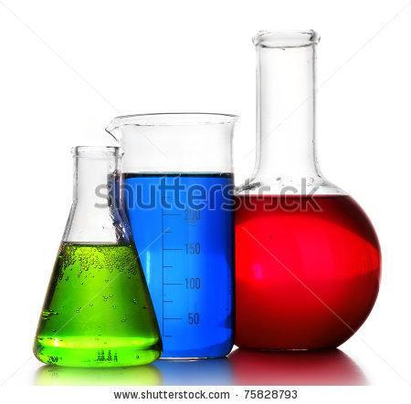
3 graduated cylinder Funnel Flask beaker Dropping bottle Watch glass Petri dish Reagent bottle Microscope slides Staining boxes Coplin jars Vials Dissecting scissors Test tube Slide boxes Cover slips pipette Forceps Dissecting needle Scalpel


4 Necessary equipment in preparation microscopy Rotary Microtome Ultra-Microtome Freezing Microtome


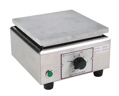
5 Paraffin Oven Water bath Hot plate

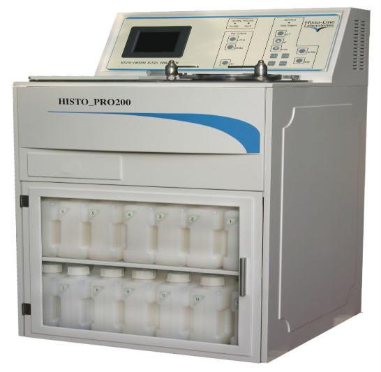

6 Automatic Tissue Processor Automatic Tissue Processor Automatic Tissue Processor Automatic Tissue Processor Balances Automatic Tissue Processor

7
8 Prepare some necessary solutions in laboratory preparations Some general rules for the preparation of solutions: 1- When preparing a 70% alcohol, for example, of the 95%: measured 70 ml of alcohol by graduated cylinder and then increase that amount to 95 ml by distilled water and in the same way can be prepared in any concentration is required. 1- When preparing 15% of the specific ways : we measure 15 ml of this liquid and add to it 85% of distilled water becomes final amount of 100 ml. 2- When preparing a 20% sodium chloride, for example: weigh 20 gm and dissolve in a certain amount of distilled water (50 ml, for example) and after completing the melting complete solution to 100 ml with distilled water. Solutions required preparation Physiological solutions Sodium chloride to wash samples focus 0.9% Sodium chloride gm. Distilled water ml. Fixatives Formal 10% Concentrated formalin. 10 ml. Distilled water ml. Dehydration water solutions (50%, 70%, 80%, 90%, ethyl alcohol) Clearing solutions Xylene Staining Hematoxylin, Eosin.
9
10
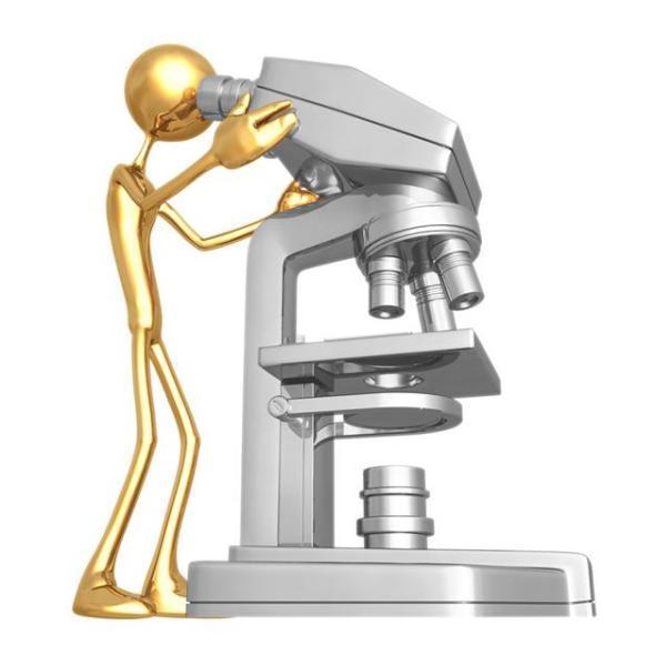
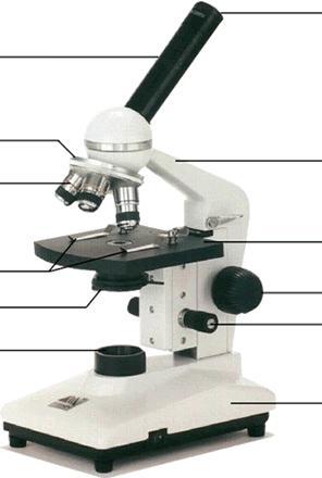
11 Part Ocular (eyepiece) Nosepiece Functions use to look at the sample. usually it's enlarge power is (10x) or more than that (20x) holds objective lenses Use to magnify the image of sample Objective Lenses High power objective lens ( 40 x) (oily lens 100x ) Low power objective lens ( 4 x) (10 x ) Arm Stage supports upper parts of the microscope, used to carry the microscope where the slide is placed Stage Clips hold slide in place on the stage Condenser Collect light and direct it to the sample Diaphragm Control the amount of light which reaching to the condenser Coarse Adjustment Knob used to focus when using the low power objective Fine Adjustment Knob used to focus when using the high power objective Light Source provides light Base supports the microscope
12 1- When moving your microscope, always carry it with both hands, hold the arm with one hand and place the other hand under the base for support 2- Turn the nosepiece so that the lowest power objective lens is "clicked" into position 3-Use the coarse adjustmetnt knob to move the stage down. Your microscope slide should be prepared with a cover glass over the sample. This will help protect the objective lenses if they touch the slide. Place slide on the stage and fasten it with the stage clips 4- Look at the objective lens and the stage from the side and turn the coarse focus knob so that the stage moves upward. Move until the image comes into focus without touching the slide! 5- Now, look through the eyepiece and adjust the diaphragm for the greatest amount of light. 6- Slowly turn the fine adjustment knob. Continue until the image comes into focus. 7- Move the slide around until the image comes in center of the field of view and adjust the diaphragm for the clearest image. 8- Now, you should be able to change to the next objective lenses with only minimal use of the focusing adjustment. Use the fine adjustment, if available. If you cannot focus on your specimen, repeat steps 4 through 7 with the higher power objective lens in place. Do not allow the objective lens to touch the slide!
13 9The proper way to use a microscope is to look through the eyepiece with both eyes (this helps avoid eye strain). If you want close one eye when looking into the microscope, it's ok. Do not touch the glass part of the lenses with your fingers. Use only special lens paper to clean the lenses. 10- When finished, raise the tube (or lower the stage), click the low power lens into position and remove the slid. 11- Always keep your microscope covered when not in use. Dust is the number 1 enemy! Microscopes allow us to see micro organisms which are too small for the naked eye to see them such as bacteria,viruses,fungi and many important parts of the body like individual cells. Magnification power= multiply the objective lens (the one facing your specimen) with the ocular lens (the one through which you look). Most ocular lenses are 10x Example: If the eyepiece power x10,objective lens power x40 power magnification = 10 x 40 = 400 times
14
 A- Prepare plant cells in the skin of the onion: Method: Clean the slide and cover slip. Cut a piece of onion. Use Forceps to pull off a very thin piece of onion skin.")
15 Preparing temporary samples by loading Devices and tools required : Light microscope - anatomy Tools - toothpick - slides - Cover slip distilled water- Forceps - dropper - vegetable samples (onion tomato potato) A- Prepare plant cells in the skin of the onion: Method: Clean the slide and cover slip. Cut a piece of onion. Use Forceps to pull off a very thin piece of onion skin. Put drop of water on slide using dropper Place a small piece of onion skin on water drop. Carefully place cover slip on onion skin (figure1) Press the cover slip down carefully to remove any air bubbles. Place prepared slide onto stage of microscope. Look at onion cells under low power (4 x), then use medium power (10 x) and high power (40 x) and write the notes. x10
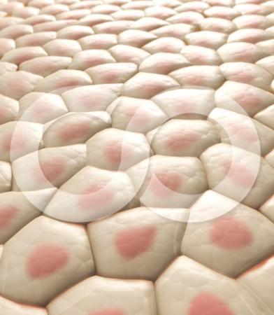
16 B - Prepare a Chromoplasts cells in tomato : Method: Clean the slide and cover slip. Cut a piece of tomato. Use Forceps to pull off a very thin piece of tomato pulp. put drop of water on slide using dropper Put a small piece of tomato pulp on water drop. Carefully place cover slip on sample. Carefully Press the cover slip down to remove any air bubbles.. Put prepared slide onto stage of microscope. Look at Chromoplasts in tomato cells under low power (4 x), then use medium power (10 x) and high power (40 x) and write the notes X40
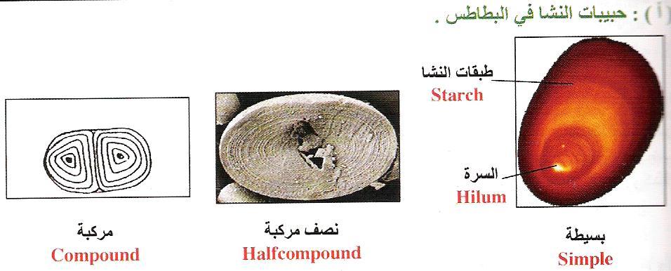
17 C - Prepare starch granules in potato: Method Clean the slide and cover slip. Cut pieces of potato. Put potato pieces in a cup contained water for 10 minutes. Take a drop from solution and put it on slide using dropper Carefully place cover slip on drop. Carefully Press the cover slip down to remove any air bubbles. Put prepared slide onto stage of microscope. Look at starch granules under low power (4 x), then use medium power (10 x) and high power (40 x) and write the notes. X40

18 D- Prepare squamous cell epithelial lining of the mouth: Method: 1 - Put a small drop of distilled water in the center of clean microscopic slide. 2 - Rubbed several times lining of the oral cavity by the end transverse toothpick. (figure 1) 3 - Move the end of the toothpick in a small drop of water on a microscopic slide, even cells spread completely in the drop of water.. (figure 2) 4 - Placed a cover slide and examine the sample by using a microscope. (figure 3) X40


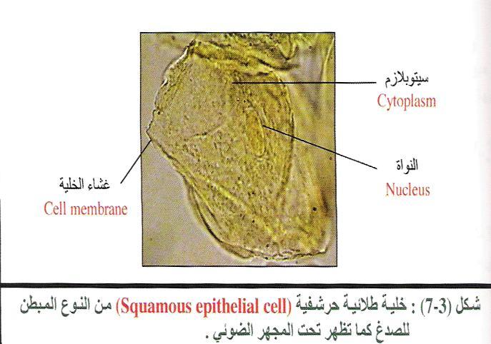
19 خاليا طالئية حرشفية مصبوغة بأزرق الميثلين كما تظهر تحت المجهر
20


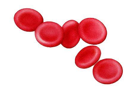
21 Tools required : Slides - medical swab - sterile needle - Light microscope Method: 1. Clean your slide by water then alcohol 95% 2. Cleans your finger by medical swab. 3. Make a puncture on a fingertip on sterile needle. 4. Wipe away the first drop of blood with clean gauze. 5.Touch the next drop of blood with a clean slide. 6. Bring a clean spreader slide, held at a 45 angle, toward the drop of blood on the specimen slide. 7. Wait until the blood spreads along the entire width of the spreader slide. 8. While holding the spreader slide at the same angle, push it forward rapidlyand smoothly. Then leave the blood smear to dry by air.

22 9- Stain the blood smear by using Leishman dye for 2-3 minute. 10- Remove the stain with distilled water then leave slid until dry. 11- examine the sample by using microscope.
23 :Tools used Bunsen flame or a candle - Slides - coverslip Bacterial culture, or yogurt Methylene blue dye Light microscope Inoculation loop Method: 1- cleans slide well and pass it on the flame before use several times 2- sterilize Loop before and after use by using the flame 3- takes Sample from yogurt, by puts point on the edge of the slide and then uniqueness and left to dry like blood smear 4- pass slide on a Bunsen flame several times to kill the bacteria and fixed it up to does not go away during the dye 5- put two drops of Methylene blue dye and leave for 3-5 minutes 6- clean the dye and rinsed by distilled water 7- dried in the air or on the flame put a drop of Cedar - wood oil on a slide and examined by the oily lens(100x) X 100

24

 9. Dry the slide, add Glycerin, cover slip.")
25 Required tools and solutions: Distilled water - Himatoxlin stain Eosin stain - fatty tissue taken from any animal 70 % alcohol - formalin - Glycerin - slides Covers Sudan III stain. Method: 1. Prepare fat tissue on the slide. 2. Fixed on formalin 5-10 min. 3. Wash by distilled water. 4. Stain by Sudan III 15 min. 5. Wash on alcohol 70% until stain removed. 6. Wash by distilled water. 7. Stain by H.X 3-5 min.(optional) 8. Wash on tap water 5 min.(optional) 9. Dry the slide, add Glycerin, cover slip. Result : the fat cells will stain by orange color, nucleus by blue. x10


26
 Method: 3- Preparation of the connective tissue (taken from a thin layer under the skin of the animal) on a glass slide 2-Fixed on formalin")
. 9-Clear in Xylene 5 min. 10- Drop of Canada balsam, mount the cover slip. Result: observe fibers and different cells.")
27 Required tools and solutions: Distilled water - Hematoxylin stain Eosin stain - connective tissue Xylene - 70% alcohol - formalin - Glycerin - slides Cover slip Canada balsam - different concentration of alcohol(70%-80%- 90%- 100%) Method: 3- Preparation of the connective tissue (taken from a thin layer under the skin of the animal) on a glass slide 2-Fixed on formalin for 5-10 min. 3-Wash by distal water. 3-Stain by H.X (Hematoxylin stain) for 15 min. 4-Wash on tap water 10 min. 5- Wash by distal water. 6-Stain by Eosin 2 min. 7- Wash by distal water. 8-Pass the slide on different concentration of alcohol (ascending). 9-Clear in Xylene 5 min. 10- Drop of Canada balsam, mount the cover slip. Result: observe fibers and different cells. x10

28

29 STEP 1 Fixation Obtaining a fresh specimen, wash by Saline to removed the blood Then Fixative by10% formalin or other fixatives for hours. Make sure you have enough fixative to cover tissues. Fixative volume should be 5-10 times of tissue volume. After that, rinsing the fixative from sample by running tap water for 12hour. STEP 2 Tissue Processing Specimen is placed in plastic cassettes, processed using Automatic Tissue Processor
30 Tissue Processing Including 1. Dehydration: (series of ethanol (alcohol) of increasing concentration until pure) 50% Ethanol, one changes, 1 hour. 70% Ethanol, two changes, 1 hour each. 80% Ethanol, one change, 1 hour. 90% Ethanol, one change, 1 hour. 95% Ethanol, one change, 1 hour. 100% Ethanol, three changes, 1hour each )Time depends on the size of the sample) 2. Clearing: By using clearing agent called Xylene. Causing we cannot infiltrate tissue with wax because wax and ethanol are largely immiscible. So we have to use intermediate solvent that is fully miscible with both ethanol and paraffin wax. Xylene, three changes, 1.5 hour each 3. Wax Infiltration : The paraffin wax based histological waxes are the most popular. Paraffin wax (58-60 ºC), two changes, 2 hours each Embedding tissues into paraffin blocks.


31 STEP 3 Embedding: By Embedding Machine STEP 4 Cutting, Trimming and Sectioning By Rotary Microtome
 for 2 min each concentration. 3. wash by tap water for 2 mint. 4.")
32 STEP 5 Floating Section is placed in floating bath.then loading on the slide. Staining : STEP 6 1. Put the slide in xylene, 2 changes for 2-5 min. to remove wax. 2. put the slide at descending series of alcohol (100%- 95%- 70%-50%-30%) for 2 min each concentration. 3. wash by tap water for 2 mint. 4. Staining by Hematoxylin for 5min. 5. Rinsing by running tap water for 2 min. 6. put the slide at ascending series of alcohol (30%-50%-70%- 95%) for 2 min each concentration. 7. Staining by Eosin for 2 min. 8.(95%-95% - 100%- 100%) for 2 min each concentration. 9. put slide at Xylene, 2 changes for 2 min.
33 STEP 7 Mounting drop canada balsam. Cover slip. Observation slide under microscope Final STEP RESULT: Hematoxylin= Nucleus (Blue color) Eosin= Cytoplasm (Pink color).


34
35 Required tools and solutions: Clean slides - ascending series of alcohol - Canada balsam - Xylene insects (ants - bee - beetle)- cover slip. Method: Prepare parts of the insect ( wings- leg - mouth parts) or complete body if the insect is a small. Put the sample at ascending series of alcohol (70%-80%- 90%- 100%) for 5-10 minute each concentration. Put sample in Xylene even become transparent. Put sample on clean slides then covering by Canada balsam. Put cover slip and dried the sample in the oven at 37 degrees. examine the sample by using a microscope.
: In order to study tissues with a microscope they must be preserved (fixed)- fixation Following fixation, blocks of tissue must be cut into thin
- fixation Following fixation, blocks of tissue must be cut into thin") : In order to study tissues with a microscope they must be preserved (fixed)- fixation Following fixation, blocks of tissue must be cut into thin sections.-microtomy Other techniques involve dehydration
: In order to study tissues with a microscope they must be preserved (fixed)- fixation Following fixation, blocks of tissue must be cut into thin sections.-microtomy Other techniques involve dehydration
Phenion FT Skin Model Histological processing Paraffin sections
 Phenion FT Skin Model Histological processing Paraffin sections Objective This Standard Operation Procedure is recommended to fix and embed Phenion FT Skin Models in order to prepare paraffin sections.
LAB 3 CHARACTERIZING YOUR UNKNOWN BACTERIA AND USING MORE COMPLEX STAINS. Part I: Isolating Your Unknown Bacteria and Describing Colony Morphology
 LAB 3 CHARACTERIZING YOUR UNKNOWN BACTERIA AND USING MORE COMPLEX STAINS Objectives In this lab you will learn how to: - describe bacteria on the basis of colony and cell morphology - isolate bacterial
What is Life? Project PART 1: Looking at Cells Lab
 What is Life? Project PART 1: Looking at Cells Lab Directions: Complete the drawings and answer the questions in the space provided. For each drawing: Title the drawing of the specimen (e.g. Cork Cells)
Surgical Gown. Tongue Depressor. A disposable gown worn by medical staff during surgery. A thin, flat, wooden stick rounded at both ends
 Tongue Depressor A thin, flat, wooden stick rounded at both ends Accidentally dropped on the floor by the doctor 16 Surgical Gown A disposable gown worn by medical staff during surgery Used by the surgeon
Lab. Elodea, Onion, and Cheek Cell Lab. Be your best. Cell Biologist s Name: Period: Date: Mrs. Bouchard -7 th Grade Science
 Purpose Ques*on: How do plant cells and animal cells differ? Cheek Cell Lab Materials: Water bo6le with dropper toothpick glass slide coverslip lens paper methylene blue safety goggles lab apron paper
Basic Microbiology and Immunology Practical Course
 Basic Microbiology and Immunology Practical Course 2 Lab # 2: Colouring the microorganisms Rules that must be followed to maintain an aseptic zone 3 For most bacterial cultures, you will use a sterile
Student Performance Guide. Student Performance Guide. Student Performance Guide
 LESSON 8-2 Collecting and Processing Specimens for Parasite Examination Student Performance Guide LESSON 8-3 Microscopic Methods for Student Performance Guide LESSON 8-4 Preparing and Staining Smears for
Bacterial smear and Staining
 Practical Microbiology 18-22/11/2018 University of Sulaimani college of Pharmacy Year2 Lab. 4: Bacterial smear and Staining Before staining and observing a microbe under a microscope, a smear must be prepared.
MOLLUSCS PROCESSING FOR DIAGNOSIS BY HISTOLOGY
 European Union Reference Laboratory for Molluscs Diseases MOLLUSCS PROCESSING FOR DIAGNOSIS BY HISTOLOGY SUMMARY 1. SCOPE...2 2. REFERENCES...2 3. GENERAL INFORMATION...2 4. EQUIPMENT AND ENVIRONMENT...2
Brazosport College Life Science Laboratory Safety Rules and Regulations
 Brazosport College Life Science Laboratory Safety Rules and Regulations Laboratory Safety Procedures for Biology Labs Permanent Link: http://bit.ly/bc-labsafety The risks incurred in the biology laboratories
STUDENT LABORATORY PACKET
 L5 Elodea-Onion-Cheek-Cell_Size Page 1 of 7 STUDENT LABORATORY PACKET Student s Full Name Lab #5: Elodea, Onion, Cheek Cells-Cell Size Lab Instructor Date Points Microscope # OBJECTIVES: a. to examine
Pelagia Research Library. Staining reactions of microwave processed tissues compared with conventional paraffin wax processed tissues
 Available online at www.pelagiaresearchlibrary.com European Journal of Experimental Biology, 2011, 1 (1): 57-62 Staining reactions of microwave processed tissues compared with conventional paraffin wax
Prisma & Film Staining Workshop. Application Specialist Mea Pelkonen
 Prisma & Film Staining Workshop Application Specialist Mea Pelkonen Tissue-Tek Prisma Tissue-Tek Prisma Always program the Prisma in the following order: 1. Edit solution names Check if desired solution
Steps of microbial smear preparation :
 Lab 4 STAINING Practical Microbiology Microbial smear : It is a very small amount of microbial growth ( broth or solid ) spreaded on a clean slide and drying by air. Fixation : The process of passing the
Staining of the clinical material or the bacteria from colonies on laboratory media provide a direct visualization of the morphology of the organisms
 COMMON STAINING PROCEDURES Staining of the clinical material or the bacteria from colonies on laboratory media provide a direct visualization of the morphology of the organisms as well as their reactions
Biomedical Laboratory Science
 Biomedical Laboratory Science!! " " New for 2015-2016 The 70% mastery for each skill has been added to the rating sheet so that competitors and advisors know what score is required for mastery. Rating
Exercise 6-A STAINING OF MICROORGANISMS DIRECT VS INDIRECT STAINING
 Exercise 6-A STAINING OF MICROORGANISMS DIRECT VS INDIRECT STAINING Introduction The morphological features of individual microorganisms may be examined either by observing living, unstained materials,
Observing Moss and Cheek Cells
 Observing Moss and Cheek Cells Name: Block: Partner(s): Date:!! SAFETY PRECAUTIONS!! Methylene blue solution will stain clothes and skin. Broken slides should be handled with paper towels, not bare hands.
BIOL 251 BASIC MICROBIOLOGY
 BIOL 251 BASIC MICROBIOLOGY CHARACTERISATION OF BACTERIA CHARACTERISATION OF BACTERIA CHARACTERISATION OF BACTERIA MICROSCOPIC To be able to examine microbes microscopically, they need to be stained
PREPARATION OF BLOOD FILMS FOR MALARIA DETECTION
 PREPARATION OF BLOOD FILMS FOR MALARIA DETECTION Materials for Preparation of Malaria Smears: Clean and wrapped slides Sterile lancets 70% ethanol and water Absorbent cotton wool Surgical gloves Lint-free
PROTOCOLS FOR ANATOMY/MICROMORPHOLOGY
 PROTOCOLS FOR ANATOMY/MICROMORPHOLOGY General dissection of spikelets... 2 Hand sections and epidermal scrapes of bamboo leaves... 2 Clearing and staining of intact plant organs... 4 Scanning electron
Exercise 6-D STAINING OF MICROORGANISMS ENDOSPORE STAINS, CAPSULE STAINS & FLAGELLA
 Exercise 6-D STAINING OF MICROORGANISMS ENDOSPORE STAINS, CAPSULE STAINS & FLAGELLA Introduction Endospore stains, capsule stains, and flagellar stains are staining techniques that allow for the differentiation
AN INTRODUCTION TO METHODS OF STUDYING THE MORBID HISTOLOGY OF DISEASE-CARRYING INSECTS.
 243 AN INTRODUCTION TO METHODS OF STUDYING THE MORBID HISTOLOGY OF DISEASE-CARRYING INSECTS. By CAPTAIN A. E. HAMERTON, D.S.O. Royal Army Medical Oorps. THE great technical improvements in modern histological
ab Trichrome Stain (Connective Tissue Stain)
") Version 3 Last updated 12 February 2019 ab150686 Trichrome Stain (Connective Tissue Stain) For the histological visualization of collagenous connective tissue fibers in tissue sections. This product is
Version 3 Last updated 12 February 2019 ab150686 Trichrome Stain (Connective Tissue Stain) For the histological visualization of collagenous connective tissue fibers in tissue sections. This product is
ab Elastic (Connective Tissue Stain)
") Version 2 Last updated 25 June 2018 ab150667 Elastic (Connective Tissue Stain) For the histological staining of Elastin in tissue sections. This product is for research use only and is not intended for
Version 2 Last updated 25 June 2018 ab150667 Elastic (Connective Tissue Stain) For the histological staining of Elastin in tissue sections. This product is for research use only and is not intended for
BOTANY Lab Manual BSc.-III Medical Semester V
 BOTANY Lab Manual BSc.-III Medical Semester V 212 Experiment 1 Aim: Determine Water Potential of Vacuolar Sap by Plasmolytic Method. Requirements: Leaves of Tradescantia solutions of different concentrations,
Lab Six:- Medical Microbiology Prepared by: Luma J. Witwit. Staining
 Staining Even with the microscope, bacteria are difficult to see unless they are treated in a way that increases contrast between the organisms and their background. The most common method to increase
Laboratory Exercise # 8: Other Staining Techniques
 Laboratory Exercise # 8: Other Staining Techniques Purpose: The purpose of this laboratory exercise is to acquaint the student with staining techniques other than the Gram stain that are routinely used
UNIT 7 BASIC TECHNIQUES OF SLIDE PREPARATION
 UNIT 7 BASIC TECHNIQUES OF SLIDE PREPARATION Structure 7.1 Introduction Objectives 7.2 Cleaning, Care and Storage of Slides Washing up Used Slides Cleaning Routine for New Slides Storage of Prepared Slides
WHAT IS GEL ELECTROPHORESIS?
 Getting Started With Gel Electrophoresis a world of learning Presented by Peter J Ball, Southern Biological. For further information, please contact the author by phone (03) 9877-4597 or by email peterjball@southernbiological.com.
EXPERIMENT. Bacterial Morphology and Staining Techniques
 EXPERIMENT Bacterial Morphology and Staining Techniques Hands-On Labs, Inc. Version 42-0240-00-02 Review the safety materials and wear goggles when working with chemicals. Read the entire exercise before
ROUTINE TECHNIC FOR SURGICAL SPECIMENS. Fixation, Dehydration and Embedding
 A TRICHROME STAINING METHOD FOR ROUTINE USE SERGIO A. BENCOSME, M.D. Department of Pathology, University of Ottawa, and the Ottawa General Hospital, Ottawa, Ontario, Canada Despite the added information
Exercise 6-C STAINING OF MICROORGANISMS ACID-FAST STAIN
 Exercise 6-C STAINING OF MICROORGANISMS ACID-FAST STAIN Introduction The acid-fast stain is a differential stain that separates bacteria on the basis of the lipid content of their cell walls. Bacteria
SOP BIO-002 FOR SHARPS USAGE AND DISPOSAL
 ENVIRONMENTAL AND EMERGENCY MANAGEMENT Environmental Health and Safety University Crossing Suite 140 Lowell MA 01854 http://www.uml.edu/eem/ SOP BIO-002 FOR SHARPS USAGE AND DISPOSAL SCOPE This policy
Stains and Solutions Used in Hematology and Cytology
 Stains and Solutions Used in Hematology and Cytology A APPENDIX Acid-Fast Stain Commercially prepared acid-fast stains are available 1. Ziehl Neelsen carbolfuchsin: Dissolve 3.0 g basic fuchsin in 100
ANALYSIS OF FINGERPRINTS, LIPSTICK 2 ND HAIR
 ANALYSIS OF FINGERPRINTS, LIPSTICK 2 ND HAIR LAB FORENSICS.3 From Sourcebook, National Science Foundation, 1997 INTRODUCTION PART A. OBTAINING A FINGERPRINT Black ink stamp pad Tissue paper 4 x 4 cm Card
for Stool Examination Issued by: LABORATORY MANAGER Original Date: March 13, 2000 Approved by: Laboratory Director Hematoxylin Stain
 Section: Page 28 Policy # MI\PAR\05\06\v01 Page 1 of 5 Subject Title: Laboratory Procedures for Stool Examination Issued by: LABORATORY MANAGER Original Date: March 13, 2000 Approved by: Laboratory Director
Plant Microtechnique Part Two
 Plant Microtechnique Part Two Clearing Clearing is the process of removing depositions suspended in water or other liquids by using natural or chemical means. Clearing means transfer plant sample from
Biomedical Laboratory Science
 Biomedical Laboratory Science New for 2018-2019 At ILC, photo ID must be presented prior to competing in each round. Biotechnology: Science for the New Millennium Edition 2 has been released. For information
Franklin Regional School District SCIENCE MIDDLE SCHOOL Bid #16 Vendor Specifications
 SCIENCE MIDDLE SCHOOL Bid #16 Quotation number: (Number you want reflected on the Purchase Order) Company: Address: Telephone: Email: Date: Authorized name (print): Authorized Signature: 1 GLASSWARE/TUBING/CYLINDERS
Mt. San Antonio College: Spring 2018 MICR 22 Lab Orientation. Welcome to the Microbiology 22 Laboratory!
 Mt. San Antonio College: Spring 2018 MICR 22 Lab Orientation Welcome to the Microbiology 22 Laboratory! Laboratory Objectives: To teach concepts of microbiological techniques using critically selected
Crime Busters. Lin Wozniewski
 Crime Busters Lin Wozniewski lwoz@iun.edu Safety Students must wear: Closed shoes All skin from neck to toes covered Lab coat or lab apron Indirect vent or unvented chemical splash proof goggles. All skin
Study Guide-Forensic Science Chapter 5- Hair Name:
 1. Is hair class or individual evidence? Class 2. Does hair only absorb substances from inside the body? _No, Inside and out_ 3. From hair, one can determine the following: _Human or animal Race Origin
Updated by S. McNew, March Deborah Jung Microbiology Preparation Technician
 Southeast Missouri State University PROTOCOL FOR SCIENCE EQUIPMENT USAGE AT REGIONAL CAMPUSES WITH EMPHASIS ON BS240/BS242 MICROORGANISMS AND THEIR HUMAN HOSTS Updated by S. McNew, March 2018 Personnel
Procedure 30 Collecting A Blood Specimen Using The Vacuum-Tube System. Procedure 31 Collecting A Blood Specimen Using A Needle And Syringe
 Chapter 6 Phlebotomy Procedure 29 Performing A Venipuncture Procedure 30 Collecting A Blood Specimen Using The Vacuum-Tube System Procedure 31 Collecting A Blood Specimen Using A Needle And Syringe Procedure
Wake Forest Institute for Regenerative Medicine
 Evaluation of Algeness as a Dermal Filler Material November 2014 Final Report John D. Jackson, PhD, Associate Professor, Institute for Regenerative Medicine Institute, Wake Forest Baptist Medical Center
Student Performance Guide. Student Performance Guide. Student Performance Guide. Student Performance Guide. LESSON 3-3 Bleeding Time
 LESSON 3-3 Bleeding Time Student Performance Guide LESSON 3-4 Prothrombin Time Student Performance Guide LESSON 3-5 Activated Partial Thromboplastin Time Student Performance Guide LESSON 3-6 Rapid Tests
ORTON and Post (1932) and Cutler (1935) investigated the use of diethylene
 and Cutler (1935) investigated the use of diethylene") 593 A Modified Ester Wax for Embedding Tissues By W. CHESTERMAN AND E. H. LEACH {From the University Laboratory of Physiology, Oxford) SUMMARY I. A modification of Steedman's ester wax embedding method
593 A Modified Ester Wax for Embedding Tissues By W. CHESTERMAN AND E. H. LEACH {From the University Laboratory of Physiology, Oxford) SUMMARY I. A modification of Steedman's ester wax embedding method
How to Give a Subcutaneous (SC) Injection to Your Child
 Injection to Your Child") How to Give a Subcutaneous (SC) Injection to Your Child Supplies: Needles and syringes Alcohol swabs and gauze Vial with the drug solution Sharps container (Health Facts for You #4587) Band-Aids Distraction
How to Give a Subcutaneous (SC) Injection to Your Child Supplies: Needles and syringes Alcohol swabs and gauze Vial with the drug solution Sharps container (Health Facts for You #4587) Band-Aids Distraction
Some Common Prosection Techniques
 Some Common Prosection Techniques Contrary to a common misconception, dissection is not just cutting into a specimen. Dissection is a term for the techniques used to carefully separate and expose internal
Biohazardous Waste. 1. Solid Biohazardous Waste (non-sharps) Storage
 Storage") Biohazardous Waste There are 4 general categories of biohazardous wastes based on the physical form of the waste. Each form must be segregated, identified, decontaminated and disposed of in an appropriate
Biohazardous Waste There are 4 general categories of biohazardous wastes based on the physical form of the waste. Each form must be segregated, identified, decontaminated and disposed of in an appropriate
DZC Marketing 15, Jalan Radin Anum 2, Bandar Baru Sri Petaling Kuala Lumpur, Malaysia. Tel : Fax :
 DZC Marketing 15, Jalan Radin Anum 2, Bandar Baru Sri Petaling 57000 Kuala Lumpur, Malaysia. Tel : 03-9057 1316 Fax : 03-90578925 sales@dzcmarketing.com WhatsApp +6018-666 8794 MEDICAL DISPOSABLE LIST
Preparing the Gel Box and Pouring the Agarose Gel
 Preparing the Gel Box and Pouring the Agarose Gel Student Workstation Quantity Plastic chamber 1 8-well comb 1 Ruler 1 Molten agarose 50 ml Marking pen 1 Protocol 1. Using a ruler, measure the length,
COLLECTION INSTRUCTIONS - SAMPLES (VARIOUS TYPES)
") COLLECTION INSTRUCTIONS - SAMPLES (VARIOUS TYPES) You need to complete the necessary forms for each sample collected. To avoid any possible contamination, please ensure that gloves are worn during the
COLLECTION INSTRUCTIONS - SAMPLES (VARIOUS TYPES) You need to complete the necessary forms for each sample collected. To avoid any possible contamination, please ensure that gloves are worn during the
International Journal of Science, Environment and Technology, Vol. 7, No 5, 2018,
 International Journal of Science, Environment and Technology, Vol. 7, No 5, 2018, 1726 1730 ISSN 2278-3687 (O) 2277-663X (P) Review Article HISTOLOGICAL STUDY OF HAIR FOLLICLES OF CATTLE BREEDS OF MAHARASHTRA
Name: Date: Period: Can I eat that? Lab
 Name: Date: Period: Can I eat that? Lab Objective Part 1 Engage Your teacher is allergic to blue dye that is used in many foods and products. Your objective is to determine what colors of M&Ms or skittles
ab Gram Stain Kit (Microorganism Stain)
") Version 2 Last updated 27 June 2018 ab150672 Gram Stain Kit (Microorganism Stain) For the histological differentiation of Gram-Positive and Gram- Negative bacteria. This product is for research use only
Version 2 Last updated 27 June 2018 ab150672 Gram Stain Kit (Microorganism Stain) For the histological differentiation of Gram-Positive and Gram- Negative bacteria. This product is for research use only
KERATIN CONTAMINATION
 KERATIN CONTAMINATION Keratin contamination is almost always observed as a background protein. Wear only nitrile gloves and rinse with HPLC grade water all trays, containers and surfaces that contact the
EXERCISE 8C - Lab Procedures
 EXERCISE 8C - Lab Procedures SAFETY WARNING: Acrylamide in the unpolymerized form is a skin irritant and a potential neurotoxin. Fortunately, the acrylamide in your gels is polymerized, so it should not
Paper Chromatography and Steam Distillation EVERY STUDENT MUST BRING AT LEAST 3 ORANGES TO LAB FOR THIS EXPERIMENT! Equipment
 Paper Chromatography and Steam Distillation EVERY STUDENT MUST BRING AT LEAST 3 ORANGES TO LAB FOR THIS EXPERIMENT! Equipment You will need a 600 ml beaker, a 50 ml graduated cylinder, 4 Expo Wet Erase
Introductory Chemistry
 Introductory Chemistry Lab 1: Introduction and Safety Objectives Learn how work to safely in the chemical laboratory Learn when and how to use the safety equipment in the chemical laboratory Learn the
MEDICATION AND INJECTION ADMINISTRATION EDUCATIONAL BOOKLET FOR OVULATION INDUCTION
 MEDICATION AND INJECTION ADMINISTRATION EDUCATIONAL BOOKLET FOR OVULATION INDUCTION Comprehensive Guide to Understanding, Mixing, and Administering Fertility Medications IMPORTANT PHONE NUMBERS: WEEKEND
AUTOPSY OF A DILL PICKLE
 AUTOPSY OF A DILL PICKLE BACKGROUND Notes for teacher: This activity serves as an introduction to dissection. It also reinforces concepts of anatomical directions, planes, and body cavities. Some imagination
ab Papanicolaou (PAP) Red Stain Kit (Cytology Stain)
 Red Stain Kit (Cytology Stain)") Version 2 Last updated 27 June 2018 ab150679 Papanicolaou (PAP) Red Stain Kit (Cytology Stain) For the Differentiation of Cells in Vaginal Smears for the Detection of Vaginal, Uterine and Cervical Cancer.
Version 2 Last updated 27 June 2018 ab150679 Papanicolaou (PAP) Red Stain Kit (Cytology Stain) For the Differentiation of Cells in Vaginal Smears for the Detection of Vaginal, Uterine and Cervical Cancer.
COMMON STAINING TECHNIQUE
 2 COMMON STAINING TECHNIQUE 2.1 INTRODUCTION Staining is technique used in microscopy to enhance contrast in the microscopic image. Stains and dyes are frequently used in biological tissues for viewing,
BSL-2 Emergency Plan
 BSL-2 Emergency Plan Spills General Spill Cleanup Guidelines: Know how to get the HVAC unit servicing the lab space shut down in order to limit the spread of contamination. Wear gloves and lab coat. Use
PENTAMYCETIN is an antibiotic that: Stops the growth of bacteria Kills bacteria
 READ THIS FOR SAFE AND EFFECTIVE USE OF YOUR MEDICINE PATIENT MEDICATION INFORMATION Read this carefully before you start taking PENTAMYCETIN each time you get a refill. This leaflet is a summary and will
Figure A. Figure B To prevent premature activation of the needle safety guard, do not touch the NEEDLE GUARD ACTIVATION CLIPS at any time during use.
 INSTRUCTIONS FOR USE STELARA (stel ar a) (ustekinumab) injection, for subcutaneous use Instructions for injecting STELARA using a prefilled syringe. Read this Instructions for Use before you start using
E-Blotter Operation. Technical Bulletin E-03 MATERIAL PROCEDURE
 E-Blotter Operation MATERIAL BSA (1 mg/ml; 0.05 g BSA (Sigma-Aldrich Ltd., St Louis, MO; U.S.A.) dissolve in 50 ml ddh 2 O, aliquot to 1 ml in micro-centrifuge tubes and freeze in -20 C) NK92 cell lysate
2. Mix the plant material with 5 ml of rubbing alcohol and let it soak for a few minutes. Swirl the container to mix it as you wait.
 Spa Day Perfume 1. Grate, grind, or chop up about 1 teaspoon of plant material of your choice (e.g. nutmeg, citrus peel, cinnamon, cloves, scented leaves or flowers, and herbal teabags are all good sources
Safety Rules for Laboratory
 Safety Rules for Laboratory These protocols are intended to protect you and make your laboratory experience enjoyable and productive. Section I: CVM General Laboratory Protocols (these rules apply to all
State of Kuwait Ministry of Health Infection Control Directorate SAFE INJECTION
 State of Kuwait Ministry of Health Infection Control Directorate SAFE INJECTION May 2010 Contents I. Introduction II. Prevention strategies III. Best practices for injection A. General safety practices
SELYE and McKeown (1935) and Baker (1948) have noted the presence of
 and Baker (1948) have noted the presence of") A Pigment in the Rat's Uterus By ]. G. WARBRICK {From the Department of Anatomy, University of Glasgow, Glasgow, W. 2) With one plate (fig. i) SUMP4ARY 1. A yellowish-brown pigment was found at the old
A Pigment in the Rat's Uterus By ]. G. WARBRICK {From the Department of Anatomy, University of Glasgow, Glasgow, W. 2) With one plate (fig. i) SUMP4ARY 1. A yellowish-brown pigment was found at the old
MANICURE. Before preparing client for manicure, look for any nail disorders or infections like:
 Practical 1 MANICURE Objectives At the end of this session, you will be able to: clean and shape the nails using manicure tools clean and massage the hands apply nail polish Tools, Equipments and Materials
LAB 5 Blood Collection
 LAB 5 Blood Collection Purpose The Purpose of this lab is to learn how to collect a sample of blood properly. From this blood sample disease such as anemia, viral infections, iron deficiency, spherocytosis,
Experiment 11 Identification of Food Colors in Candies
 Experiment 11 Identification of Food Colors in Candies Pre-lab Assignment Before coming to lab: Read the lab thoroughly. Answer the pre-lab questions that appear at the end of this lab exercise. Purpose
Lenis Needle-free Safety Syringe Device User Manual
 Lenis Needle-free Safety Syringe Device User Manual 1 Table of Contents Welcome.3 Lenis Kit Components.4 Instructions 5-9 Maintenance and Care..10 Troubleshooting. 11 Warranty.12 Precautions 13 Return
Dextrose 25%solution or equivalent. Dextrose 5%+normal saline.9% solution Adhesive fabric tape,stretchable,easily cut roll
 Item no. Item description Unit Qty.. 3. 4.. 6. 7. 8. 9. 0... 3. 4.. 6. 7. 8. 9. 0... 3. 4.. 6. 7. 8. 9. 30. 3. 3. 33. 34. 3. 36. 37. 38. 39. 40. 4. 4. Dextrose %solution or equivalent Dextrose %solution
Tips On Proper Instrument Cleaning, Handling and Maintenance!
 304 304 Rinsing Tips On Proper Instrument Cleaning, Handling and Maintenance! *Immediately after instrument use thoroughly rinse off all blood, tissue and other fluids. *Using filtered water to rinse and
CHM111 Lab Physical Separations Grading Rubric
 CHM111 Lab Physical Separations Grading Rubric Name Team Name Criteria Points possible Points earned Lab Performance Printed lab handout and rubric was brought to lab 3 Safety and proper waste disposal
Experiment #3. Physical Separations Candy Chromatography
 Experiment #3. Physical Separations Candy Chromatography Goals 1. To physically separate and identify dyes in candy by comparison to commercial food dyes using paper chromatography. 2. To become familiar
Migraine Attack Abortive Treatment Medication Overuse Protocol Treatment Refractory Cluster Headache Treatment
 D i h y d r o e r g o t a m i n e ( D H E ) S u b c u t a n e o u s I n j e c t i o n G u i d e Migraine Attack Abortive Treatment Medication Overuse Protocol Treatment Refractory Cluster Headache Treatment
Single Source For Your Histology Reagents. Protecting Every Life Story In Your Lab.
 A Division of General Data Healthcare Histology Innovation for a NEW Generation Ready-To-Use Reagents, Dyes & Stains Routine Product Stains Name & Special Here Stains Single Source For Your Histology Reagents.
A GUIDE TO HEAD LICE PREVENTION AND TREATMENT
 A GUIDE TO HEAD LICE PREVENTION AND TREATMENT May 2018 What are Head Lice? Head lice are very small, tan-colored insects about the size of a sesame seed which live only on human heads. They live and lay
Title. Author(s)KONDO, Keiji; Vanek, Milan; Rasmussen, Palle V.; Blo. CitationJournal of the Graduate School of Agriculture, Hokka. Issue Date
KONDO, Keiji; Vanek, Milan; Rasmussen, Palle V.; Blo. CitationJournal of the Graduate School of Agriculture, Hokka. Issue Date") Title The beauty of mink pelage observea with SEM Author(s)KONDO, Keiji; Vanek, Milan; Rasmussen, Palle V.; Blo CitationJournal of the Graduate School of Agriculture, Hokka Issue Date 2001-03 Doc URL http://hdl.handle.net/2115/13157
Title The beauty of mink pelage observea with SEM Author(s)KONDO, Keiji; Vanek, Milan; Rasmussen, Palle V.; Blo CitationJournal of the Graduate School of Agriculture, Hokka Issue Date 2001-03 Doc URL http://hdl.handle.net/2115/13157
Lab 6.1: Animal histology & microtechnique
 Purpose The purpose of this lab is to introduce cytology students to basic methods of tissue processing, staining, and histology. Successful completion of this lab should produce high quality microscope
ROBOT PIN TOOL CLEANING AND LIQUID SAMPLE TRANSFER
 OVERVIEW TECHNICAL NOTE 67B ROBOT PIN TOOL CLEANING AND LIQUID SAMPLE TRANSFER There are several key steps in the successful use of pin tools: 1. The first and most important step is to start with clean
BIO Lab 12: Bacteria. Materials Required:
 Bacteria We look not at the things which are seen, but at the things which are not seen. For the things which are seen are temporal; but the things which are not seen are eternal. 2 Cor 4:18 Introduction
LIQUID SOAP INTRODUCTION
 LIQUID SOAP The hygiene is important for people to keep clean and at the same time to avoid infections and other kind of diseases. This soap is used exclusively by hands. Objectives Drawing a simple of
Comparative Proteomics Kit II: Western Blot Module Quick Guide
 Comparative Proteomics Kit II: Western Blot Module Quick Guide Lesson 1 1 Label one 1.5 ml fliptop micro tube for each of five fish samples. Also label one screw-cap micro tube for each fish sample. 2.
Standard Operating Procedures
 Standard Operating Procedures (V1_4/7/16) Safe Working Practices for Leica Laser Micro Dissection Microscope Table of Contents I. General Information II. Facility Orientation and Training III. Startup
RADIANT CREAM COLOR INSTRUCTIONS
 RADIANT CREAM COLOR INSTRUCTIONS Hello Beautiful Finally, beautiful, naturally healthy-looking hair is in your hands. Looking for some extra help? Our expert colorists are here to guarantee salongorgeous
LYMPHOCYTE ISOLATION
 Blood tubes to be processed for Lymphocyte Isolation and/or Lymphocyte Transformation should be placed in the rack located on top of the black cabinet and stored at room temperature. A. WARM WASH MEDIUM
Table 6: Detailed Infection Prevention and Control Procedures for Tattooing and Micropigmentation. Use During Tattooing
 FACT SHEET Table 6: Detailed Infection Prevention and Control Procedures for and Micropigmentation 1. Skin Preparation Spray bottle with a solution of soap and water Single use disposable razor The skin
GETTING COMFORTABLE WITH TAKING STELARA
 GETTING COMFORTABLE WITH TAKING STELARA Your easy-to-follow overview of self-injection This guide is a supplement to the full Instructions for Use. This guide is not intended to replace those instructions.
Fingerprinting 2005, 2004, 2002, 1993 by David A. Katz. All rights reserved.
 Fingerprinting 2005, 2004, 2002, 1993 by David A. Katz. All rights reserved. Fingerprinting is the most cost effective method of suspect identification. The fingers of a person contain small ridges which
Standard Laboratory Practice for Consumer Applied Pet Stain and Odor Removal Chemical Evaluation on Pile Yarn Floor Coverings
 P.O. Box 2048 Dalton Georgia 30722-2048 706.278.3176 carpet-rug.org CRI Test Method - 116 Technical Bulletin Standard Laboratory Practice for Consumer Applied Pet Stain and Odor Removal Chemical Evaluation
ACTIVITY 3-1 TRACE EVIDENCE: HAIR
 ACTIVITY 3-1 TRACE EVIDENCE: HAIR Objectives: By the end of this activity, you will be able to: 1. Describe the external structure of hair. 2. Distinguish between different hair samples based on color,
Crime Busters. Safety. What To Bring. What To Bring. Prep the Team. The Story Arc
 Safety Crime Busters How to Be Successful at This Event Presented By Scott A Holdren Regional Director State Event Supervisor National Event Supervisor Students must wear Closed-toed shoes ANSI Z87 indirect