Parasitology PARASITOLOGY FIXATIVES, REAGENTS & STAINS FIXATIVES, REAGENTS & STAINS. Hymenolepis nana Paragonimus species Diphyllobothrium latum
|
|
|
- Giles Williamson
- 6 years ago
- Views:
Transcription


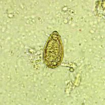
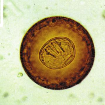
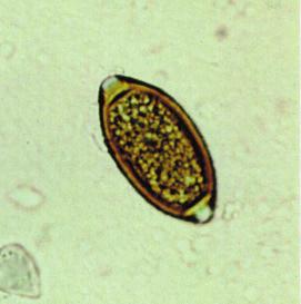
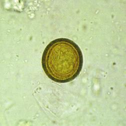
1 FIXATIVES, REAGENTS & STAINS Hymenolepis nana Paragonimus species Diphyllobothrium latum FIXATIVES, REAGENTS & STAINS Clonorchis Hymenolepis diminuta Trichuris trichiura Taenia species Hookworm Fasciola species PARASITOLOGY
2 1 Fixatives 10% Formalin in Buffered Saline This preserves the morphology of cysts and ova. Trophozoites do not preserve well in Formalin-Saline and parasite morphology is not maintained adequately for a permanently stained faecal smear. Can be used in the Parasep (Ridley Allen) method. - It is recommended that 1 part of stool be mixed with 3 parts of Formalin/Formalin-Saline preservative for the storage of bulk specimens. 10% Formalin Fixative in Water Part No This preservative is a good overall fixative and will fix both ova and cysts although it only preserves the internal morphology of the cysts for up to 6 months, after which the cytoplasm of the organism becomes granular with poor nuclear definition. Trophozoites do not preserve well in formalin and parasite morphology is not maintained adequately for a permanently stained faecal smear. Preferred to 10% Formalin in Saline when to be used in Parasep (Ridley Allen concentration method). Sodium Acetate Acetic Acid Formalin (SAF) Part No Fresh stools may be preserved using SAF especially when a delay may occur between the passage of the faeces and its delivery for subsequent laboratory examination. SAF fixed material is suitable for direct examination, concentration (Formalin/Ethyl Acetate) and permanent staining. - A pea sized sample (0.5g) of faeces (include external and internal portions) or 1ml of loose stool should be added to about 5ml of SAF fixative. Mix thoroughly and if necessary break up solid material to ensure it is well dispersed in SAF. Preparation of SAF preserved smears for permanent staining 1. Wash specimen at least twice and preferably three times in Saline to remove all traces of SAF. Centrifuge after each wash at 1000g for at least 1 minute. (10 minutes for Cryptosporidia and Isospora). 2. Shake the Mayer s Glycerin/Albumin to ensure the albumin and glycerin are well mixed. Place a small drop on the centre of a prelabelled slide. 3. Transfer a drop of the washed sediment onto the Mayer s Glycerin/Albumin and mix the two together well with an applicator stick. 4. Using the applicator stick, spread the mixture over the slide so there are alternating thick and thin bands. The thick areas are made with a rolling motion of the wrist. Holding the applicator stick in the middle, closer to the slide, may provide more control when making the smear. If the smear looks too thick or is improperly made, the specimen can easily be redistributed on the slide. 5. Allow the slides to dry thoroughly before staining. The smear should not appear shiny or have a wet appearance, otherwise it may flake off during the staining procedure. 6. Proceed to Staining. Preparation of smears for staining from SAF preserved stool specimens. The Formalin/Ethyl Acetate concentration method for ova and cysts should be employed e.g. using Parasep. 1. Each stool specimen should be treated on an individual basis depending on its composition. Although straining is appropriate for most specimens, mucous strands containing Cryptosporidia, other coccidia, or Giardia lamblia may become trapped in the gauze. Therefore, mucoid specimens and strands of mucous are processed without staining and the mucous itself should be used to make the smear. In these cases, the specimen is still washed in Saline to remove the SAF. Mucoid specimens are also processed without straining, and the mucous itself is applied as the smear. 2. Centrifugation for 10 minutes when processing the stool specimen increases the likelihood of detecting organisms such as Cryptosporidia and Isospora particularly in fatty stool specimens. Smears are made from the washed sediment, not the concentrate, since the organisms are often caught in the ethyl acetate plug. 3. When using SAF as a preservative, it is essential to wash the specimen two or three times to remove the SAF otherwise the specimen may flake off the slide. Removal of the excess debris provides a cleaner specimen. 4. Preparing slides for haematoxylin staining requires some practice before ideal slides are made. Albumin is used as a protein fixative to ensure the specimen adheres to the slide. When a slide is properly made, thin and thick bands are formed. The end product should be a smear thin enough to read a newspaper through. The thick areas aid in the detection of the trophozoites and cysts as well as ova such as unfertilised Ascaris and Hymenolepisnana which do not always concentrate well. With acknowledgements to J.Williams, LSH & TM & A.Moody, LHTD.
3 Fixatives Reagents Schaudinn's Fixative Add mercuric chloride to the water and place in a water bath, preferably in an extraction hood, until it is dissolved. Allow to cool and form crystals. * Health hazard - POISON, harmful by ingestion and skin contact. Danger of cumulative effects. Irritating to eyes and skin. Stock solution Saturated mercuric chloride 600ml 95% ethyl alcohol 300ml - Add 5ml glacial acetic acid to 95ml of the stock solution. Prepare faecal smears without allowing the smears to dry and place them immediately in Schaudinn s fixative for 1 hour. The reagents used in the preparation of this fixative are hazardous and should be handled with care. The working reagents should be prepared fresh for use. This fixative is particularly good for making permanent stained faecal smears of protozoan trophozoites Mayer's Glycerin/Albumin Mayer s Glycerin/Albumin is used when preparing slides for staining. The Albumin helps to ensure that the specimen adheres to the slide and the Glycerin retains sufficient moisture to prevent distortion or disruption of organisms on drying. 1. Shake the Mayer s Glycerin/Albumin to ensure it is well mixed. Place a drop in the centre of the slide. Add a drop of washed sediment to the Mayer s Glycerin/ Albumin and mix well, using an applicator stick. 2. Using the same applicator stick spread the mixture on the slide ensuring that there are alternative thick and thin bands of mixture. 3. Allow the slides to dry well at room temperature before staining. Physiological Saline Bayers Solution This following technique is useful for the preservation of cyst morphology. - Dilute stock solution 1 in 10 with distilled water before use. Mix 1 part of faeces with 1 part of Bayer s solution. Merthiolate-Iodine Formalin (MIF) Part No For use Stock solution Lugol s iodine (see below) 4.7ml 0.3ml - Add approximately 1 gram of faeces to the 4.7ml of MIF/0.3ml Iodine solution and emulsify well. Ova, cysts and larvae can be preserved in MIF for several months. Formol- Ethyl Acetate concentration methods can be performed on samples preserved by any of the above. Polyvinyl Alcohol (PVA) - To use, emulsify 1 part of faeces in 3 parts of PVA solution. This method will preserve ova, larvae and trophozoites well, but cysts may show some distortion. However some ova and cysts do not concentrate well when preserved in PVA. Faecal smears made from the faeces/pva mixture and allowed to dry can be used for the permanent staining of trophozoites. Before staining, the slides must be placed in 70% ethyl alcohol containing 5 10 drops of Lugol s Iodine (see below) to remove the mercuric chloride. Buffered saline solution. Triton X-100 Solution Part No Used to emulsify parasites in faeces, for use in standard Parasep protocols. Ethyl Acetate Part No A solvent that removes fat from faeces, for use in standard Parasep protocols. Acetone Part No Solvent. Neutral Red Aqueous solution and dye, can be used in the Gram s technique. DPX Mounting Media Immersion Oil For use in immersion microscopy. 2
4 3 Stains Eosin/Saline This stain is useful for the detection of motile trophozoites of Entamoeba species. - Emulsify faeces directly in a warm 37 C solution of Eosin in Saline. The amoebae are easily seen unstained against a pink background. The coarse, granular endoplasm can be differentiated from the clear, colourless ectoplasm. Acridine Orange (Acetate Buffered) The addition of Acridine Orange to a faecal concentrate highlights the chromidial bars of Entamoeba coli, Entamoeba histolytica/dispar and Entamoeba hartmanni, which fluoresce bright green. Working solution Stock Acridine Orange Glacial acetic acid Buffered water ph 6.8 1ml 0.5ml 8.5ml - Add an equal volume of stain to the concentrate. Examine the deposit under UV light after 30 minutes. Auramine Phenol (Lempert) 1. Make faecal smears as for ZN and fix in methanol. 2. Stain with Auramine-Phenol (Lempert) for 10-15min. 3. Rinse thoroughly in tap water. 4. Decolourise in acid alcohol (as for ZN). 5. Rinse thoroughly in tap water. 6. Counterstain with 0.1% potassium permanganate for 30 sec. 7. Rinse thoroughly in tap water, allow to air dry. Do not blot dry, many brands of blotting paper will fluoresce! Results Oocysts appear as bright yellow discs against a dark background. Condensed from ACP broadsheet 128, June 1991, Laboratory methods for diagnosing Cryptosporidiosis. Field's Stain - Solution A and Solution B Part No. 1482/83 N.B. Both solutions are ready for use and should not be diluted. This technique is a rapid Field s Stain method, which enables rapid staining of fixed thin films of various clinical samples. This particular method is very useful for staining films of unformed faeces, faecal exudates, duodenal aspirates etc. 1. Make a thin film of faeces/exudate and allow to dry. 2. Fix in methanol for 1 min. 3. Flood slide with 1ml of Field s Stain B (Code: 1483). 4. Immediately add an equal volume of Field s Stain A (Code: 1482) mix well on slide and allow to Stain for 1 min. 5. Rinse well in tap water and drain dry. 6. Examine the film using the oil immersion objective and Immersion Oil. Results Parasite nuclei and structures containing Chromatin - Red Cytoplasm - Bluish-grey Leucocyte nuclei - Purple Yeasts and bacteria - Dark blue Reference: Moody A.H. and Fleck S.L. (1985) Versatile Field s Stain. J.Clin. Pathol. 38(7), Giemsa Stain Rapid Part No Giemsa Stain can also be used to stain films of unformed faeces, faecal exudate, duodenal aspirates etc. 1. Make a thin film of faeces/exudate. Allow to dry. 2. Fix in methanol for 1 min. 3. Tip off the methanol and flood the slide with Giemsa Stain (Code: 1484) diluted 1:10 with buffered distilled water ph 7.2. The diluted stain must be freshly prepared each time. 4. Stain for min. 5. Run tap water on to the slide to float off the stain and prevent deposition of precipitate on to the film. Allow to drain dry. 6. Examine the film using the oil immersion objective and Immersion Oil. Results Parasite nuclei and structures containing Chromatin - Red Cytoplasm - Bluish-grey Leucocyte nuclei - Purple Yeasts and bacteria - Dark blue Lugol's Iodine - Aqueous Part No Temporary Stain for Protozoa. 1. Make wet preparations following concentration by the formol-ether method. 2. Add an equal volume of Lugol s Iodine to 25% glacial acetic acid. 3. Place a drop of the wet preparation on a slide and add a drop of the Iodine/acetic acid mixture prepared in (2) above. 4. Cover slip and examine. Result Iodine Stains: Glycogen - Brown Nuclear Chromatin of Amoebic Cysts - Brown/Black.
5 Stains Iron Haematoxylin Solution A and Solution B Part No. 1487/1488 Preparation of Working Iron Haematoxylin Solution - Notes 1. Mix equal volumes of the two solutions and filter. 2. Allow to stand at least two hours (preferably overnight) before use in order that the chemical reaction is complete. Staining is optimum 3-5 days after preparation. If the Iron Haematoxylin Solution is used immediately after preparation the parasites may be stained an intense blue with little nuclear detail differentiation. When the Iron Haematoxylin Solution is mature (usually 3-5 days after preparation) the background should stain grey with the protozoa light blue, and the nuclei blue-black. The background staining of the slide depends on the composition of the specimen. The stain is normally useable for a week. Shelf life can be extended by storage in a stoppered bottle in the dark after each use. The Picric Acid removes more stain from non-parasitic matter than parasitic matter, and more from the cytoplasm of protozoa than the nucleus. Some cells retain Picric Acid. Complete removal of the picric acid will result in an understained slide. Smears such as these containing Picric Acid will fade with time. If a slide appears cloudy, then dehydration has been inadequate. Agitation in the final alcohols can improve the clarity of the smear. Thick areas may contain more water. Never examine microscopic slides when the DPX is still wet, since the resolution is affected. Drying the smears at 37 C is acceptable but care must be taken that air bubbles do not form during drying. Iron Haematoxylin Staining Procedure 1. Prepare smear using Mayers Albumin. 2. Treat with 95% ethanol or IMS for at least 10 minutes to coagulate albumin, fixing smear to slide. Glycerin is removed at this point and care should be taken to ensure slides do not dry out from this stage on. 3. Slides should be brought to water stepwise through 70% and 25% alcohols. The slides should be treated for at least 10 minutes at each step. Running tap water is best for removal of all residual alcohol. An additional 2 minutes in alkalinised water (by addition of a drop of ammonium hydroxide) is recommended. 4. Stain slides for 10 minutes in Iron Haematoxylin Solution (Codes 1487, 1488) - see notes on preparation. 5. Wash for 30 seconds in distilled or de-ionised water. 6. Treat for 10 minutes using 50% saturated Picric Acid which preferentially destains background. 7. Wash slides in running tap water for at least 10 minutes to remove Picric Acid, stopping destaining. 8. Transfer to alkalinised tap water for 2 minutes. 9. Treat for 10 minutes in alkalinised 70% alcohol (+ 2 drops ammonium hydroxide). 10. Bring to 100% alcohol stepwise through 95% and absolute alcohol (twice) leaving slides in each alcohol for at least 10 minutes. 11. Clear in Xylene for at least 10 minutes, twice. 12. Mount in DPX or any similar neutral mounting medium. 13. Examine using Immersion Oil under oil immersion lens. Trichrome for Microsporidia Part No Make smears from unconcentrated stool specimens in 10% Formalin (1:3 ratio). NOTE: ensure that the smears are extremely thin. 2. Fix in methanol for 5 mins. 3. Stain in modified Trichrome stain (Code: 1489) for 90mins. 4. Rinse in acid alcohol for 10 secs. 5. Rinse briefly in 95% alcohol. 6. Place in 95% alcohol for 5 mins. 7. Place in 100% alcohol for 10 mins. 8. Clear in Xylene for 10 mins. 9. Examine under X 100 oil immersion objective, using Immersion Oil. Interpretation Microsporidial spores are ovoid and refractile and the spore wall stains bright pinkish-red. Occasionally the cellular content of some spores does not stain and appears transparent, others show a pinkish-red stained belt girding the spores either diagonally or equatorially. The spores are approximately 1.5 by 0.9µ. The background debris and bacteria are counterstained faint green. References Weber R et al (1992) Improved light-microscopical detection of Microsporidia spores in stool and duodenal aspirates. New Eng. J. Med. 326(3): With acknowledgements to J.Williams, LSH & TM & A. Moody, LHTD GRAM STAIN PROTOCOL 4
6 Stains Trichrome for Protozoa Part No May be used to stain fresh faeces, prefixed faeces (only certain fixatives) or cultured organisms. The method varies slightly depending on the sample preparation used. 1. Make thin smears of fresh stool or cultured amoebae on a clean microscope slide and immediately fix in Schaudinn s Solution* - do not allow slide to dry. Leave for at least 30mins, preferably overnight. *Schaudinn s Fixative contains Mercuric Chloride which is very poisonous via mouth and skin contact. 2. Transfer to 70% ethanol for 5 mins (omit this step if using PVA fixed material). 3. Transfer to alcoholic-iodine for 2 mins. 4. Transfer to 70% ethanol for 5 mins. 5. Transfer to 70% ethanol for 2-5 mins. 6. Transfer to Trichrome stain (Code: 1490) for 10 mins. 7. Differentiate in 0.5% acetic acid/alcohol for 12 secs. 8. Transfer to 100% ethanol for 1 sec only. 9. Transfer to 100% ethanol for 2-5 mins. 10. Transfer to 100% ethanol for 2.5 mins. 11. Transfer to xylene for 2-5 mins. 12. Transfer to xylene for 2 mins. 13. Mount with DPX or similar neutral mountant. NB: Do not allow the slide to dry at any time during processing. With acknowledgments to J. Williams, LSH & TM & A.Moody, LHTD. Methylene Blue Methylene Blue is a blue counterstain used to differentiate bacteria. Modified Z/N Stain Pack (Cold Kinyoun) Part No Suggested 1. Flood the heat fixed smears with Carbol Fuchsin (ZN) and steam gently for 5 minutes. Add more stain if necessary to prevent drying. 2. Wash with water and decolourise with acid-alcohol until the dye no longer runs off the slide. 3. Wash with water and counterstain for seconds with Methylene Blue or Malachite Green. 4. Wash, blot dry and examine. Acid fast organisms stain red, the background and other organisms stain blue Carbol Fuchsin for Modified Z/N Stain (Cold Kinyoun) Suggested 1. Flood the fixed smears with Carbol Fuchsin (Kinyoun) and stain for 2 minutes without heating. 2. Wash with tap water and decolourise with acid-alcohol until the dye no longer runs off the slide. 3. Wash with tap water and counterstain for seconds with Methylene Blue or Malachite Green. 4. Wash, blot dry and examine. Acid fast organisms stain red, the background and other organisms stain blue. Malachite Green Part No Malachite Green is a green counterstain used to differentiate bacteria.
7 General Microbiology Stain Reagents Gram Stain Protocol Gram's Iodine Crystal Violet Part No Part No Safranin O (Gram) Part No Neutral Red Carbol Fuchsin (Gram) Gram's Decolouriser Part No Part No Introduction Gram s Stain distinguishes between the two major classes of bacteria due to the differences in cell wall structure; Gram-positive bacteria, remain coloured after the staining procedure, and gram-negative bacteria, which do not retain dye. In the staining technique, cells on a microscope slide are heat-fixed and stained with a basic dye, Crystal Violet (Code: 1492), which stains all bacterial cells blue. Iodine solution (Code: 1491) is then added that allows the Iodine to enter the cells and form a water-insoluble complex with the Crystal Violet dye. The preparation is then treated with a decolourise solvent, in which the Iodine-Crystal Violet complex is soluble. Following solvent treatment, only gram-positive cells remain stained, possibly because of their thick cell wall, which is not permeable to solvent. After the staining procedure, cells are treated with a counterstain which may be Safranin O (Gram) (Code: 1493), Carbol Fuchsin (Gram) (Code: 1495) or Neutral Red. Counterstained gram-negative cells appear red, and gram-positive cells remain blue. Although the cell walls of gram-negative and gram-positive bacteria are similar in chemical composition, the cell wall of gram-negative bacteria is a thin layer sandwiched between an outer lipid-containing cell envelope and the inner cell membrane, whereas the gram-positive cell wall is much thicker, lacks the cell envelope, and contains additional substances, such as teichoic acids, polymers composed of glycerol or ribitol. 6. Decolourise with Gram s Decolouriser (Code: 1476) (typically 3-5 seconds) 7. Wash with tap water 8. Counterstain with one of the following: 9. Safranin O (Gram) (Code: 1493) for 30 seconds or 10. Neutral Red for 1 minute or 11. Carbol Fuchsin (Gram) (Code: 1495) for 1 minute 12. Flush with tap water until no further stain can be flushed out 13. Air dry and examine Examination The Gram s staining procedure will stain all Gram +ve cells violet. The Neutral Red counterstain will stain all cells, but will be masked by the violet colour of the Gram s stain. Hence all Gram ve cells will appear red stained. Gram's Stain Pack Part No Contents A Crystal Violet B Gram's Iodine Diluent C Gram's Iodine Concentrate D Gram's Decolouriser E Counterstain: choice of (Safranin O (Gram), Neutral Red, or Carbol Fuchsin (Gram)) * * Please specify on order which counterstain required. Note The Gram's Iodine Concentrate (C) should be added to the Diluent (B) and mixed well before use. All solutions are now ready to use in dropper bottles. Suggested 1. Flood the heat fixed smears with Crystal Violet (A) and allow to stain for up to 1 minute. 2. Wash with tap water 3. Stain with Gram s Iodine (B+C) for 1 minute. 4. Wash with tap water 5. Decolourise (D) until no further stain runs from the slide. 6. Wash thoroughly with tap water 7. Counterstain (E) with Safranin O (Gram), Neutral Red, or Carbol Fuchsin (Gram) for up to 1 minute. 8. Wash, air dry and examine. Expected result Gram positive organisms - purple Gram negative organisms - red The difference in reactivity between gram-positive and gram-negative bacteria is linked with differences in physiological properties of the two groups. Gram-positive bacteria are generally more sensitive to growth inhibition by dyes, halogens, many antibiotics, and to attack by phagocytosis and are more resistant to digestion by the enzymes pepsin and trypsin and enzymes in animal sera. Suggested 1. Heat fix sample 2. Flood heat fixed sample with Crystal Violet (Code: 1492) solution for 1 minute 3. Rinse with tap water 4. Stain with Gram s Iodine (Code: 1491) for 1 minute 5. Rinse with tap water Lugol's Iodine Part No The addition of iodine to a stool concentrate highlights the internal inclusions of cysts; e.g. the nuclei and glycogen mass, thus aiding their identification. For example, the addition of iodine enhances refraction of the nuclei of Endolimax nana, stains the peripheral chromatin of the nuclei of Entamoeba species and demonstrates the well-defined glycogen mass which is a feature of pre-cysts or immature cysts of E. coli and cysts of Iodamoeba butschlii. Iodine does not stain the body of Entamoeba species. 6
 is a red stain used to differentiate bacteria, used in the Gram s technique. Carbol Fuchsin (Gram) Part No. 1495 Used in the Gram s technique Lugol's Iodine Part No.")
8 General Microbiology Stain Reagents Crystal Violet Part No Primary gram stain Crystal Violet Solution is a violet stain used to differentiate bacteria. Safranin O (Gram) Part No Safranin O (Gram) is a red stain used to differentiate bacteria, used in the Gram s technique. Carbol Fuchsin (Gram) Part No Used in the Gram s technique Lugol's Iodine Part No Used in the Gram s technique Gram's Decolouriser Part No Used in the Gram s technique Products can be ordered direct from Apacor or from an appointed distributor Visit our website for all the latest information or on: sales@apacor.com 7 UNIT 5, SAPPHIRE CENTRE, FISHPONDS ROAD, WOKINGHAM, BERKSHIRE, RG41 2QL, ENGLAND TEL: +44 (0) FAX: +44 (0) APA004 - V5 07/2014
Staining of the clinical material or the bacteria from colonies on laboratory media provide a direct visualization of the morphology of the organisms
 COMMON STAINING PROCEDURES Staining of the clinical material or the bacteria from colonies on laboratory media provide a direct visualization of the morphology of the organisms as well as their reactions
for Stool Examination Issued by: LABORATORY MANAGER Original Date: March 13, 2000 Approved by: Laboratory Director Hematoxylin Stain
 Section: Page 28 Policy # MI\PAR\05\06\v01 Page 1 of 5 Subject Title: Laboratory Procedures for Stool Examination Issued by: LABORATORY MANAGER Original Date: March 13, 2000 Approved by: Laboratory Director
Lab Six:- Medical Microbiology Prepared by: Luma J. Witwit. Staining
 Staining Even with the microscope, bacteria are difficult to see unless they are treated in a way that increases contrast between the organisms and their background. The most common method to increase
Bacterial smear and Staining
 Practical Microbiology 18-22/11/2018 University of Sulaimani college of Pharmacy Year2 Lab. 4: Bacterial smear and Staining Before staining and observing a microbe under a microscope, a smear must be prepared.
Basic Microbiology and Immunology Practical Course
 Basic Microbiology and Immunology Practical Course 2 Lab # 2: Colouring the microorganisms Rules that must be followed to maintain an aseptic zone 3 For most bacterial cultures, you will use a sterile
BIOL 251 BASIC MICROBIOLOGY
 BIOL 251 BASIC MICROBIOLOGY CHARACTERISATION OF BACTERIA CHARACTERISATION OF BACTERIA CHARACTERISATION OF BACTERIA MICROSCOPIC To be able to examine microbes microscopically, they need to be stained
Steps of microbial smear preparation :
 Lab 4 STAINING Practical Microbiology Microbial smear : It is a very small amount of microbial growth ( broth or solid ) spreaded on a clean slide and drying by air. Fixation : The process of passing the
Student Performance Guide. Student Performance Guide. Student Performance Guide
 LESSON 8-2 Collecting and Processing Specimens for Parasite Examination Student Performance Guide LESSON 8-3 Microscopic Methods for Student Performance Guide LESSON 8-4 Preparing and Staining Smears for
COMMON STAINING TECHNIQUE
 2 COMMON STAINING TECHNIQUE 2.1 INTRODUCTION Staining is technique used in microscopy to enhance contrast in the microscopic image. Stains and dyes are frequently used in biological tissues for viewing,
: In order to study tissues with a microscope they must be preserved (fixed)- fixation Following fixation, blocks of tissue must be cut into thin
- fixation Following fixation, blocks of tissue must be cut into thin") : In order to study tissues with a microscope they must be preserved (fixed)- fixation Following fixation, blocks of tissue must be cut into thin sections.-microtomy Other techniques involve dehydration
: In order to study tissues with a microscope they must be preserved (fixed)- fixation Following fixation, blocks of tissue must be cut into thin sections.-microtomy Other techniques involve dehydration
Exercise 6-D STAINING OF MICROORGANISMS ENDOSPORE STAINS, CAPSULE STAINS & FLAGELLA
 Exercise 6-D STAINING OF MICROORGANISMS ENDOSPORE STAINS, CAPSULE STAINS & FLAGELLA Introduction Endospore stains, capsule stains, and flagellar stains are staining techniques that allow for the differentiation
Exercise 6-C STAINING OF MICROORGANISMS ACID-FAST STAIN
 Exercise 6-C STAINING OF MICROORGANISMS ACID-FAST STAIN Introduction The acid-fast stain is a differential stain that separates bacteria on the basis of the lipid content of their cell walls. Bacteria
Laboratory Exercise # 8: Other Staining Techniques
 Laboratory Exercise # 8: Other Staining Techniques Purpose: The purpose of this laboratory exercise is to acquaint the student with staining techniques other than the Gram stain that are routinely used
Laboratory technique and preparations
 Laboratory technique and preparations Bio 381 written by : Hind Alzaylaee Alshareef_ Maryam Alzayn Alshareef 9/17/2012 graduated cylinder Funnel Flask beaker Dropping bottle Watch glass Petri dish Reagent
Stains and Solutions Used in Hematology and Cytology
 Stains and Solutions Used in Hematology and Cytology A APPENDIX Acid-Fast Stain Commercially prepared acid-fast stains are available 1. Ziehl Neelsen carbolfuchsin: Dissolve 3.0 g basic fuchsin in 100
LAB 3 CHARACTERIZING YOUR UNKNOWN BACTERIA AND USING MORE COMPLEX STAINS. Part I: Isolating Your Unknown Bacteria and Describing Colony Morphology
 LAB 3 CHARACTERIZING YOUR UNKNOWN BACTERIA AND USING MORE COMPLEX STAINS Objectives In this lab you will learn how to: - describe bacteria on the basis of colony and cell morphology - isolate bacterial
KERATIN CONTAMINATION
 KERATIN CONTAMINATION Keratin contamination is almost always observed as a background protein. Wear only nitrile gloves and rinse with HPLC grade water all trays, containers and surfaces that contact the
ab Gram Stain Kit (Microorganism Stain)
") Version 2 Last updated 27 June 2018 ab150672 Gram Stain Kit (Microorganism Stain) For the histological differentiation of Gram-Positive and Gram- Negative bacteria. This product is for research use only
Version 2 Last updated 27 June 2018 ab150672 Gram Stain Kit (Microorganism Stain) For the histological differentiation of Gram-Positive and Gram- Negative bacteria. This product is for research use only
EXPERIMENT. Bacterial Morphology and Staining Techniques
 EXPERIMENT Bacterial Morphology and Staining Techniques Hands-On Labs, Inc. Version 42-0240-00-02 Review the safety materials and wear goggles when working with chemicals. Read the entire exercise before
Pelagia Research Library. Staining reactions of microwave processed tissues compared with conventional paraffin wax processed tissues
 Available online at www.pelagiaresearchlibrary.com European Journal of Experimental Biology, 2011, 1 (1): 57-62 Staining reactions of microwave processed tissues compared with conventional paraffin wax
Exercise 6-A STAINING OF MICROORGANISMS DIRECT VS INDIRECT STAINING
 Exercise 6-A STAINING OF MICROORGANISMS DIRECT VS INDIRECT STAINING Introduction The morphological features of individual microorganisms may be examined either by observing living, unstained materials,
A New Method for Staining Connective Tissue Fibres, with a Note on Liang's Method for Nerve-fibres. By G. OWEN
 421 A New Method for Staining Connective Tissue Fibres, with a Note on Liang's Method for Nerve-fibres By G. OWEN (From the Department of Zoology, The University, Glasgow) With two plates (figs, i and
WHAT IS GEL ELECTROPHORESIS?
 Getting Started With Gel Electrophoresis a world of learning Presented by Peter J Ball, Southern Biological. For further information, please contact the author by phone (03) 9877-4597 or by email peterjball@southernbiological.com.
GIVD TO GMDN CORRELATION TABLE
 GIVD TO GMDN CORRELATION TABLE GIVD code: 00 00 00 00 Not for IVD use 0 32931 Bath, flotation, tissue Not for IVD use 0 32938 Forlin, neutral buffered EDMA code: 13 01 03 01 Histo/Cyto stains 1891 17080
fully a good result. However, it was not until addition of
 A METHOD OF STAINING BACTERIAL FLAGELLA AND CAPSULES TOGETHER WITH A STUDY OF THE ORIGIN OF FLAGELLA EINAR LEIFSON From the Department of Pathology and Bacterioogy, Johns Hopkins University, Baltimore
AN INTRODUCTION TO METHODS OF STUDYING THE MORBID HISTOLOGY OF DISEASE-CARRYING INSECTS.
 243 AN INTRODUCTION TO METHODS OF STUDYING THE MORBID HISTOLOGY OF DISEASE-CARRYING INSECTS. By CAPTAIN A. E. HAMERTON, D.S.O. Royal Army Medical Oorps. THE great technical improvements in modern histological
VisPRO 5 Minutes Protein Stain Kit
 Manual VisPRO 5 Minutes Protein Stain Kit VP01-125/VP01-500/VP05-125/VP05-500 V2.0 Store at room temperature For Research Use Only Introduction VisPRO 5 Minutes Protein Stain Kit (1 nanogram grade) provides
PREPARATION OF BLOOD FILMS FOR MALARIA DETECTION
 PREPARATION OF BLOOD FILMS FOR MALARIA DETECTION Materials for Preparation of Malaria Smears: Clean and wrapped slides Sterile lancets 70% ethanol and water Absorbent cotton wool Surgical gloves Lint-free
PROTOCOLS FOR ANATOMY/MICROMORPHOLOGY
 PROTOCOLS FOR ANATOMY/MICROMORPHOLOGY General dissection of spikelets... 2 Hand sections and epidermal scrapes of bamboo leaves... 2 Clearing and staining of intact plant organs... 4 Scanning electron
PET Barrier Test PET- R- 02
 PET Barrier Test PET- R- 02 The following protocol is designed to provide a procedure for identifying and quantifying residual amounts of three barrier materials, EVOH, MXD6 nylon, and epoxy diamine, in
Optiblot Non-Reducing Electrophoresis Kit
 Optiblot Non-Reducing Electrophoresis Kit Instructions for Use For the use in non-reducing SDS-PAGE with precast gels This product is for research use only and is not intended for diagnostic use. 1 Table
ROUTINE TECHNIC FOR SURGICAL SPECIMENS. Fixation, Dehydration and Embedding
 A TRICHROME STAINING METHOD FOR ROUTINE USE SERGIO A. BENCOSME, M.D. Department of Pathology, University of Ottawa, and the Ottawa General Hospital, Ottawa, Ontario, Canada Despite the added information
Prisma & Film Staining Workshop. Application Specialist Mea Pelkonen
 Prisma & Film Staining Workshop Application Specialist Mea Pelkonen Tissue-Tek Prisma Tissue-Tek Prisma Always program the Prisma in the following order: 1. Edit solution names Check if desired solution
Optiblot SDS-PAGE Gel
 Optiblot SDS-PAGE Gel Instructions for Use For the use in SDS-PAGE with precast gels This product is for research use only and is not intended for diagnostic use. 1 Table of Contents 1. Introduction 3
ABOUT US. Mission. Keeping prices competitive: We take pride in making sure our products arrive at your lab with the fairest price possible.
 STAIN KITS ABOUT US Mission Keeping it real: Our job is to help you get your job done! Everything we do is designed with your needs out front. Our goal is to make sure your goals are met. Keeping the lines
Copper Stain & Destain Kit for Electrophoresis Instruction Manual. Catalog Number
 Copper Stain & Destain Kit for Electrophoresis Instruction Manual Catalog Number 161-0470 Table of Contents Page Section 1 Introduction 1 1.1 Introduction and Principle 1 1.2 Product Description 2 1.3
Optiblot SDS-PAGE Gel
 Optiblot SDS-PAGE Gel Instructions for Use For the use in SDS-PAGE with precast gels This product is for research use only and is not intended for diagnostic use. 1 Table of Contents 1. Introduction 3
UNIT 7 BASIC TECHNIQUES OF SLIDE PREPARATION
 UNIT 7 BASIC TECHNIQUES OF SLIDE PREPARATION Structure 7.1 Introduction Objectives 7.2 Cleaning, Care and Storage of Slides Washing up Used Slides Cleaning Routine for New Slides Storage of Prepared Slides
Staining Criteria Handbook
 Staining Criteria Handbook General Pathology (Routine Histopathology) Neuropathology Edition 4 November 2015 Index Page Haematoxylin and Eosin Assessment Criteria 3 Special Stains A & B Assessment Criteria
ab Trichrome Stain (Connective Tissue Stain)
") Version 3 Last updated 12 February 2019 ab150686 Trichrome Stain (Connective Tissue Stain) For the histological visualization of collagenous connective tissue fibers in tissue sections. This product is
Version 3 Last updated 12 February 2019 ab150686 Trichrome Stain (Connective Tissue Stain) For the histological visualization of collagenous connective tissue fibers in tissue sections. This product is
Optiblot SDS-PAGE Gel
 Instructions for Use For the use in SDS-PAGE with precast gels This product is for research use only and is not intended for diagnostic use. www.abcam.com 1 Table of Contents Optiblot SDS-PAGE Gel 1. Introduction
tech 66 General Statements Regarding the Chemical Resistance of nora Floor Coverings
 This document is written as a general statement to address nora s stain resistance to chemical contamination however facilities must realize that it does not address all the chemicals and chemical compounds
ab Elastic (Connective Tissue Stain)
") Version 2 Last updated 25 June 2018 ab150667 Elastic (Connective Tissue Stain) For the histological staining of Elastin in tissue sections. This product is for research use only and is not intended for
Version 2 Last updated 25 June 2018 ab150667 Elastic (Connective Tissue Stain) For the histological staining of Elastin in tissue sections. This product is for research use only and is not intended for
Western Blotting Systems CAT NO:EPS-B0015 & EPS-B0016
 Western Blotting Systems CAT NO:EPS-B0015 & EPS-B0016 TABLE OF CONTENTS Important User Information Section 1 General Information 1.1 Introduction 1.2 Standard supply 1.3 Specifications Section 2 Instructions
On Double Staining Nucleated Blood-Corpuscles with Anilin Dyes.
 292 DE. VINCENT HAEBIS. On Double Staining Nucleated Blood-Corpuscles with Anilin Dyes. Vincent Harris, M.D., Demonstrator of Physiology at St. Bartholomew's Hospital. THE usefulness of the process of
Carpet Cleaning Guide. Carpet Cleaning 101: An Overview
 Carpet Cleaning Guide Carpet Cleaning 101: An Overview Step One: Identify Carpet Fiber Type Cotton Wool, Silk Nylon Olefin Polyester Acrylic Synthetic Composition Cellulosic Cotton Seed, Jute Protein Based-
TECHNICAL INFORMATION
 TECHNICAL INFORMATION TURCO DY-CHEK INDUSTRIAL PENETRANT (STEP 2) BY THE HAND WIPE OR SOLVENT REMOVAL METHOD DESCRIPTION: TURCO DY-CHEK INDUSTRIAL PENETRANT INSPECTION is a method of non-destructive testing
Maintenance Guidelines Scuba
 Maintenance Code W/B-Clean with water-based cleanser or diluted household bleach. Regular Maintenance Vacuum regularly using the proper attachment to avoid pilling. For non-woven textiles, wipe regularly
Single Source For Your Histology Reagents. Protecting Every Life Story In Your Lab.
 A Division of General Data Healthcare Histology Innovation for a NEW Generation Ready-To-Use Reagents, Dyes & Stains Routine Product Stains Name & Special Here Stains Single Source For Your Histology Reagents.
STUDENT LABORATORY PACKET
 L5 Elodea-Onion-Cheek-Cell_Size Page 1 of 7 STUDENT LABORATORY PACKET Student s Full Name Lab #5: Elodea, Onion, Cheek Cells-Cell Size Lab Instructor Date Points Microscope # OBJECTIVES: a. to examine
E-Blotter Operation. Technical Bulletin E-03 MATERIAL PROCEDURE
 E-Blotter Operation MATERIAL BSA (1 mg/ml; 0.05 g BSA (Sigma-Aldrich Ltd., St Louis, MO; U.S.A.) dissolve in 50 ml ddh 2 O, aliquot to 1 ml in micro-centrifuge tubes and freeze in -20 C) NK92 cell lysate
EXERCISE 8C - Lab Procedures
 EXERCISE 8C - Lab Procedures SAFETY WARNING: Acrylamide in the unpolymerized form is a skin irritant and a potential neurotoxin. Fortunately, the acrylamide in your gels is polymerized, so it should not
ab Papanicolaou (PAP) Red Stain Kit (Cytology Stain)
 Red Stain Kit (Cytology Stain)") Version 2 Last updated 27 June 2018 ab150679 Papanicolaou (PAP) Red Stain Kit (Cytology Stain) For the Differentiation of Cells in Vaginal Smears for the Detection of Vaginal, Uterine and Cervical Cancer.
Version 2 Last updated 27 June 2018 ab150679 Papanicolaou (PAP) Red Stain Kit (Cytology Stain) For the Differentiation of Cells in Vaginal Smears for the Detection of Vaginal, Uterine and Cervical Cancer.
Phenion FT Skin Model Histological processing Paraffin sections
 Phenion FT Skin Model Histological processing Paraffin sections Objective This Standard Operation Procedure is recommended to fix and embed Phenion FT Skin Models in order to prepare paraffin sections.
Instrument Cleaning. Dental Clinics
 1 CLEANING FOR INFECTION CONTROL Dental Clinics 2 This presentation is designed to reflect the recommendations given in the following Australian Standards and Guidelines. Throughout this discussion we
Lab. Elodea, Onion, and Cheek Cell Lab. Be your best. Cell Biologist s Name: Period: Date: Mrs. Bouchard -7 th Grade Science
 Purpose Ques*on: How do plant cells and animal cells differ? Cheek Cell Lab Materials: Water bo6le with dropper toothpick glass slide coverslip lens paper methylene blue safety goggles lab apron paper
Experiment 11 Identification of Food Colors in Candies
 Experiment 11 Identification of Food Colors in Candies Pre-lab Assignment Before coming to lab: Read the lab thoroughly. Answer the pre-lab questions that appear at the end of this lab exercise. Purpose
BOTANY Lab Manual BSc.-III Medical Semester V
 BOTANY Lab Manual BSc.-III Medical Semester V 212 Experiment 1 Aim: Determine Water Potential of Vacuolar Sap by Plasmolytic Method. Requirements: Leaves of Tradescantia solutions of different concentrations,
ROBOT PIN TOOL CLEANING AND LIQUID SAMPLE TRANSFER
 OVERVIEW TECHNICAL NOTE 67B ROBOT PIN TOOL CLEANING AND LIQUID SAMPLE TRANSFER There are several key steps in the successful use of pin tools: 1. The first and most important step is to start with clean
Comparative Proteomics Kit II: Western Blot Module Quick Guide
 Comparative Proteomics Kit II: Western Blot Module Quick Guide Lesson 1 1 Label one 1.5 ml fliptop micro tube for each of five fish samples. Also label one screw-cap micro tube for each fish sample. 2.
Glycoprotein Staining Kit
 193PR 02 G-Biosciences 1-800-628-7730 1-314-991-6034 technical@gbiosciences.com A Geno Technology, Inc. (USA) brand name Glycoprotein Staining Kit With Rapidstain for Enhanced Glycoprotein & Non Glycoproteins
Preparation of Ink. Abstract
 1 Preparation of Ink Abstract To Study the Preparation of Ink. This project throws a light on types of inks from manufacturing point of view and includes method for preparing them either in chemistry laboratory
SPECIAL STAINS IN HISTOPATHOLOGY
 SPECIAL STAINS IN HISTOPATHOLOGY VAN GIESON MOVAT S PENTACHROME STAIN DR RAZANA MOHD ALI SPECIAL STAINS IN HISTOLOGY STAINS FOR MICROORGANISM CONNECTIVE TISSUE STAINS STAINS FOR PIGMENTS AND MINERAL INTRODUCTION
SELYE and McKeown (1935) and Baker (1948) have noted the presence of
 and Baker (1948) have noted the presence of") A Pigment in the Rat's Uterus By ]. G. WARBRICK {From the Department of Anatomy, University of Glasgow, Glasgow, W. 2) With one plate (fig. i) SUMP4ARY 1. A yellowish-brown pigment was found at the old
A Pigment in the Rat's Uterus By ]. G. WARBRICK {From the Department of Anatomy, University of Glasgow, Glasgow, W. 2) With one plate (fig. i) SUMP4ARY 1. A yellowish-brown pigment was found at the old
Antiseptics, Disinfectants, and Sterilants (Germicides)
") Antiseptics, Disinfectants, and Sterilants (Germicides) This information is provided for the benefit of health-care workers who may not have access to the wide variety of products available in modern health
Antiseptics, Disinfectants, and Sterilants (Germicides) This information is provided for the benefit of health-care workers who may not have access to the wide variety of products available in modern health
ANALYSIS OF FINGERPRINTS, LIPSTICK 2 ND HAIR
 ANALYSIS OF FINGERPRINTS, LIPSTICK 2 ND HAIR LAB FORENSICS.3 From Sourcebook, National Science Foundation, 1997 INTRODUCTION PART A. OBTAINING A FINGERPRINT Black ink stamp pad Tissue paper 4 x 4 cm Card
Maintenance Guidelines Bluff
 Maintenance Code W/B-Clean with water-based cleanser or diluted household bleach. Regular Maintenance Vacuum regularly using the proper attachment to avoid pilling. For non-woven textiles, wipe regularly
What is Life? Project PART 1: Looking at Cells Lab
 What is Life? Project PART 1: Looking at Cells Lab Directions: Complete the drawings and answer the questions in the space provided. For each drawing: Title the drawing of the specimen (e.g. Cork Cells)
Tips On Proper Instrument Cleaning, Handling and Maintenance!
 304 304 Rinsing Tips On Proper Instrument Cleaning, Handling and Maintenance! *Immediately after instrument use thoroughly rinse off all blood, tissue and other fluids. *Using filtered water to rinse and
American Cleaning Institute Development of Exposure Assessments Glossary of Functional Classes
 American Cleaning Institute Development of Exposure Assessments Glossary of Functional Classes Abrasive: Abrasive ingredients are materials that are used to polish, buff, or scour away soils such as dirt
Material Safety Data Sheet
 Section 1: Product and Company Identification Product Name Catalog Number Company Cyclic GMP CLIA Kit (High-Sensitivity) SKT-210 StressMarq Biosciences Inc. PO Box 55036 Cadboro Bay Victoria BC V8N4G0
The Effects of Shear on Neutralized Carbomers in Aqueous Conditions
 The Effects of Shear on Neutralized Carbomers in Aqueous Conditions Lyndel Speedy 18/07/2014 With thanks to Ensign Laboratories 1 Abstract Carbomer is the generic name for a class of high molecular weight
The Specialist's Free Guide to Stain Removal
 The Specialist's Free Guide to Stain Removal Red Wine, Coffee, and Pet Stains No More! The Specialist Complete Carpet Care Expert Carpet, Upholstery & Area Rug Cleaning (714) 350-9290 info@thespecialistccc.com
Maintenance Guidelines Ellipsis
 Maintenance Code W/B-Clean with water-based cleanser or diluted household bleach. Regular Maintenance Vacuum regularly using the proper attachment to avoid pilling. For non-woven textiles, wipe regularly
MIGLYOL 840. Excellent light emollient. Alternative to IPM. INCI: Propylene Glycol Dicaprylate/Dicaprate. Excellent light and dry emollient
 Excellent light emollient Excellent light and dry emollient Alternative to IPM 1. Description: MIGLYOL 840 is a propylene glycol diester of saturated plant fatty acids with chain lengths of C 8 and C 10.
Safety Data Sheet BDH5088 BDH5088-4L. VWR 100 Matsonford Road Suite 200 Radnor, PA USA
 SECTION 1: Identification 1.1. Product Identifier Trade Name or Designation: Buffer, Reference Standard, ph 12.00 ± 0.02 at 25 C Product Number: Other Identifying Product Numbers: BDH5088 BDH5088-4L 1.2.
Tape Strip. Year Group: BVSc4 + Document number: CSL_P03
 Year Group: BVSc4 + Document number: CSL_P03 Equipment list: Equipment for this station: Sellotape Glass microscope slides Wax marker crayon Gloves Diff-Quik booklet ( CSL_L06 Diff-Quik Staining ) Diff-Quik
MATERIAL SAFETY DATA SHEET Description: SAF POWDERKLENZ Revision Number: 01
 NON-CLOGGING HEAVY DUTY POWDER DEGREASER & CLEANER GENERAL SAF-POWDERKLENZ is a versatile, safe and easy to use, multi-purpose water soluble degreaser and cleaner that fizzes in contact with water resulting
ORTON and Post (1932) and Cutler (1935) investigated the use of diethylene
 and Cutler (1935) investigated the use of diethylene") 593 A Modified Ester Wax for Embedding Tissues By W. CHESTERMAN AND E. H. LEACH {From the University Laboratory of Physiology, Oxford) SUMMARY I. A modification of Steedman's ester wax embedding method
593 A Modified Ester Wax for Embedding Tissues By W. CHESTERMAN AND E. H. LEACH {From the University Laboratory of Physiology, Oxford) SUMMARY I. A modification of Steedman's ester wax embedding method
SECTION 1. Identification of the substance/mixture and of the company/undertaking. Synonym(s) Colloidal Coomassie blue; Coomassie brilliant blue G 250
 Colloidal Coomassie blue; Coomassie brilliant blue G 250") National Centre for Biotechnology Education Coomassie blue stain Safety data sheet Prepared in accordance with Regulation (EC) No. 1907/2006 (REACH) Version 2.0 Created: 1 June 2015 Revised: 26 June 2015
National Centre for Biotechnology Education Coomassie blue stain Safety data sheet Prepared in accordance with Regulation (EC) No. 1907/2006 (REACH) Version 2.0 Created: 1 June 2015 Revised: 26 June 2015
Unit 3 Hair as Evidence
 Unit 3 Hair as Evidence A. Hair as evidence a. Human hair is one of the most frequently pieces of evidence at the scene of a violent crime. Unfortunately, hair is not the best type of physical evidence
TEO ClearPAGE Precast Gel Running Instructions for SDS PAGE and DNA/Native Gels. Table of Contents
 TEO ClearPAGE Precast Gel Running Instructions for SDS PAGE and DNA/Native Gels Table of Contents General Information... 1 Running buffer preparation... 1 Sample preparation...2 Gel preparation...2 Loading
Uniperol Bleach IT. Technical Information. Europe
 Technical Information TIe/ EU July 2011/I (5/2011)(WJA) Page 1 of 7 First Edition Europe = Registered trade mark of BASF in several countries Uniperol Bleach IT Basic bleaching agent, without optical brightener
MOLLUSCS PROCESSING FOR DIAGNOSIS BY HISTOLOGY
 European Union Reference Laboratory for Molluscs Diseases MOLLUSCS PROCESSING FOR DIAGNOSIS BY HISTOLOGY SUMMARY 1. SCOPE...2 2. REFERENCES...2 3. GENERAL INFORMATION...2 4. EQUIPMENT AND ENVIRONMENT...2
The Many Uses of Hydrogen Peroxide. 1. Whiten your clothes with HP instead of bleach
 The Many Uses of Hydrogen Peroxide Hydrogen Peroxide is an active agent composed of nothing more than water and oxygen. The oxidation process of this substance is incredibly good at killing disease organisms.
zenithinteriors.com Epic MELBOURNE SYDNEY CANBERRA BRISBANE PERTH ADELAIDE AUCKLAND WELLINGTON SHANGHAI HONG KONG
 zenithinteriors.com Epic Epic An affordable solution of workstation when organisations are undergoing growth, change and re-structure. FEATURES The slim formed lines of EPIC incorporate the essentials
ARGANisme cosmetics 55. bd Anoual. Casablanca MOROCCO Tel: Fax:
 Page 1 sur 5 ARGANisme cosmetics 55. bd Anoual. Casablanca MOROCCO Tel: +212 522 860 518 Fax: +212 522 860 715 SAFETY EVALUATION OF "ARGANisme ARGAN OIL" 1. Qualitative and Quantitative Composition of
STANDARDIZED CLINICAL PROTOCOLS. Sterilization Protocols. Aravind Eye Care System 1, Anna Nagar, Madurai , Tamilnadu, India
 STANDARDIZED CLINICAL PROTOCOLS Sterilization Protocols Aravind Eye Care System 1, Anna Nagar, Madurai - 625 020, Tamilnadu, India i.exe Standardized Sterilization Protocol STANDARDIZED STERILIZATION PROCEDURE
Rittel s EZ-100 TANNING INSTRUCTIONS
 Rittel s EZ-100 TANNING INSTRUCTIONS RITTEL S EZ-2000 Kit Using EZ-100 the newest and highest quality tanning agent available and only from RITTEL S and our authorized Distributors! This is a powdered
Maintenance Guidelines Compound
 6696 Maintenance Code W/B-Clean with water-based cleanser or diluted household bleach. Regular Maintenance Vacuum regularly using the proper attachment to avoid pilling. For non-woven textiles, wipe regularly
Gafquat 440, 755N, 755N-P, 755N-O and HS-100, HS-100-O polymers Cationic conditioning copolymers
 PRODUCT DATA Consumer Specialties ashland.com NUMBER 4817-1 (Supersedes 4817) Page 1 of 8 Gafquat 440, 755N, 755N-P, 755N-O and HS-100, HS-100-O polymers Cationic conditioning copolymers Introduction Gafquat
SAFETY DATA SHEET Description: SAF L.F. STRIPPER Revision Number: 00
 HEAVY DUTY FLOOR STRIPPER AND CLEANER GENERAL SAF EA LF STRIPPER is a powerful acrylic polymer based polish remover/stripper suitable for automatic scrubbing machines. SAF EA LF STRIPPER can be used on
Spot-Cleaning Tips and Remedies
 The best and most convenient way to remove spots and spills is to have your carpet serviced by an Aladdin Professional. However, the following tips and home remedies should help remove most spots and odors.
SAFETY DATA SHEET according to 1907/2006/EC, Article 31
 SAFETY DATA SHEET according to 197/26/EC, Article 31 14185 NatureCare FRC Universal Print Replacement Cartridge Part A (P1) T/M SECTION 1: Identification of the substance/mixture and of the company/undertaking
were made by the National Physical Laboratory, were collected into EDTA-K2 anticoagulant (1-5 films were made shortly after blood collection.
 J. clin. Path., 97, 8, 8-8 An evaluation of some commercial Romanowsky stains P. N. MARSHALL, S. A. BENTLEY, AND S. M. LEWIS From the Department ofhaematology, Royal Postgraduate Medical School, Hammersmith
Item/Package Details Size Item Bottle ph Shelf Life 1.0 oz/29.6 ml 1101 Lucite Matte Silver Pump months
 B-Kleer 10 Lotion Use of mild to moderate acne. Gently exfoliates encrustation and acne pustules. Penetrates pores to eliminate most acne pimples. Reduces the severity of acne blemishes. Help prevent new
STAIN-PROOF TEST RESULTS
 Test Results STAIN-PROOF TEST RESULTS Contents University of New South Wales, Australia: STAIN RESISTANCE oil on granite. University of New South Wales, Australia: STAIN RESISTANCE iodine on granite. Bemac
StainEase Gel Staining Tray
 USER GUIDE StainEase Gel Staining Tray Catalog Number NI2400 Revision date 19 March 2012 Publication Part number IM-2400 MAN0000731 For Research Use Only. Not intended for any animal or human therapeutic
It Works! Multi Stain Remover Kit with 2 Tubes & 2 Sponges
 It Works! Multi Stain Remover Kit with 2 Tubes & 2 Sponges USE: It Works for clothing stains, carpets, upholstery, car seats, head liners, all types of over garments, leather, vinyl, aluminum rims, jewelry,
Bio-compatibility Requirements
 Bio-compatibility Requirements Printing Bio-compatible Parts on PolyJet 3D Printers with MED610 TM The methods and conditions described in this document were tested at Stratasys for printing parts from