COMMON STAINING TECHNIQUE
|
|
|
- Charlene Barber
- 6 years ago
- Views:
Transcription
1 2 COMMON STAINING TECHNIQUE 2.1 INTRODUCTION Staining is technique used in microscopy to enhance contrast in the microscopic image. Stains and dyes are frequently used in biological tissues for viewing, often with the aid of different microscopes. Stains may be used to define and examine bulk tissues (highlighting, for example, muscle fibers or connective tissue), cell populations (classifying different blood cells, for instance), or organelles within individual cells. Bacteria have nearly the same refractive index as water, therefore, when they are observed under a microscope they are opaque or nearly invisible to the naked eye. Different types of staining methods are used to make the cells and their internal structures more visible under the light microscope. Microscopes are of little use unless the specimens for viewing are prepared properly. Microorganisms must be fixed & stained to increase visibility, accentuate specific morphological features, and preserve them for future use OBJECTIVES After reading this lesson, you will be able to: describe the need for staining techniques explain terms related to staining techniques discuss the substances used as stain enlist various staining techniques classify & explain various stains 20
2 2.2 TERMS RELATED TO STAINING MODULE Stain A stain is a substance that adheres to a cell, giving the cell color. The presence of color gives the cells significant contrast so they are much more visible. Different stains have different affinities for different organisms, or different parts of organisms. They are used to differentiate different types of organisms or to view specific parts of organisms Staining Staining is an auxiliary technique used in microscopy to enhance contrast in the microscopic image. Stains and dyes are frequently used in biology and medicine to highlight structures in biological tissues for viewing, often with the aid of different microscopes. Fixation Fixation by itself consists of several steps aims to preserve the shape of the cells or tissue involved as much as possible. Sometimes heat fixation is used to kill, adhere, and makes them permeable so it will accept stains What can be used as stain The substance be used as a stain must be colored or it should react in the system to give a colored product, because of which some portion of the system becomes colored and the rest remains colorless. Staining renders the organism more visible, it displays the structure and finer details of bacteria and it helps to differentiate between organisms Staining techniques Direct staining - The organism is stained and background is left unstained Negative staining - The background is stained and the organism is left unaltered INTEXT QUESTIONS 2.1 Fill in the blanks: 1. Staining is primarily used to enhance... in the image 2. Substances that adhere to cell giving colour to cell are called as aims to preserve the shape of the cells 4. The organism is stained and background is left unstained in... staining technique 5. Background is stained and the organism is left unchanged in... staining technique 21
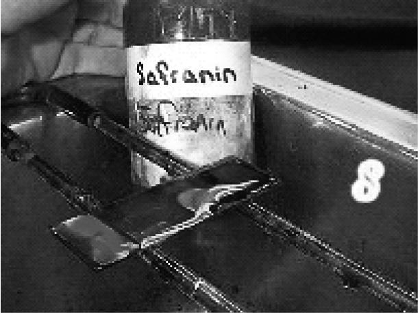
3 2.3 KINDS OF STAINS Stains are classified as Simple stain Differential stain Structural or special stains Simple Staining The staining process involves immersing the sample (before or after fixation and mounting) in dye solution, followed by rinsing and observation. Many dyes, however, require the use of a mordant, a chemical compound that reacts with the stain to form an insoluble, coloured precipitate. When excess dye solution is washed away, the mordanted stain remains. Simple staining is one step method using only one dye. Basic dyes are used in direct stain and acidic dye is used in negative stain. Simple staining techniques is used to study the morphology better, to show the nature of the cellular contents of the exudates and also to study the intracellular location of the bacteria Commonly used simple stains are Methylene blue Dilute carbol fuchsin Polychrome methylene blue Loeffler s Methylene Blue Method of Staining Flood the smear with methylene blue, allow for 2 minutes, pour off the stain and allow the air to dry by keeping in a slanting position and by this the organism will retain the methylene blue stain Use Methylene blue staining is used to make out clearly the morphology of the organisms eg. H.influenzae in CSF, Gonococci in urethral pus Polychrome Methylene Blue Preparation Allow Loeffler s Methylene blue to ripen slowly. Methylene blue stain is kept in half filled bottles, aerate the content by shaking at intervals, Slow oxidation of methylene blue forms a violet compound and Stain gets polychrome property. The ripening nearly takes 12 months and this is hastened by addition of 1% potassium carbonate 22
4 Use Polychrome Methylene Blue is used to demonstrate Mc Fadyean reaction of B.anthracis and in this the blue bacilli Is surrounded by purple capsular material MODULE Dilute Carbol Fuchsin Preparation Prepare carbol fuchsin and dilute it to 1/15 using distilled water Method of staining Flood the smear and let stand for 30 seconds, wash with tap water and blot gently to dry Use To stain throat swab from patients of suspected Vincent s angina, (Borrelia are better stained), it is used as a counter stain in Gram stain and to demonstrate the morphology of Vibrio cholerae (comma shaped) INTEXT QUESTIONS 2.2 Fill in the blanks: 1. Chemicals that reacts with stain to form precipitates are called as dyes is used in direct stain dyes is used in negative stain 4. Commonly used simple stains are...,... & DIFFERENTIAL STAINS Gram Staining Differential Stains use two or more stains and allow the cells to be categorized into various groups or types. Both the techniques allow the observation of cell morphology, or shape, but differential staining usually provides more information about the characteristics of the cell wall (Thickness). Gram staining (or Gram s method) is an emprical method of differentiating bacterial species into two large groups (Gram-positive and Gram-negative) based on the chemical and physical properties of their cell wall. The Gram stain is almost always the first step in the identification of a bacterial organism, While Gram staining is a valuable diagnostic tool in both clinical and research settings, not all bacteria can be definitively classified by this technique, thus forming Gram variable and Gram 23
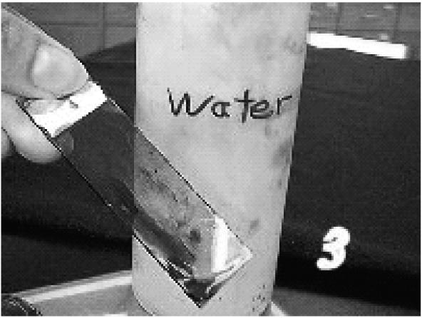
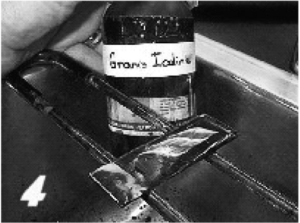
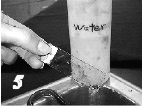
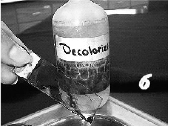
5 indeterminate groups as well. The word Gram is always spelled with a capital, referring to Hans Christian Gram, the inventor of Gram staining Gram staining Principles Gram staining is used to determine gram status to classify bacteria broadly. It is based on the composition of their cell wall. Gram staining uses crystal violet to stain cell walls, iodine as a mordant, and a fuchsin or safranin counterstain to mark all bacteria. Gram status is important in medicine; the presence or absence of a cell wall will change the bacterium s susceptibility to some antibiotics. Gram-positive bacteria stain dark blue or violet. Their cell wall is typically rich with peptidoglycan and lacks the secondary membrane and lipopolysaccharide layer found in Gram-negative bacteria Gram Staining Technique 1. Crystal violet acts as the primary stain. Crystal violet may also be used as a simple stain because it dyes the cell wall of any bacteria. 2. Gram s iodine acts as a mordant (Helps to fix the primary dye to the cell wall). 3. Decolorizer is used next to remove the primary stain (crystal violet) from Gram Negative bacteria (those with LPS imbedded in their cell walls). Decolorizer is composed of an organic solvent, such as, acetone or ethanol or a combination of both.) 4. Finally, a counter stain (Safranin), is applied to stain those cells (Gram Negative) that have lost the primary stain as a result of decolorization Gram Reaction Gram-positive bacteria are those that are stained dark blue or violet by Gram staining. This is in contrast to Gram-negative bacteria, which cannot retain the crystal violet stain, instead taking up the counter stain (safranin or fuchsine) and appearing red or pink. Gram-positive organisms are able to retain the crystal violet stain because of the high amount of peptidoglycan in the cell wall. Grampositive cell walls typically lack the outer membrane found in Gram-negative bacteria. Gram-negative bacteria are those bacteria that do not retain crystal violet dye in the Gram staining protocol. In a Gram stain test, a counter stain (commonly safranin) is added after the crystal violet, coloring all Gram-negative bacteria with a red or pink color. The test itself is useful in classifying two distinct types of bacteria based on the structural differences of their cell walls. On the other hand, Gram-positive bacteria will retain the crystal violet dye when washed in a decolorizing solution. 24


6 MODULE Fig. 2.1: Gram Reaction. 25
7 INTEXT QUESTIONS 2.3 Fill in the blanks: 1. Gram staining uses... to stain cell walls,... as mordant &... as counter stain 2. Gram positive bacteria stains... colour 3. Gram negative bacteria stains... colour 4. Gram positive organism are able to retain crystal violet stain because of high amount of... in the cell wall 2.5 ACID-FAST STAINING The Ziehl Neelsen stain, also known as the acid-fast stain, widely used differential staining procedure. The Ziehl Neelsen stain was first described by two German doctors; Franz Ziehl (1859 to 1926), a bacteriologist and Friedrich Neelsen (1854 to 1894) a pathologist. In this type some bacteria resist decolourization by both acid and alcohol and hence they are referred as acidfast organisms. This staining technique divides bacteria into two groups namely acid-fast and non acid-fast. This procedure is extensively used in the diagnosis of tuberculosis and leprosy. Mycobacterium tuberculosis is the most important of this group, as it is responsible for the disease called tuberculosis (TB) along with some others of this genus Principle Mycobacterial cell walls contain a waxy substance composed of mycolic acids. These are β-hydroxy carboxylic acids with chain lengths of up to 90 carbon atoms. The property of acid fastness is related to the carbon chain length of the mycolic acid found in any particular species Ziehl- Neelsen Procedure 1. Make a smear. Air Dry. Heat Fix. 2. Flood smear with Carbol Fuchsin stain Carbol Fuchsin is a lipid soluble, phenolic compound, which is able to penetrate the cell wall 3. Cover flooded smear with filter paper 4. Steam for 10 minutes. Add more Carbol Fuchsin stain as needed 5. Cool slide 6. Rinse with DI water 26
 until the red color stops streaming from the smear 9. Rinse with DI water 10.")
8 7. Flood slide with acid alcohol (leave 15 seconds). The acid alcohol contains 3% HCl and 95% ethanol, or you can declorase with 20% H 2 SO 4 8. Tilt slide 45 degrees over the sink and add acid alcohol drop wise (drop by drop) until the red color stops streaming from the smear 9. Rinse with DI water 10. Add Loeffler s Methylene Blue stain (counter stain). This stain adds blue color to non-acid fast cells. Leave Loeffler s Blue stain on smear for 1 minute 11. Rinse slide. Blot dry. 12. Use oil immersion objective to view. MODULE Fig. 2.2: Ziehi-Neelsen acid fast staining procedure Fig
9 INTEXT QUESTIONS 2.4 Fill in the blanks: 1. Organisms that resist decolourisation by acid and alcohol are called as staining is used in the diagnosis of Tuberculosis and Leprosy 3. Acid fastness is related to... length of mycolic acid 4. Acid fast staining groups bacteria into two groups namely... & SPECIAL STAINS Stains for Metachromatic granules Stain for spores Stain for capsules Stain for spirochetes Stain for flagella Albert s Staining for C. diphtheriae In all cases of suspected Diphtheria, stain one of the smears with Gram stain. If Gram stained smear shows morphology suggestive of C.diphtheriae, proceed to do Albert staining which demonstrates the presence or absence of metachromatic granules. C.diphtheriae are thin Gram positive bacilli, straight or slightly curved and often enlarged (clubbing) at one or both ends and are arranged at acute angles giving shapes of Chinese letters or V shape which is characteristic of these organisms. Present in the body of the bacillus are numerous metachromatic granules which give the bacillus beaded or barred appearance. These granules are best demonstrated by Albert s stain. Albert staining Albert stain I Toluidine blue 0.15 gm Malachite green 0.20 gm Glacial acetic acid 1.0 ml Alcohol(95%) 2.0 ml Distilled water 100 ml 28
10 Albert stain II Iodine 2.0 gm Potassium iodide 3.0 gm Distilled water 300 ml Albert staining Procedure Cover the heat-fixed smear with Albert stain I. Let it stand for two minutes. Wash with water. Cover the smear with Albert stain II. Let it stand for two minutes. Wash with water, blot dry and examine. To demonstrate metachromatic granules in C.diphtheriae. These granules appear bluish black whereas the body of bacilli appear green or bluish green. MODULE Capsule staining The purpose of the capsule stain is to reveal the presence of the bacterial capsule, the water-soluble capsule of some bacterial cells is often difficult to see by standard simple staining procedures or after the Gram stain. The capsule staining methods were developed to visualize capsules and yield consistent and reliable results Capsule may appear as clear halo when a fresh sample is stained by Grams or Leishman stain, Negative staining- using - India ink, Nigrosin India ink Commercially available India ink is used undiluted Procedure Place a loop full of India ink on the slide A small portion of the culture is emulsified in the drop of ink Place a clean cover slip over the preparation without bubbles. Press down gently Examine under dry objective Uses India ink is used to demonstrate capsule which is seen as unstained halo around the organisms distributed in a black background eg. Cryptococcus Endospore Staining Bacterial endospores are metabolically inactive, highly resistant structures produced by some bacteria as a defensive strategy against unfavorable 29
11 environmental conditions. Primary stain - is malachite green, which stains both vegetative cells and endospores and heat is applied to help the primary stain penetrate the endospore. Decolorized with water, which removes the malachite green from the vegetative cell but not the endospore, Safranin - counterstain any cells which have been decolorized, At the end of the staining process, vegetative cells will be pink, and endospores will be dark green Flagella stain Flagella are fragile appendages Cannot be seen under ordinary microscope Hence the surface is coated with a precipitate to form a colloidal substance This precipitate serves as a layer of stainable material Components 1. 1% Osmic acid 2. Mordant 10% Tannic acid Sat.potassium alum 10% Ferric chloride 3. Fontana s silver solution Use This is used to demonstrate the flagella and the organisms stain black and flagella appear light brown INTEXT QUESTIONS 2.5 Fill in the blanks: 1. Presence or absence of metachromatic granules is demonstrated by... staining chemical is used in capsule staining is used as primary stain in Endospore staining is the counter stain in endospore staining 30
12 MODULE WHAT YOU HAVE LEARNT Staining is technique used in microscopy to enhance contrast in the microscopic image. Stain is a substance that adheres to a cell, giving the cell colour. Strains are classified as Simple stain, Differential stain and Special stains. Gram staining is used to differentiate bacterial species as Gram-positive and Gram-negative based on the chemical and physical properties of cell wall. Acid Fast staining technique or Ziehl Neelsen stain divides bacteria into acid fast and non-acid-fast and this is used in diagnosis of tuberculosis and Leprosy. Albert staining technique demonstrates the presence or absence of metachromatic grannules which is used in identification of C. diphtheria bacilli. TERMINAL QUESTIONS 1. List staining techniques 2. Describe different kinds of stains 3. Explain gram staining 4. Explain Acid fast staining ANSWERS TO INTEXT QUESTIONS Contrast 2. Stain 3. Fixation 4. Direct 5. Negative 31
13 Mordant 2. Basic 3. Acidic 4. Methylene blue, Polychrome methylene blue & Dilute carbol fuchsin Crystal violet, Iodine & Fuchsin 2. Dark blue or violet 3. Red or pink 4. Peptidoglycan Acid fast organism 2. Acid fast staining 3. Carbon chain 4. Acid fast and Non acid fast Albert s 2. Indian ink 3. Malachite green 4. Safranin 32
Lab Six:- Medical Microbiology Prepared by: Luma J. Witwit. Staining
 Staining Even with the microscope, bacteria are difficult to see unless they are treated in a way that increases contrast between the organisms and their background. The most common method to increase
Staining of the clinical material or the bacteria from colonies on laboratory media provide a direct visualization of the morphology of the organisms
 COMMON STAINING PROCEDURES Staining of the clinical material or the bacteria from colonies on laboratory media provide a direct visualization of the morphology of the organisms as well as their reactions
BIOL 251 BASIC MICROBIOLOGY
 BIOL 251 BASIC MICROBIOLOGY CHARACTERISATION OF BACTERIA CHARACTERISATION OF BACTERIA CHARACTERISATION OF BACTERIA MICROSCOPIC To be able to examine microbes microscopically, they need to be stained
Bacterial smear and Staining
 Practical Microbiology 18-22/11/2018 University of Sulaimani college of Pharmacy Year2 Lab. 4: Bacterial smear and Staining Before staining and observing a microbe under a microscope, a smear must be prepared.
Steps of microbial smear preparation :
 Lab 4 STAINING Practical Microbiology Microbial smear : It is a very small amount of microbial growth ( broth or solid ) spreaded on a clean slide and drying by air. Fixation : The process of passing the
Exercise 6-C STAINING OF MICROORGANISMS ACID-FAST STAIN
 Exercise 6-C STAINING OF MICROORGANISMS ACID-FAST STAIN Introduction The acid-fast stain is a differential stain that separates bacteria on the basis of the lipid content of their cell walls. Bacteria
Laboratory Exercise # 8: Other Staining Techniques
 Laboratory Exercise # 8: Other Staining Techniques Purpose: The purpose of this laboratory exercise is to acquaint the student with staining techniques other than the Gram stain that are routinely used
Exercise 6-D STAINING OF MICROORGANISMS ENDOSPORE STAINS, CAPSULE STAINS & FLAGELLA
 Exercise 6-D STAINING OF MICROORGANISMS ENDOSPORE STAINS, CAPSULE STAINS & FLAGELLA Introduction Endospore stains, capsule stains, and flagellar stains are staining techniques that allow for the differentiation
LAB 3 CHARACTERIZING YOUR UNKNOWN BACTERIA AND USING MORE COMPLEX STAINS. Part I: Isolating Your Unknown Bacteria and Describing Colony Morphology
 LAB 3 CHARACTERIZING YOUR UNKNOWN BACTERIA AND USING MORE COMPLEX STAINS Objectives In this lab you will learn how to: - describe bacteria on the basis of colony and cell morphology - isolate bacterial
EXPERIMENT. Bacterial Morphology and Staining Techniques
 EXPERIMENT Bacterial Morphology and Staining Techniques Hands-On Labs, Inc. Version 42-0240-00-02 Review the safety materials and wear goggles when working with chemicals. Read the entire exercise before
Basic Microbiology and Immunology Practical Course
 Basic Microbiology and Immunology Practical Course 2 Lab # 2: Colouring the microorganisms Rules that must be followed to maintain an aseptic zone 3 For most bacterial cultures, you will use a sterile
Exercise 6-A STAINING OF MICROORGANISMS DIRECT VS INDIRECT STAINING
 Exercise 6-A STAINING OF MICROORGANISMS DIRECT VS INDIRECT STAINING Introduction The morphological features of individual microorganisms may be examined either by observing living, unstained materials,
fully a good result. However, it was not until addition of
 A METHOD OF STAINING BACTERIAL FLAGELLA AND CAPSULES TOGETHER WITH A STUDY OF THE ORIGIN OF FLAGELLA EINAR LEIFSON From the Department of Pathology and Bacterioogy, Johns Hopkins University, Baltimore
Jap. J. Leprosy 49, (1984)
") Jap. J. Leprosy 49, 183-189 (1984) The Carbol Fuchsin Staining of Mycobacteria in Leprosy Institutes of Southeast Asia and Japan, Especially the Effect of "Basic Fuchsin" used in the Carbol Fuchsin Formula
Jap. J. Leprosy 49, 183-189 (1984) The Carbol Fuchsin Staining of Mycobacteria in Leprosy Institutes of Southeast Asia and Japan, Especially the Effect of "Basic Fuchsin" used in the Carbol Fuchsin Formula
Stains and Solutions Used in Hematology and Cytology
 Stains and Solutions Used in Hematology and Cytology A APPENDIX Acid-Fast Stain Commercially prepared acid-fast stains are available 1. Ziehl Neelsen carbolfuchsin: Dissolve 3.0 g basic fuchsin in 100
Laboratory technique and preparations
 Laboratory technique and preparations Bio 381 written by : Hind Alzaylaee Alshareef_ Maryam Alzayn Alshareef 9/17/2012 graduated cylinder Funnel Flask beaker Dropping bottle Watch glass Petri dish Reagent
: In order to study tissues with a microscope they must be preserved (fixed)- fixation Following fixation, blocks of tissue must be cut into thin
- fixation Following fixation, blocks of tissue must be cut into thin") : In order to study tissues with a microscope they must be preserved (fixed)- fixation Following fixation, blocks of tissue must be cut into thin sections.-microtomy Other techniques involve dehydration
: In order to study tissues with a microscope they must be preserved (fixed)- fixation Following fixation, blocks of tissue must be cut into thin sections.-microtomy Other techniques involve dehydration
ab Gram Stain Kit (Microorganism Stain)
") Version 2 Last updated 27 June 2018 ab150672 Gram Stain Kit (Microorganism Stain) For the histological differentiation of Gram-Positive and Gram- Negative bacteria. This product is for research use only
Version 2 Last updated 27 June 2018 ab150672 Gram Stain Kit (Microorganism Stain) For the histological differentiation of Gram-Positive and Gram- Negative bacteria. This product is for research use only
Parasitology PARASITOLOGY FIXATIVES, REAGENTS & STAINS FIXATIVES, REAGENTS & STAINS. Hymenolepis nana Paragonimus species Diphyllobothrium latum
 FIXATIVES, REAGENTS & STAINS Hymenolepis nana Paragonimus species Diphyllobothrium latum FIXATIVES, REAGENTS & STAINS Clonorchis Hymenolepis diminuta Trichuris trichiura Taenia species Hookworm Fasciola
for Stool Examination Issued by: LABORATORY MANAGER Original Date: March 13, 2000 Approved by: Laboratory Director Hematoxylin Stain
 Section: Page 28 Policy # MI\PAR\05\06\v01 Page 1 of 5 Subject Title: Laboratory Procedures for Stool Examination Issued by: LABORATORY MANAGER Original Date: March 13, 2000 Approved by: Laboratory Director
ANALYSIS OF FINGERPRINTS, LIPSTICK 2 ND HAIR
 ANALYSIS OF FINGERPRINTS, LIPSTICK 2 ND HAIR LAB FORENSICS.3 From Sourcebook, National Science Foundation, 1997 INTRODUCTION PART A. OBTAINING A FINGERPRINT Black ink stamp pad Tissue paper 4 x 4 cm Card
SELYE and McKeown (1935) and Baker (1948) have noted the presence of
 and Baker (1948) have noted the presence of") A Pigment in the Rat's Uterus By ]. G. WARBRICK {From the Department of Anatomy, University of Glasgow, Glasgow, W. 2) With one plate (fig. i) SUMP4ARY 1. A yellowish-brown pigment was found at the old
A Pigment in the Rat's Uterus By ]. G. WARBRICK {From the Department of Anatomy, University of Glasgow, Glasgow, W. 2) With one plate (fig. i) SUMP4ARY 1. A yellowish-brown pigment was found at the old
A NEW METHOD FOR STAINING LEPROSY BACILLI. V. H ALLBERG From the Institute of H ygiene and B acteriology at the University of Upsala, Swed6n
 A NEW METHOD FOR STAINING LEPROSY BACILLI by V. H ALLBERG From the Institute of H ygiene and B acteriology at the University of Upsala, Swed6n To a paper on a new method for staining tubercle bacilli with
Student Performance Guide. Student Performance Guide. Student Performance Guide
 LESSON 8-2 Collecting and Processing Specimens for Parasite Examination Student Performance Guide LESSON 8-3 Microscopic Methods for Student Performance Guide LESSON 8-4 Preparing and Staining Smears for
ROUTINE TECHNIC FOR SURGICAL SPECIMENS. Fixation, Dehydration and Embedding
 A TRICHROME STAINING METHOD FOR ROUTINE USE SERGIO A. BENCOSME, M.D. Department of Pathology, University of Ottawa, and the Ottawa General Hospital, Ottawa, Ontario, Canada Despite the added information
A New Method for Staining Connective Tissue Fibres, with a Note on Liang's Method for Nerve-fibres. By G. OWEN
 421 A New Method for Staining Connective Tissue Fibres, with a Note on Liang's Method for Nerve-fibres By G. OWEN (From the Department of Zoology, The University, Glasgow) With two plates (figs, i and
SPECIAL STAINS IN HISTOPATHOLOGY
 SPECIAL STAINS IN HISTOPATHOLOGY VAN GIESON MOVAT S PENTACHROME STAIN DR RAZANA MOHD ALI SPECIAL STAINS IN HISTOLOGY STAINS FOR MICROORGANISM CONNECTIVE TISSUE STAINS STAINS FOR PIGMENTS AND MINERAL INTRODUCTION
WHAT IS GEL ELECTROPHORESIS?
 Getting Started With Gel Electrophoresis a world of learning Presented by Peter J Ball, Southern Biological. For further information, please contact the author by phone (03) 9877-4597 or by email peterjball@southernbiological.com.
Pelagia Research Library. Staining reactions of microwave processed tissues compared with conventional paraffin wax processed tissues
 Available online at www.pelagiaresearchlibrary.com European Journal of Experimental Biology, 2011, 1 (1): 57-62 Staining reactions of microwave processed tissues compared with conventional paraffin wax
Staining Criteria Handbook
 Staining Criteria Handbook General Pathology (Routine Histopathology) Neuropathology Edition 4 November 2015 Index Page Haematoxylin and Eosin Assessment Criteria 3 Special Stains A & B Assessment Criteria
ABOUT US. Mission. Keeping prices competitive: We take pride in making sure our products arrive at your lab with the fairest price possible.
 STAIN KITS ABOUT US Mission Keeping it real: Our job is to help you get your job done! Everything we do is designed with your needs out front. Our goal is to make sure your goals are met. Keeping the lines
Prisma & Film Staining Workshop. Application Specialist Mea Pelkonen
 Prisma & Film Staining Workshop Application Specialist Mea Pelkonen Tissue-Tek Prisma Tissue-Tek Prisma Always program the Prisma in the following order: 1. Edit solution names Check if desired solution
AN INTRODUCTION TO METHODS OF STUDYING THE MORBID HISTOLOGY OF DISEASE-CARRYING INSECTS.
 243 AN INTRODUCTION TO METHODS OF STUDYING THE MORBID HISTOLOGY OF DISEASE-CARRYING INSECTS. By CAPTAIN A. E. HAMERTON, D.S.O. Royal Army Medical Oorps. THE great technical improvements in modern histological
PET Barrier Test PET- R- 02
 PET Barrier Test PET- R- 02 The following protocol is designed to provide a procedure for identifying and quantifying residual amounts of three barrier materials, EVOH, MXD6 nylon, and epoxy diamine, in
Antiseptics, Disinfectants, and Sterilants (Germicides)
") Antiseptics, Disinfectants, and Sterilants (Germicides) This information is provided for the benefit of health-care workers who may not have access to the wide variety of products available in modern health
Antiseptics, Disinfectants, and Sterilants (Germicides) This information is provided for the benefit of health-care workers who may not have access to the wide variety of products available in modern health
GIVD TO GMDN CORRELATION TABLE
 GIVD TO GMDN CORRELATION TABLE GIVD code: 00 00 00 00 Not for IVD use 0 32931 Bath, flotation, tissue Not for IVD use 0 32938 Forlin, neutral buffered EDMA code: 13 01 03 01 Histo/Cyto stains 1891 17080
PREPARATION OF BLOOD FILMS FOR MALARIA DETECTION
 PREPARATION OF BLOOD FILMS FOR MALARIA DETECTION Materials for Preparation of Malaria Smears: Clean and wrapped slides Sterile lancets 70% ethanol and water Absorbent cotton wool Surgical gloves Lint-free
What is Life? Project PART 1: Looking at Cells Lab
 What is Life? Project PART 1: Looking at Cells Lab Directions: Complete the drawings and answer the questions in the space provided. For each drawing: Title the drawing of the specimen (e.g. Cork Cells)
ab Elastic (Connective Tissue Stain)
") Version 2 Last updated 25 June 2018 ab150667 Elastic (Connective Tissue Stain) For the histological staining of Elastin in tissue sections. This product is for research use only and is not intended for
Version 2 Last updated 25 June 2018 ab150667 Elastic (Connective Tissue Stain) For the histological staining of Elastin in tissue sections. This product is for research use only and is not intended for
DO DIFFERENT WOUND DRESSINGS PROMOTE WOUND HEALING?
 DO DIFFERENT WOUND DRESSINGS PROMOTE WOUND HEALING? A MUGANZA MD, FCS (SA), FRCSI Head, Burns Unit, Chris Hani Baragwanath Academic Hospital and University of Witwatersrand Wound healing is a complex and
EXTRACTION OF ANTHOCYANIN PIGMENTS FROM RED APPLE SKIN, EGGPLANT SKIN, RADISH SKIN, AND
 LEARNING ABOUT PLANT COLORS AND PIGMENTATION Developed by Julia Dupin, 2017 This activity was designed to showcase the diversity of pigment types in plants and show how they can be extracted from plant
-hairs grows out of a follicle (has cells with DNA for analysis) - hair extends from here (in the follicle) has cells with DNA
 - hair extends from here (in the follicle) has cells with DNA") Name _ period Unit 4: Hair and Fibers Anatomy and Use in Forensic Science Objectives You will understand that: Hair is. Hair can be used to back up. Hair absorbs and adsorbs substances both from within
Name _ period Unit 4: Hair and Fibers Anatomy and Use in Forensic Science Objectives You will understand that: Hair is. Hair can be used to back up. Hair absorbs and adsorbs substances both from within
Unit 3 Hair as Evidence
 Unit 3 Hair as Evidence A. Hair as evidence a. Human hair is one of the most frequently pieces of evidence at the scene of a violent crime. Unfortunately, hair is not the best type of physical evidence
Cleaning and Disinfection Protocol for Emergency Services Fire, Ambulance, Police, Search & Rescue
 This document has been developed in accordance with current applicable infection control and regulatory guidelines. It is intended for use as a guideline only. At no time should this document replace existing
KERATIN CONTAMINATION
 KERATIN CONTAMINATION Keratin contamination is almost always observed as a background protein. Wear only nitrile gloves and rinse with HPLC grade water all trays, containers and surfaces that contact the
Single Source For Your Histology Reagents. Protecting Every Life Story In Your Lab.
 A Division of General Data Healthcare Histology Innovation for a NEW Generation Ready-To-Use Reagents, Dyes & Stains Routine Product Stains Name & Special Here Stains Single Source For Your Histology Reagents.
Examination of Milk BENJAMIN S. LEVINE, PH.D., F.A.P.H.A., AND. were previously studied by us and a re-
 Sept., 1949 Comparative Analysis of the Standard Methods Methylene Blue Stain and Advantages of the Polychrome and Acid-and-Water-Free Stains in the Direct Microscopic Examination of Milk BENJAMIN S. LEVINE,
UNIT 7 BASIC TECHNIQUES OF SLIDE PREPARATION
 UNIT 7 BASIC TECHNIQUES OF SLIDE PREPARATION Structure 7.1 Introduction Objectives 7.2 Cleaning, Care and Storage of Slides Washing up Used Slides Cleaning Routine for New Slides Storage of Prepared Slides
Candidate. Number Other Names
 Centre Number Surname Candidate Number Other Names For Examiner s Use Total EMPA mark Notice to Candidate. The work you submit for assessment must be your own. If you copy from someone else or allow another
found identity rule out corroborate
 Hair as Evidence Human hair is one of the most frequently found pieces of evidence at the scene of a violent crime. Unfortunately, hair is not the best type of physical evidence for establishing identity.
Biology of Hair. Hair is composed of the protein, which is also the primary component of finger and toe.
 Prof. J. Dodd Forensic Science http://media.popularmechanics.com/images/pmx0706forensicshairsmall.jpg Biology of Hair Hair is composed of the protein, which is also the primary component of finger and
On Double Staining Nucleated Blood-Corpuscles with Anilin Dyes.
 292 DE. VINCENT HAEBIS. On Double Staining Nucleated Blood-Corpuscles with Anilin Dyes. Vincent Harris, M.D., Demonstrator of Physiology at St. Bartholomew's Hospital. THE usefulness of the process of
Medical Forensics Notes
 Medical Forensics Notes The Biology of Hair Hair is composed of the protein keratin, which is also the primary component of finger and toe nails. The Biology of Hair Hair is produced from a structure called
BIO Lab 12: Bacteria. Materials Required:
 Bacteria We look not at the things which are seen, but at the things which are not seen. For the things which are seen are temporal; but the things which are not seen are eternal. 2 Cor 4:18 Introduction
Preparation of Ink. Abstract
 1 Preparation of Ink Abstract To Study the Preparation of Ink. This project throws a light on types of inks from manufacturing point of view and includes method for preparing them either in chemistry laboratory
Contents. I. Sweaty and Smelly Feet... 3 II. How Can We Prevent Smelly Feet... 5 III. 10 Simple Cures for Smelly Feet... 8 IV. Final thoughts...
 ~ 1 ~ Contents I. Sweaty and Smelly Feet... 3 II. How Can We Prevent Smelly Feet... 5 III. 10 Simple Cures for Smelly Feet... 8 IV. Final thoughts... 17 ~ 2 ~ I. Sweaty and Smelly Feet Every foot has its
The Many Uses of Hydrogen Peroxide. 1. Whiten your clothes with HP instead of bleach
 The Many Uses of Hydrogen Peroxide Hydrogen Peroxide is an active agent composed of nothing more than water and oxygen. The oxidation process of this substance is incredibly good at killing disease organisms.
VGH Laboratory Guidelines Positive blood cultures from patients with suspect Ebola Virus Disease or other Viral Hemorrhagic Fevers
 Page 1 of 13 VGH Laboratory Guidelines Positive blood cultures from patients with suspect Ebola Virus Disease or other Viral Hemorrhagic Fevers Blood Culture technologist: 1. BACTEC FX signals positive
Forensic Science. Presentation developed by T. Trimpe 2006
 Forensic Science http://media.popularmechanics.com/images/pmx0706forensicshairsmall.jpg Presentation developed by T. Trimpe 2006 http://sciencespot.net/ Biology of Hair Hair is composed of the protein
I Colouring and Perming I
 I Colouring and Perming I 14.1 Introduction Color is dynamic, it can be used to express personality, mood, fashion and time. Hair coloring is being done since ages. In todays world coloring has became
Forensic Science. Presentation developed by T. Trimpe
 Forensic Science http://media.popularmechanics.com/images/pmx0706forensicshairsmall.jpg Presentation developed by T. Trimpe 2006 http://sciencespot.net/ Biology of Hair Hair is composed of the protein
Phenion FT Skin Model Histological processing Paraffin sections
 Phenion FT Skin Model Histological processing Paraffin sections Objective This Standard Operation Procedure is recommended to fix and embed Phenion FT Skin Models in order to prepare paraffin sections.
Tips On Proper Instrument Cleaning, Handling and Maintenance!
 304 304 Rinsing Tips On Proper Instrument Cleaning, Handling and Maintenance! *Immediately after instrument use thoroughly rinse off all blood, tissue and other fluids. *Using filtered water to rinse and
STUDENT LABORATORY PACKET
 L5 Elodea-Onion-Cheek-Cell_Size Page 1 of 7 STUDENT LABORATORY PACKET Student s Full Name Lab #5: Elodea, Onion, Cheek Cells-Cell Size Lab Instructor Date Points Microscope # OBJECTIVES: a. to examine
HAIR MINERAL ANALYSIS, AN INTRODUCTION by Lawrence Wilson, MD
 HAIR MINERAL ANALYSIS, AN INTRODUCTION by Lawrence Wilson, MD MARCH 2013, THE CENTER FOR DEVELOPMENT HAIR MINERAL ANALYSIS, AN INTRODUCTION Hair tissue mineral analysis or HTMA is a soft tissue mineral
Biology of Hair. Hair is composed of the protein keratin, which is also the primary component of finger and toe nails.
 Forensic Science http://media.popularmechanics.com/images/pmx0706forensicshairsmall.jpg Presentation developed by T. Trimpe 2006 http://sciencespot.net/ Biology of Hair Hair is composed of the protein
Student Performance Guide. Student Performance Guide. Student Performance Guide. Student Performance Guide. LESSON 3-3 Bleeding Time
 LESSON 3-3 Bleeding Time Student Performance Guide LESSON 3-4 Prothrombin Time Student Performance Guide LESSON 3-5 Activated Partial Thromboplastin Time Student Performance Guide LESSON 3-6 Rapid Tests
PROTOCOLS FOR ANATOMY/MICROMORPHOLOGY
 PROTOCOLS FOR ANATOMY/MICROMORPHOLOGY General dissection of spikelets... 2 Hand sections and epidermal scrapes of bamboo leaves... 2 Clearing and staining of intact plant organs... 4 Scanning electron
Crime Busters. Lin Wozniewski
 Crime Busters Lin Wozniewski lwoz@iun.edu Safety Students must wear: Closed shoes All skin from neck to toes covered Lab coat or lab apron Indirect vent or unvented chemical splash proof goggles. All skin
tech 66 General Statements Regarding the Chemical Resistance of nora Floor Coverings
 This document is written as a general statement to address nora s stain resistance to chemical contamination however facilities must realize that it does not address all the chemicals and chemical compounds
What is paraphenylenediamine and where is it found? What are the reactions to PPD allergy?
 What is paraphenylenediamine and where is it found? Paraphenylenediamine (PPD) is a chemical substance that is widely used as a permanent hair dye. It may also been found in textile or fur dyes, dark coloured
Tape Strip. Year Group: BVSc4 + Document number: CSL_P03
 Year Group: BVSc4 + Document number: CSL_P03 Equipment list: Equipment for this station: Sellotape Glass microscope slides Wax marker crayon Gloves Diff-Quik booklet ( CSL_L06 Diff-Quik Staining ) Diff-Quik
Copper Stain & Destain Kit for Electrophoresis Instruction Manual. Catalog Number
 Copper Stain & Destain Kit for Electrophoresis Instruction Manual Catalog Number 161-0470 Table of Contents Page Section 1 Introduction 1 1.1 Introduction and Principle 1 1.2 Product Description 2 1.3
OBSERVATIONS ON THE FLUORESCENT MATERIAL IN HAIRS
 OBSERVATIONS ON THE FLUORESCENT MATERIAL IN HAIRS INFECTED BY MICROSPORON IN TINEA CAPITIS* ZACHARY FELSHER, M.D., B.S. The greenish fluorescence of children's hair infected by M. audouni and M. lanosum
HI-TEC Alginate Wound Care Dressing
 Alginate HI-TEC Alginate Wound Care Dressing It is a fabric form. Alginate Dressing is derived from pure seaweed. It is used for bedsore wounds and diabetic wounds. Keeps the wound the moist and accelerate
Standard Laboratory Practice for Consumer Applied Pet Stain and Odor Removal Chemical Evaluation on Pile Yarn Floor Coverings
 P.O. Box 2048 Dalton Georgia 30722-2048 706.278.3176 carpet-rug.org CRI Test Method - 116 Technical Bulletin Standard Laboratory Practice for Consumer Applied Pet Stain and Odor Removal Chemical Evaluation
The development of surgery
 Surgical Hygiene The development of surgery Development of anaesthetic techniques Development of surgical techniques Rehabilitation, postoperative treatment Development of surgical hygiene Dates from the
Sterilization A Training Module
 Sterilization A Training Module In This Training Module, You Will Learn: Definition of sterilization and disinfection What needs to be sterilized and disinfected What Personal Protective Equipment is required
Dressings Range Healthcare Ltd
 Dressings Range 365 Healthcare Ltd Unit 1 West Bank Berry Hill Industrial Estate Droitwich Spa Worcestershire WR9 9AX Phone: 01905 778365 Fax: 01905 826110 E mail: info@365healthcare.com Wound Closure
Activity 27 WHOSE HAIR IS IT?
 Activity 27 WHOSE HAIR IS IT? STUDY QUESTION: THE ACTIVITY: CURRICULUM FIT: AGRICULTURE CONCEPTS: What differences are there between human and animal hair? How do forensic science laboratories use this
Locard s Exchange Principle
 Forensic Science http://media.popularmechanics.com/images/pmx0706forensicshairsmall.jpg Presentation developed by T. Tomm 2006 http://sciencespot.net/ Locard s Exchange Principle "Every Contact Leaves
ROBOT PIN TOOL CLEANING AND LIQUID SAMPLE TRANSFER
 OVERVIEW TECHNICAL NOTE 67B ROBOT PIN TOOL CLEANING AND LIQUID SAMPLE TRANSFER There are several key steps in the successful use of pin tools: 1. The first and most important step is to start with clean
Surgical Gown. Tongue Depressor. A disposable gown worn by medical staff during surgery. A thin, flat, wooden stick rounded at both ends
 Tongue Depressor A thin, flat, wooden stick rounded at both ends Accidentally dropped on the floor by the doctor 16 Surgical Gown A disposable gown worn by medical staff during surgery Used by the surgeon
Optiblot SDS-PAGE Gel
 Optiblot SDS-PAGE Gel Instructions for Use For the use in SDS-PAGE with precast gels This product is for research use only and is not intended for diagnostic use. 1 Table of Contents 1. Introduction 3
HAIRS. Morphology of Hair dermis 5/5/2017. Chapter 8 HAIR, FIBERS, AND PAINT. cortex medulla Sebaceous gland
 Chapter 8 HAIR, FIBERS, AND PAINT HAIRS 1 2 Introduction Hair is encountered as physical evidence in a wide variety of crimes. Although it is not yet possible to individualize a human hair to any single
What Am I? A featured mystery object of the month
 What Am I? A featured mystery object of the month Print a What Am I? sign. Add a "clue" in the textbox, print, and post on your bulletin board. Post a different clue every day or so and see who can identify
DNS REVO (DNS80) User Manual EACH DNS REVO WITH ONE LED LIGHT EACH PACKAGE MATCH TWO ROLLERS
 User Manual EACH DNS REVO WITH ONE LED LIGHT EACH PACKAGE MATCH TWO ROLLERS") DNS REVO (DNS80) User Manual EACH DNS REVO WITH ONE LED LIGHT EACH PACKAGE MATCH TWO ROLLERS 1 Instruction Micro-needle therapy or Derma roller is used for skin micro roller "minimally invasive" stimulation,
DNS REVO (DNS80) User Manual EACH DNS REVO WITH ONE LED LIGHT EACH PACKAGE MATCH TWO ROLLERS 1 Instruction Micro-needle therapy or Derma roller is used for skin micro roller "minimally invasive" stimulation,
EXERCISE 8C - Lab Procedures
 EXERCISE 8C - Lab Procedures SAFETY WARNING: Acrylamide in the unpolymerized form is a skin irritant and a potential neurotoxin. Fortunately, the acrylamide in your gels is polymerized, so it should not
SKC-S CLEANER and SKD-S2 DEVELOPER
 SPOTCHECK SKL-SP2 SOLVENT REMOVABLE PENETRANT, SKC-S CLEANER and SKD-S2 DEVELOPER SPOTCHECK SKL-SP2 SOLVENT REMOVABLE PENETRANT CLASSIFICATION Type 2, Methods B, C and D (Solvent Removable or Post Emulsifiable).
BOTANY Lab Manual BSc.-III Medical Semester V
 BOTANY Lab Manual BSc.-III Medical Semester V 212 Experiment 1 Aim: Determine Water Potential of Vacuolar Sap by Plasmolytic Method. Requirements: Leaves of Tradescantia solutions of different concentrations,
Comparative Proteomics Kit II: Western Blot Module Quick Guide
 Comparative Proteomics Kit II: Western Blot Module Quick Guide Lesson 1 1 Label one 1.5 ml fliptop micro tube for each of five fish samples. Also label one screw-cap micro tube for each fish sample. 2.
Addition of Benzylic Bromines to Ethyl Paraben. been found in studies to bind to the estrogen receptors. Because of this binding site, many
 Harrison Kish and Caitlin Harris December 1, 2017 Organic Chemistry II Addition of Benzylic Bromines to Ethyl Paraben Abstract Parabens are chemical compounds that have antibacterial properties and are
Forensic Science
 http://media.popularmechanics.com/images/pmx0706forensicshairsmall.jpg Forensic Science 1 Central Focus Students will understand how forensic scientists use observational, physical, chemical, and biological
Lab. Elodea, Onion, and Cheek Cell Lab. Be your best. Cell Biologist s Name: Period: Date: Mrs. Bouchard -7 th Grade Science
 Purpose Ques*on: How do plant cells and animal cells differ? Cheek Cell Lab Materials: Water bo6le with dropper toothpick glass slide coverslip lens paper methylene blue safety goggles lab apron paper
Fingerprinting 2005, 2004, 2002, 1993 by David A. Katz. All rights reserved.
 Fingerprinting 2005, 2004, 2002, 1993 by David A. Katz. All rights reserved. Fingerprinting is the most cost effective method of suspect identification. The fingers of a person contain small ridges which
ab Trichrome Stain (Connective Tissue Stain)
") Version 3 Last updated 12 February 2019 ab150686 Trichrome Stain (Connective Tissue Stain) For the histological visualization of collagenous connective tissue fibers in tissue sections. This product is
Version 3 Last updated 12 February 2019 ab150686 Trichrome Stain (Connective Tissue Stain) For the histological visualization of collagenous connective tissue fibers in tissue sections. This product is
Determining the Effects of Age of Stain on Stain Removal Annabel Winterberg, Skye Murray October 3rd Introduction
 Determining the Effects of Age of Stain on Stain Removal Annabel Winterberg, Skye Murray October 3rd 2014 Introduction The purpose of this experiment was to determine the effect of the age of a stain on
Optiblot SDS-PAGE Gel
 Optiblot SDS-PAGE Gel Instructions for Use For the use in SDS-PAGE with precast gels This product is for research use only and is not intended for diagnostic use. 1 Table of Contents 1. Introduction 3
Hair Microscopy The comparison microscope is integral to trace evidence examinations. Two matching hairs identified with the comparison microscope
 Hairs, which are composed primarily of the protein keratin, can be defined as slender outgrowths of the skin of mammals. Each species of animal possesses hair with characteristic length, color, shape,
Forensic Value of Hair
 Forensic Value of Hair Hair is class evidence morphorlogy (how it looks), index (how thick the medula is), color Mitochondrial DNA from shaft. All mitochondria comes from egg only, not sperm. So ONLY maternal